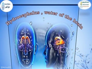
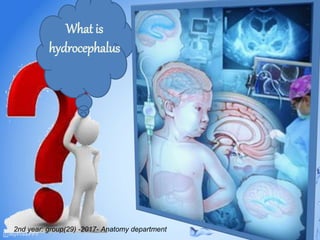
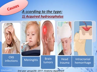
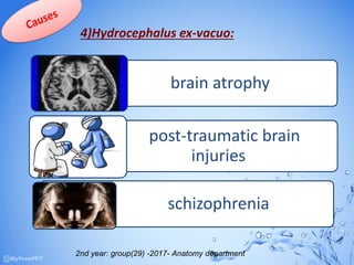
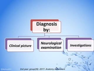
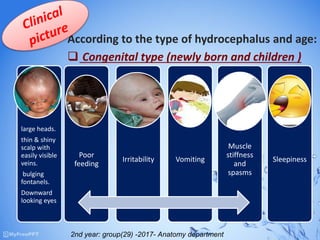
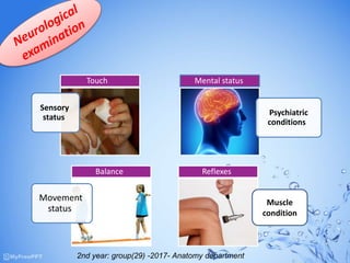
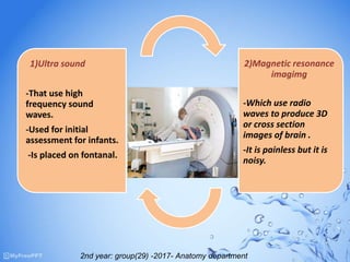
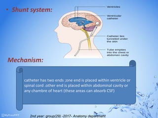
Presented by
Under supervision of
 Heba salah allam.
 Heba rabea abdelrahman.
 Heba-Allah mohammed
hasanen.
 Nermeen tarek shalaby.
 Nermeen nassar
abdelhady.
 Nada ahmed elsayed.
 Nada ashraf salman.
 Nada ehab elkholy.
 Nancy ayman sultan.
 Wafaa bahaa sherif.
2nd year: group(29) -2017- Anatomy department
m2nd year: group(29) -2017- Anatomy
department
1- Anatomy of ventricles of the brain.
2- Physiology of CSF circulation.
3- Definition of hydrocephalus.
4- Causes.
5- Investigation.
6- Clinical picture and manifestation.
7- Treatment.
2nd year: group(29) -2017- Anatomy department
Lateral ventricle
between 2 cerebral hemisphere
3rd ventricle
between the 2 thalami
4th ventricle
within the brain stem.
There are 4
ventricles within
the brain
2nd year: group(29) -2017- Anatomy department
 It is produced by the choroid
plexus which is located within
the ventricular system.
 Volume :- 150 ml
 pressure :- 60-150 ml of water
2nd year: group(29) -2017- Anatomy department
What is
hydrocephalus
2nd year: group(29) -2017- Anatomy department
 it’s a condition that occurs when fluid builds up in the skull and
causes the brain to swell.
.
 The first clinical description of an operative procedure for
hydrocephalus appears in Al-Tasrif (1000 AB )by the Arab surgeons ,
,who clearly described the evacuation of superficial intracranial fluid
in hydrocephalic children.They described it as being caused by
mechanical compression.
Definition
pathogenesis
History
they are usually unknown
but hydrocephalus may be a result of
a brain defect restricting the flow of CSF.
• Obstruvtive:
1)communicating (extraventricular).
2)non-communicating(intraventricular).
• Non-obstructive:
.Over production of CSF(rare).
• Normal pressure hydrocephalus:
Buildup of CSF puts pressure on the brain
(due to aging)
• Acquired
hydrocephal
us
• develops at
the time of
birth or at
some point
afterward.
• This type of
hydrocephalu
s can affect
individuals of
all ages
• Congenital
hydrocephalus
• it occurs in
babies born
prematurely
• before week 37
of the
pregnancy .
• They may also
have bleeding
in the brain
• which can
block the flow
of CSF.
• 4)Hydrocepha
lus ex-vacuo:
• It refers to an
enlargement
of cerebral
ventricles and
subarachnoid
spaces.
2nd year: group(29) -2017- Anatomy department
A ccording to the type:
1) Acquired hydrocephalus
.
CNS
infections Meningitis Brain
tumors
Head
trauma
Intracranial
hemorrhage
2nd year: group(29) -2017- Anatomy department
CONGENITAL type
- X linked hydrocephalus.
- Rare genital disorder.
- Arachnoid cysts.
Viscosity of CSF
NPH type
2nd year: group(29) -2017- Anatomy department
4)Hydrocephalus ex-vacuo:
brain atrophy
post-traumatic brain
injuries
schizophrenia
2nd year: group(29) -2017- Anatomy department
Diagnosis
by:
Clinical picture
Neurological
examination
Investigations
2nd year: group(29) -2017- Anatomy department
According to the type of hydrocephalus and age:
 Congenital type (newly born and children )
large heads.
thin & shiny
scalp with
easily visible
veins.
bulging
fontanels.
Downward
looking eyes
Poor
feeding
Irritability Vomiting
Muscle
stiffness
and
spasms
Sleepiness
2nd year: group(29) -2017- Anatomy department
Headach
neck
pain
inability
to
control
bladder
difficulty
walking blurred
vision
sleepiness
feeling
sick
Acquired type (children and adults )
2nd year: group(29) -2017- Anatomy department
NPH has 3 sets of distinctive symptoms
it affects:-
Urinary system
Mental ability
Mobility
Normal pressure hydrocephalus (NPH)
2nd year: group(29) -2017- Anatomy department
Sensory
status
Touch
Psychiatric
conditions
Mental status
Movement
status
Balance Reflexes
Muscle
condition
2nd year: group(29) -2017- Anatomy department
1)Ultra sound
-That use high
frequency sound
waves.
-Used for initial
assessment for infants.
-Is placed on fontanal.
2)Magnetic resonance
imagimg
-Which use radio
waves to produce 3D
or cross section
images of brain .
-It is painless but it is
noisy.
2nd year: group(29) -2017- Anatomy department
• Shunt system:
Mechanism:
catheter has two ends ;one end is placed within ventricle or
spinal cord .other end is placed within abdominal cavity or
any chambre of heart (these areas can absorb CSF)
2nd year: group(29) -2017- Anatomy department
• Types of shunt:
ventriculo-
peritoneal
shunting .
ventriculo-
atrial
shunting.
lumbo-
peritoneal
shunting.
2nd year: group(29) -2017- Anatomy department
“Hydrocephalus is an increase amount of CSF
inside the cranium, make it to swell, due to :
obstruction of pathway of CSF circulation or
Abnormal increase in CSF formation or impaired
absorption.”
Home message
2nd year: group(29) -2017- Anatomy department

Hydrocehalus

  • 2.
    Presented by Under supervisionof  Heba salah allam.  Heba rabea abdelrahman.  Heba-Allah mohammed hasanen.  Nermeen tarek shalaby.  Nermeen nassar abdelhady.  Nada ahmed elsayed.  Nada ashraf salman.  Nada ehab elkholy.  Nancy ayman sultan.  Wafaa bahaa sherif. 2nd year: group(29) -2017- Anatomy department
  • 3.
    m2nd year: group(29)-2017- Anatomy department 1- Anatomy of ventricles of the brain. 2- Physiology of CSF circulation. 3- Definition of hydrocephalus. 4- Causes. 5- Investigation. 6- Clinical picture and manifestation. 7- Treatment. 2nd year: group(29) -2017- Anatomy department
  • 4.
    Lateral ventricle between 2cerebral hemisphere 3rd ventricle between the 2 thalami 4th ventricle within the brain stem. There are 4 ventricles within the brain 2nd year: group(29) -2017- Anatomy department
  • 5.
     It isproduced by the choroid plexus which is located within the ventricular system.  Volume :- 150 ml  pressure :- 60-150 ml of water 2nd year: group(29) -2017- Anatomy department
  • 6.
    What is hydrocephalus 2nd year:group(29) -2017- Anatomy department
  • 7.
     it’s acondition that occurs when fluid builds up in the skull and causes the brain to swell. .  The first clinical description of an operative procedure for hydrocephalus appears in Al-Tasrif (1000 AB )by the Arab surgeons , ,who clearly described the evacuation of superficial intracranial fluid in hydrocephalic children.They described it as being caused by mechanical compression. Definition pathogenesis History they are usually unknown but hydrocephalus may be a result of a brain defect restricting the flow of CSF.
  • 8.
    • Obstruvtive: 1)communicating (extraventricular). 2)non-communicating(intraventricular). •Non-obstructive: .Over production of CSF(rare). • Normal pressure hydrocephalus: Buildup of CSF puts pressure on the brain (due to aging)
  • 9.
    • Acquired hydrocephal us • developsat the time of birth or at some point afterward. • This type of hydrocephalu s can affect individuals of all ages • Congenital hydrocephalus • it occurs in babies born prematurely • before week 37 of the pregnancy . • They may also have bleeding in the brain • which can block the flow of CSF. • 4)Hydrocepha lus ex-vacuo: • It refers to an enlargement of cerebral ventricles and subarachnoid spaces. 2nd year: group(29) -2017- Anatomy department
  • 10.
    A ccording tothe type: 1) Acquired hydrocephalus . CNS infections Meningitis Brain tumors Head trauma Intracranial hemorrhage 2nd year: group(29) -2017- Anatomy department
  • 11.
    CONGENITAL type - Xlinked hydrocephalus. - Rare genital disorder. - Arachnoid cysts. Viscosity of CSF NPH type 2nd year: group(29) -2017- Anatomy department
  • 12.
    4)Hydrocephalus ex-vacuo: brain atrophy post-traumaticbrain injuries schizophrenia 2nd year: group(29) -2017- Anatomy department
  • 13.
    2nd year: group(29)-2017- Anatomy department
  • 14.
  • 15.
    According to thetype of hydrocephalus and age:  Congenital type (newly born and children ) large heads. thin & shiny scalp with easily visible veins. bulging fontanels. Downward looking eyes Poor feeding Irritability Vomiting Muscle stiffness and spasms Sleepiness 2nd year: group(29) -2017- Anatomy department
  • 16.
  • 17.
    NPH has 3sets of distinctive symptoms it affects:- Urinary system Mental ability Mobility Normal pressure hydrocephalus (NPH) 2nd year: group(29) -2017- Anatomy department
  • 18.
  • 19.
    1)Ultra sound -That usehigh frequency sound waves. -Used for initial assessment for infants. -Is placed on fontanal. 2)Magnetic resonance imagimg -Which use radio waves to produce 3D or cross section images of brain . -It is painless but it is noisy. 2nd year: group(29) -2017- Anatomy department
  • 20.
    2nd year: group(29)-2017- Anatomy department
  • 21.
    • Shunt system: Mechanism: catheterhas two ends ;one end is placed within ventricle or spinal cord .other end is placed within abdominal cavity or any chambre of heart (these areas can absorb CSF) 2nd year: group(29) -2017- Anatomy department
  • 22.
    • Types ofshunt: ventriculo- peritoneal shunting . ventriculo- atrial shunting. lumbo- peritoneal shunting. 2nd year: group(29) -2017- Anatomy department
  • 23.
    “Hydrocephalus is anincrease amount of CSF inside the cranium, make it to swell, due to : obstruction of pathway of CSF circulation or Abnormal increase in CSF formation or impaired absorption.” Home message 2nd year: group(29) -2017- Anatomy department
  • 24.
    2nd year: group(29)-2017- Anatomy department