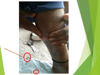
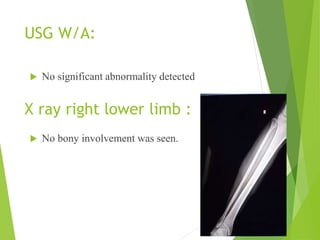
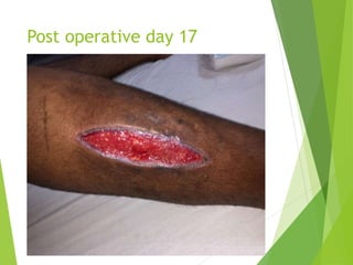
INTERESTING CASE OF LEG
SWELLING WITH
DISCHARGING MULTIPLE
SMALL CYSTS
DR SHUBH NARAYAN
GHANGHORIYA
UNDER GUIDANCE OF:
DR M. KELA
PROFESSOR AND HEAD OF
DEPARTMENT
DEPARTMENT OF GENERAL
SURGERY,SRI AUROBINDO
MEDICAL COLLEGE AND PG
HOSPITAL
PATIENT DETAILS
 NAME : XXXX
 AGE : 29 YEARS OLD
 GENDER : MALE
 RESIDENT OF : MAHARAHTRA , AKOLA
 MARITAL STATUS : MARRIED
PRESENTATION :
29 y/o male patient came to
surgery OPD on 30/5/22
with complaints of pain and
swelling on the lateral
aspect of the right lower leg
since 1 month with a
discharging sinus since 8
days.
History of presenting illness:
 The swelling was gradually increasing in size
and extending from knee to right foot toe with
prominent point over lateral aspect of right leg
at distal 2/3 of leg.
 Patient went to local clinic at akola.
 Incision and drainage was done at an outside
hospital and e/o multiple cyst drained from the
incision site.
 Histopathology report not present with patient.
 Since then patient developed c/o continuous
drainage of cyst
Past history
 No past medical history.
 h/o and incision and drainage was done 8 days
back with e/o multiple cysts drained
 histopathological examination was sent but no
reports were present with the patient
Examination:
 Mild tenderness over calf region .
 Signs of inflammation were present.
 Tendernesss , redness , slight rise in
temperature.
 Cyst discharging sinus present over the right
leg lateral aspect
 Sinus opening of size : 2x2 cms on the lateral
aspect of right leg about 13 cms above the
lateral malleolus.
INVESTIGATIONS:
 CBC : 13.0/5.94/45.3/4500/1.36 LAC
 BLOOD GROUP : B POSITIVE
 RBS : 84
 UREA : 17
 CREATININE : 0.76
 S. ALP : 105
 SGOT : 64
 SGPT : 69
 PT/INR : 15/1.0
 S.ELECTROLYTES : 146/3.51/113
 SEROLOGY : NON REACTIVE
USG W/A:
 No bony involvement was seen.
X ray right lower limb :
 No significant abnormality detected
USG LOCAL REGION :
Evidence of an ill-defined heterogeneously hypoechoic
area of size 7.1x5.2x4.5 noted in the muscular
compartment of right lower leg region. This area contains
multiple well-defined anechoic round cystic lesions along
with multiple air foci noted within it, there is no obvious
internal vascularity noted inside these cystic lesion on
Doppler study.
A fistulous tract coursing through the skin & subcutaneous
tissues is seen communicating with the collection
.
Imaging findings
Ultrasound images
multiple well-
defined anechoic
round cystic lesions
no obvious internal
vascularity noted
Evidence of a loculated thick walled pocket of collection
noted in mid leg region in muscular compartment
(involving tibialis posterior, flexor hallucis longus) with
associated surrounding edema / myositis.
It measures 3.7 x 2.3 x7.7 cm in TR, AP, CC axis
No bony involvement seen.
MRI Brain and whole body STIR study do not reveal any
focal lesion or hydatid cyst.
MRI
Treatment :
Pre anesthetic check up was
done.
Surgical m/m : incision with
drainage of multiple cysts with
pericystectomy was chosen as
the preferred surgical treatment
done on 1/6/22.
Medical m/m : Treatment
involved 400 mg of albendazole
per day for 15 days before and
Surgical procedure :
Patient lying in supine position.
Incision made on below and above
the sinus tract and on lateral aspect
incision deepened layer by layer and
incision also made on medial aspect
and deepened.
Intraop findings :
Cavity of cyst , draining the cysts excised
and debridement of surrounding tissue
done, hemostasis achieved and wash given
with 3 % NS( hypertonic saline)
Evidence of :
Cavity present below the gastrocnemius
muscle which is draining cyst.
HISTOPATH FINDINGS:
MACROSCOPIC FINDINGS:
Multiple daughter cysts measuring 0.4
cm to 0.9 cm in diameter. Externally
glistening white, smooth cut section –
smooth and glistening white.
MICROSCOPIC :
The section shows lamellate acellular
eosinophilic cyst wall with inner
germinal layer and occasional brood
capsule.
IMPRESSION:
HYDATID CYST – RIGHT LEG
• The postoperative course of the patient was
favourable. However, 2 weeks later, the patient
complained of a slight pain on the back of the
right leg.
Despite the absence of hypereosinophilia,
pulmonary and cutaneous signs, a possible
relapse was discussed. Albendazole was
continued (400 mg twice per day) and
radiological controls were performed.
UE revealed a heterogeneous aspect of the
muscular tissue and local region use imaging
confirmed a relapse, ill defined residual
collection with internal septations of size 7.7 x
1.5 x 3 cm ~ 20 cc noted in intra muscular
compartment of right leg (within tibial posterior
muscles) with surrounding thickened edematous
• The collection was drained out by the
previous incision site by regular dressing and
was managed conservatively in ward.
Post operative day 17
DISCUSSION
Primary muscular HC is a rare
localization.
However, several cases of
isolated cyst have been reported
in upper and lower proximal
muscles.
To our knowledge, no reports of
multiple HC in the muscle have
differential diagnoses
lipoma,
calcified hematoma,
cold abscess or
soft tissue tumour.
Musculoskeletal echinococcosis should
always be suspected in patients originating
from endemic regions
CONCLUSION
 The signs and symptoms of the hydatid disease
vary according to the involved organ, the site, the
effects on the adjacent tissues, complications
after rupture, secondary infection, and
immunological reactions
 The first and most common symptom in our
patient was a slow growing painless mass, and
this symptom was compatible with the
literature.
 The helpful diagnostic and classification ways of
cysts are ultrasonography, ct, and mri
 Intramuscular hydatidosis have been
classified according to the gharbi classification
REFRENCES:
1. Safioleas M, Nikiteas N, Stamatakos M,
Safioleas C, Manti CH, Revenas C,et al.
Echinococcal cyst of the subcutaneous tissue: a
rare case report.Parasitol Int 2008; 57: 236-8.
[CrossRef]
2. Duzgun N, Esme H, Duran F. M, Calik M, Cetin,
B. Hydatid Disease of the Spine: Case Report
Turkiye Klinikleri Arch Lung 2014; 15: 79-82
3. Ormeci N, Idilman R, Akyar S, Palabiyikoğlu M,
Coban S, Erdem H, et al. Hydatid cysts in muscle:
a modified percutaneous treatment approach.Int
J Infect Dis 2007; 11: 204-8. [CrossRef]
REFRENCES:
4. Jerbi Omezzine S, Abid F, Mnif H, Hafsa C,
Thabet I, Abderrazek A, et al. Primary hydatid
disease of the thigh. A rare location. Orthop
Traumatol Surg Res 2010; 96: 90-3. [CrossRef]
5. Parray FQ, Ahmad SZ, Sherwani AY, Chowdri
NA, Wani KA. Primary paraspinal hydatid cyst:
a rare presentation of Echinococcosis. Int J
Surg 2010; 8: 404-6. [CrossRef]
6. Sogut O, Ozgonul A, Bitiren M, Kose R, Cece
H. Primary hydatid cyst in the deltoid muscle:
an unusual localization. J Infect Dis 2010; 14
Suppl3:e347-8. [CrossRef]
`
THANK YOU

hydatid revision.pptx

  • 1.
    INTERESTING CASE OFLEG SWELLING WITH DISCHARGING MULTIPLE SMALL CYSTS DR SHUBH NARAYAN GHANGHORIYA UNDER GUIDANCE OF: DR M. KELA PROFESSOR AND HEAD OF DEPARTMENT DEPARTMENT OF GENERAL SURGERY,SRI AUROBINDO MEDICAL COLLEGE AND PG HOSPITAL
  • 2.
    PATIENT DETAILS  NAME: XXXX  AGE : 29 YEARS OLD  GENDER : MALE  RESIDENT OF : MAHARAHTRA , AKOLA  MARITAL STATUS : MARRIED
  • 3.
    PRESENTATION : 29 y/omale patient came to surgery OPD on 30/5/22 with complaints of pain and swelling on the lateral aspect of the right lower leg since 1 month with a discharging sinus since 8 days.
  • 5.
    History of presentingillness:  The swelling was gradually increasing in size and extending from knee to right foot toe with prominent point over lateral aspect of right leg at distal 2/3 of leg.  Patient went to local clinic at akola.  Incision and drainage was done at an outside hospital and e/o multiple cyst drained from the incision site.  Histopathology report not present with patient.  Since then patient developed c/o continuous drainage of cyst
  • 6.
    Past history  Nopast medical history.  h/o and incision and drainage was done 8 days back with e/o multiple cysts drained  histopathological examination was sent but no reports were present with the patient
  • 7.
    Examination:  Mild tendernessover calf region .  Signs of inflammation were present.  Tendernesss , redness , slight rise in temperature.  Cyst discharging sinus present over the right leg lateral aspect  Sinus opening of size : 2x2 cms on the lateral aspect of right leg about 13 cms above the lateral malleolus.
  • 8.
    INVESTIGATIONS:  CBC :13.0/5.94/45.3/4500/1.36 LAC  BLOOD GROUP : B POSITIVE  RBS : 84  UREA : 17  CREATININE : 0.76  S. ALP : 105  SGOT : 64  SGPT : 69  PT/INR : 15/1.0  S.ELECTROLYTES : 146/3.51/113  SEROLOGY : NON REACTIVE
  • 9.
    USG W/A:  Nobony involvement was seen. X ray right lower limb :  No significant abnormality detected
  • 10.
    USG LOCAL REGION: Evidence of an ill-defined heterogeneously hypoechoic area of size 7.1x5.2x4.5 noted in the muscular compartment of right lower leg region. This area contains multiple well-defined anechoic round cystic lesions along with multiple air foci noted within it, there is no obvious internal vascularity noted inside these cystic lesion on Doppler study. A fistulous tract coursing through the skin & subcutaneous tissues is seen communicating with the collection . Imaging findings
  • 11.
    Ultrasound images multiple well- definedanechoic round cystic lesions
  • 12.
  • 13.
    Evidence of aloculated thick walled pocket of collection noted in mid leg region in muscular compartment (involving tibialis posterior, flexor hallucis longus) with associated surrounding edema / myositis. It measures 3.7 x 2.3 x7.7 cm in TR, AP, CC axis No bony involvement seen. MRI Brain and whole body STIR study do not reveal any focal lesion or hydatid cyst. MRI
  • 15.
    Treatment : Pre anestheticcheck up was done. Surgical m/m : incision with drainage of multiple cysts with pericystectomy was chosen as the preferred surgical treatment done on 1/6/22. Medical m/m : Treatment involved 400 mg of albendazole per day for 15 days before and
  • 16.
    Surgical procedure : Patientlying in supine position. Incision made on below and above the sinus tract and on lateral aspect incision deepened layer by layer and incision also made on medial aspect and deepened.
  • 17.
    Intraop findings : Cavityof cyst , draining the cysts excised and debridement of surrounding tissue done, hemostasis achieved and wash given with 3 % NS( hypertonic saline)
  • 18.
    Evidence of : Cavitypresent below the gastrocnemius muscle which is draining cyst.
  • 19.
    HISTOPATH FINDINGS: MACROSCOPIC FINDINGS: Multipledaughter cysts measuring 0.4 cm to 0.9 cm in diameter. Externally glistening white, smooth cut section – smooth and glistening white. MICROSCOPIC : The section shows lamellate acellular eosinophilic cyst wall with inner germinal layer and occasional brood capsule. IMPRESSION: HYDATID CYST – RIGHT LEG
  • 20.
    • The postoperativecourse of the patient was favourable. However, 2 weeks later, the patient complained of a slight pain on the back of the right leg. Despite the absence of hypereosinophilia, pulmonary and cutaneous signs, a possible relapse was discussed. Albendazole was continued (400 mg twice per day) and radiological controls were performed. UE revealed a heterogeneous aspect of the muscular tissue and local region use imaging confirmed a relapse, ill defined residual collection with internal septations of size 7.7 x 1.5 x 3 cm ~ 20 cc noted in intra muscular compartment of right leg (within tibial posterior muscles) with surrounding thickened edematous
  • 21.
    • The collectionwas drained out by the previous incision site by regular dressing and was managed conservatively in ward.
  • 22.
  • 23.
    DISCUSSION Primary muscular HCis a rare localization. However, several cases of isolated cyst have been reported in upper and lower proximal muscles. To our knowledge, no reports of multiple HC in the muscle have
  • 24.
    differential diagnoses lipoma, calcified hematoma, coldabscess or soft tissue tumour. Musculoskeletal echinococcosis should always be suspected in patients originating from endemic regions
  • 25.
    CONCLUSION  The signsand symptoms of the hydatid disease vary according to the involved organ, the site, the effects on the adjacent tissues, complications after rupture, secondary infection, and immunological reactions  The first and most common symptom in our patient was a slow growing painless mass, and this symptom was compatible with the literature.  The helpful diagnostic and classification ways of cysts are ultrasonography, ct, and mri  Intramuscular hydatidosis have been classified according to the gharbi classification
  • 27.
    REFRENCES: 1. Safioleas M,Nikiteas N, Stamatakos M, Safioleas C, Manti CH, Revenas C,et al. Echinococcal cyst of the subcutaneous tissue: a rare case report.Parasitol Int 2008; 57: 236-8. [CrossRef] 2. Duzgun N, Esme H, Duran F. M, Calik M, Cetin, B. Hydatid Disease of the Spine: Case Report Turkiye Klinikleri Arch Lung 2014; 15: 79-82 3. Ormeci N, Idilman R, Akyar S, Palabiyikoğlu M, Coban S, Erdem H, et al. Hydatid cysts in muscle: a modified percutaneous treatment approach.Int J Infect Dis 2007; 11: 204-8. [CrossRef]
  • 28.
    REFRENCES: 4. Jerbi OmezzineS, Abid F, Mnif H, Hafsa C, Thabet I, Abderrazek A, et al. Primary hydatid disease of the thigh. A rare location. Orthop Traumatol Surg Res 2010; 96: 90-3. [CrossRef] 5. Parray FQ, Ahmad SZ, Sherwani AY, Chowdri NA, Wani KA. Primary paraspinal hydatid cyst: a rare presentation of Echinococcosis. Int J Surg 2010; 8: 404-6. [CrossRef] 6. Sogut O, Ozgonul A, Bitiren M, Kose R, Cece H. Primary hydatid cyst in the deltoid muscle: an unusual localization. J Infect Dis 2010; 14 Suppl3:e347-8. [CrossRef] `
  • 31.