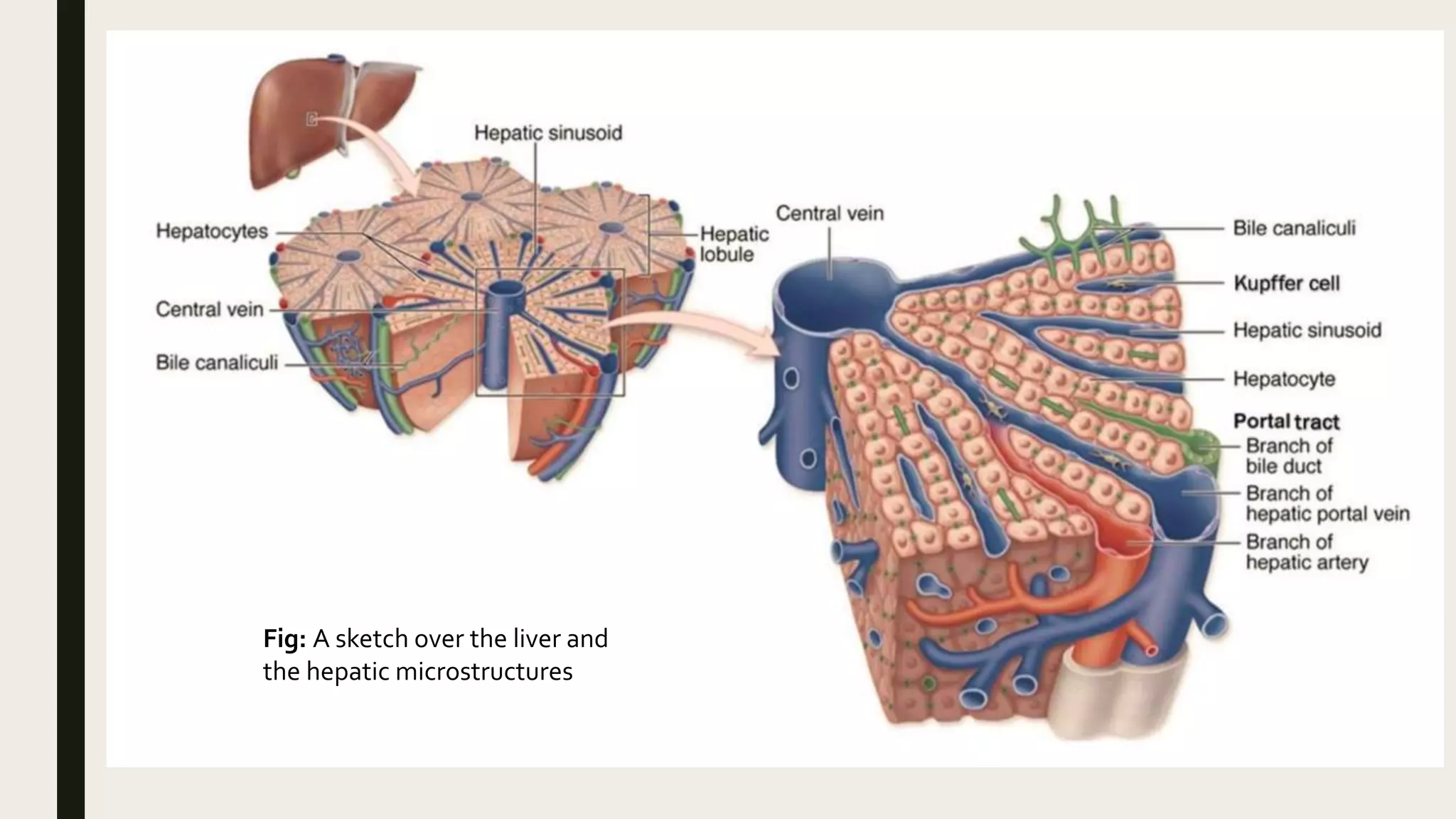
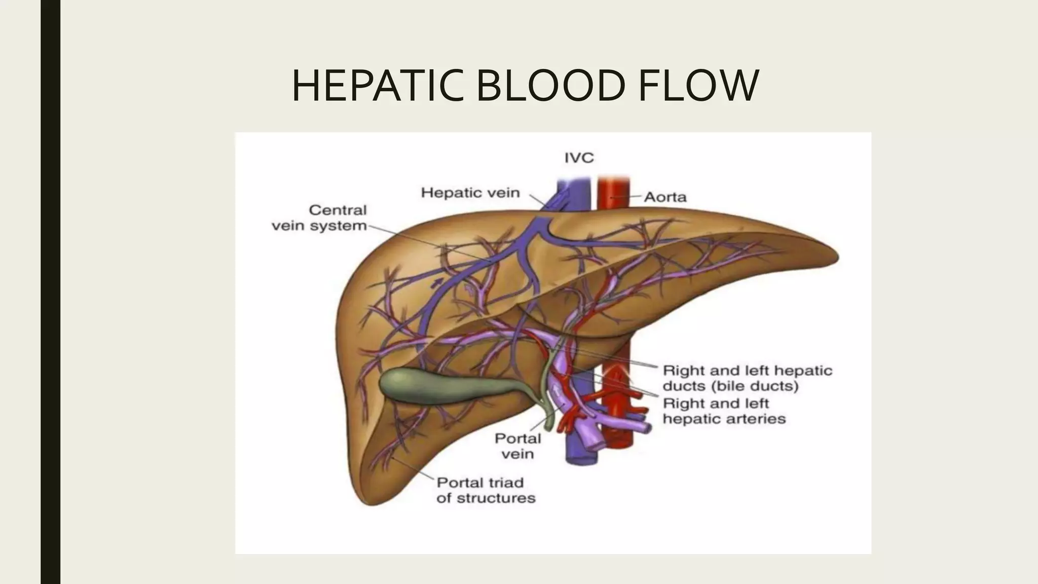
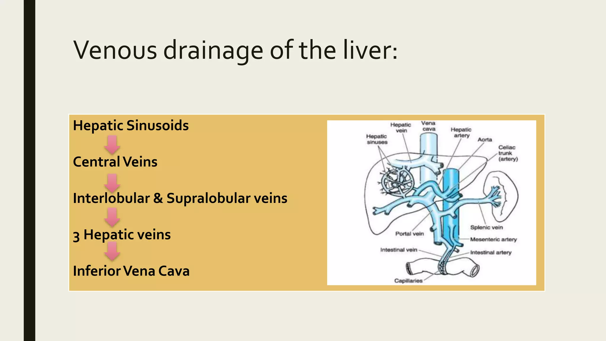
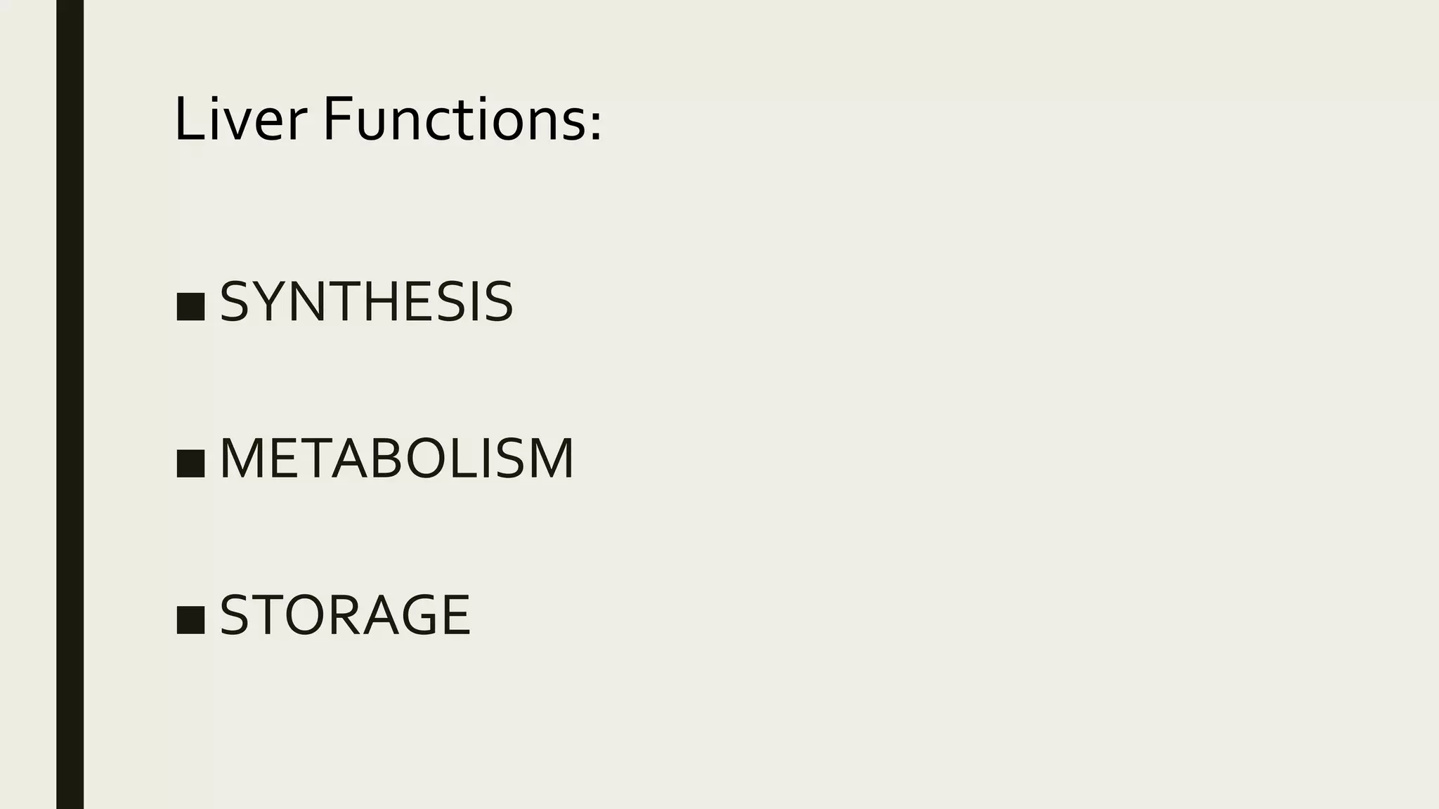
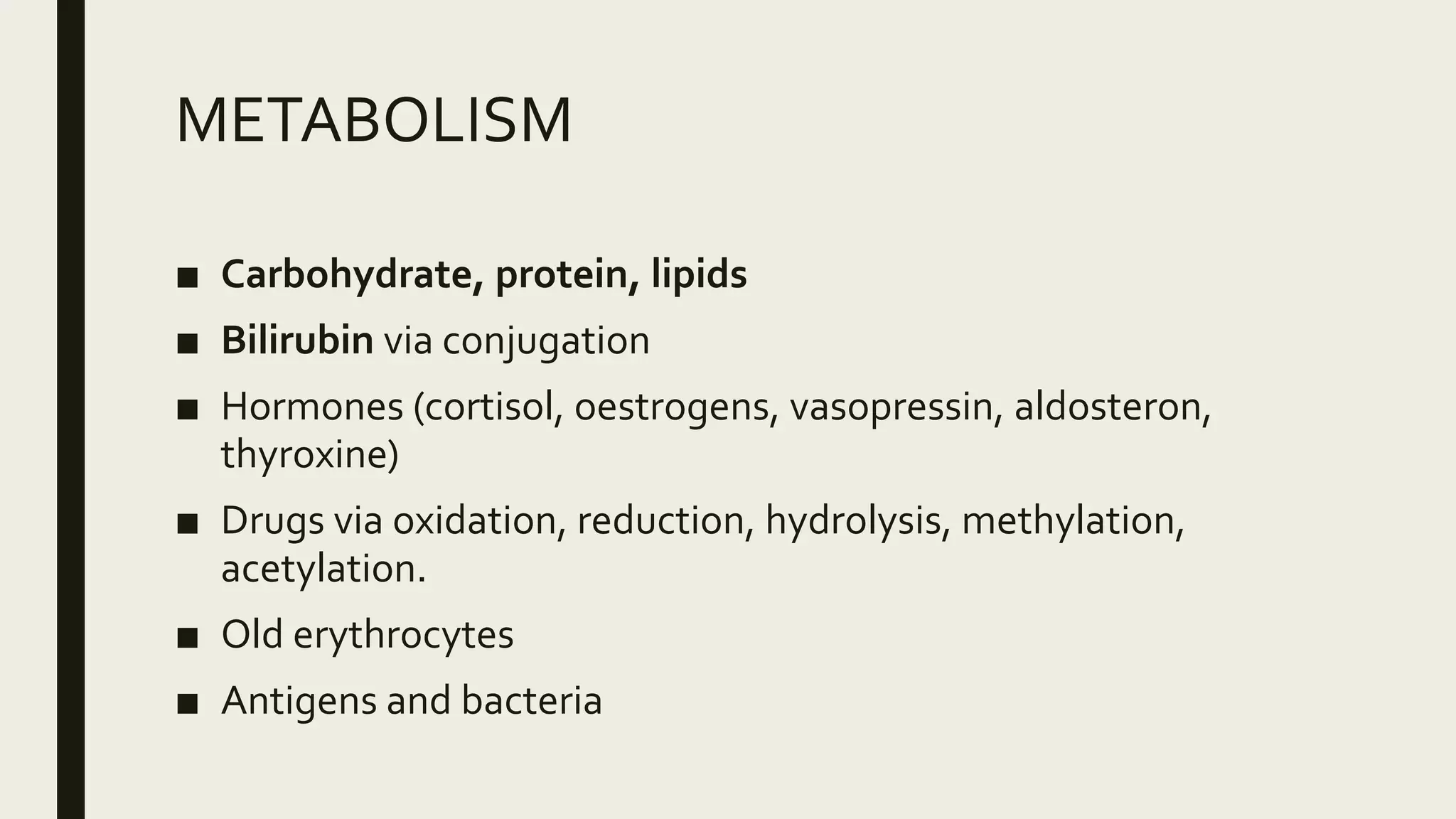
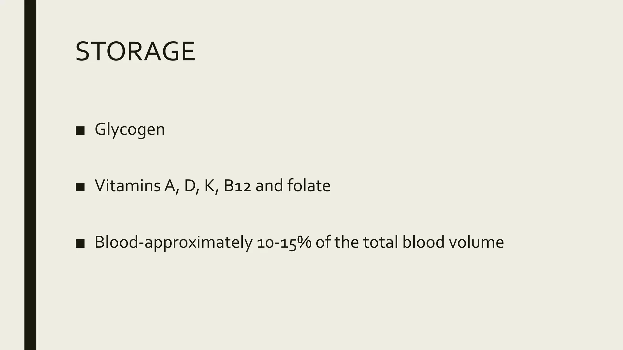
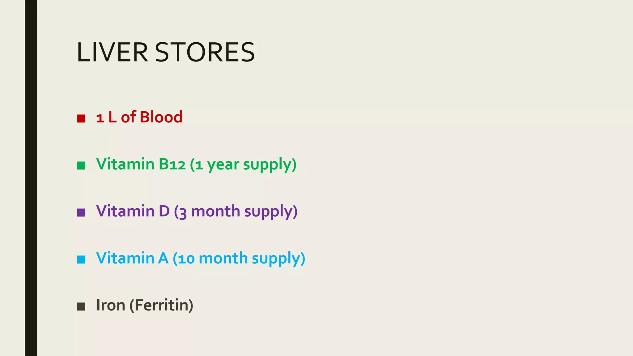
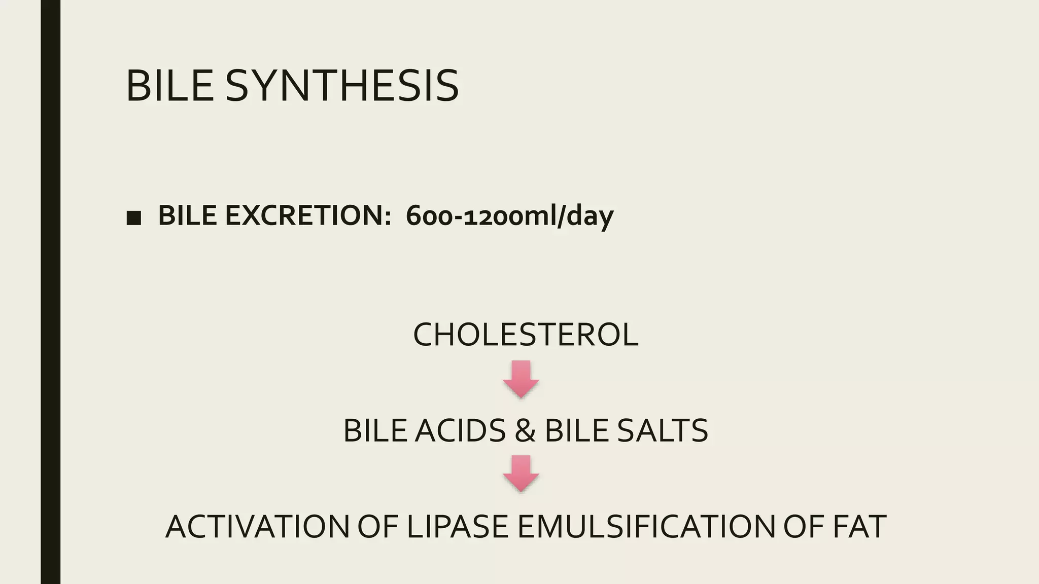
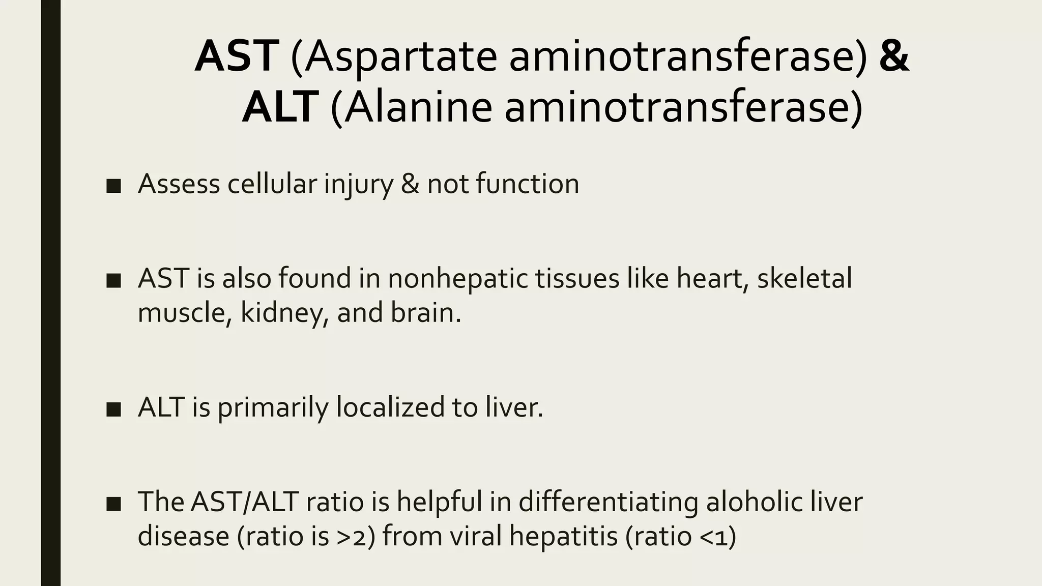
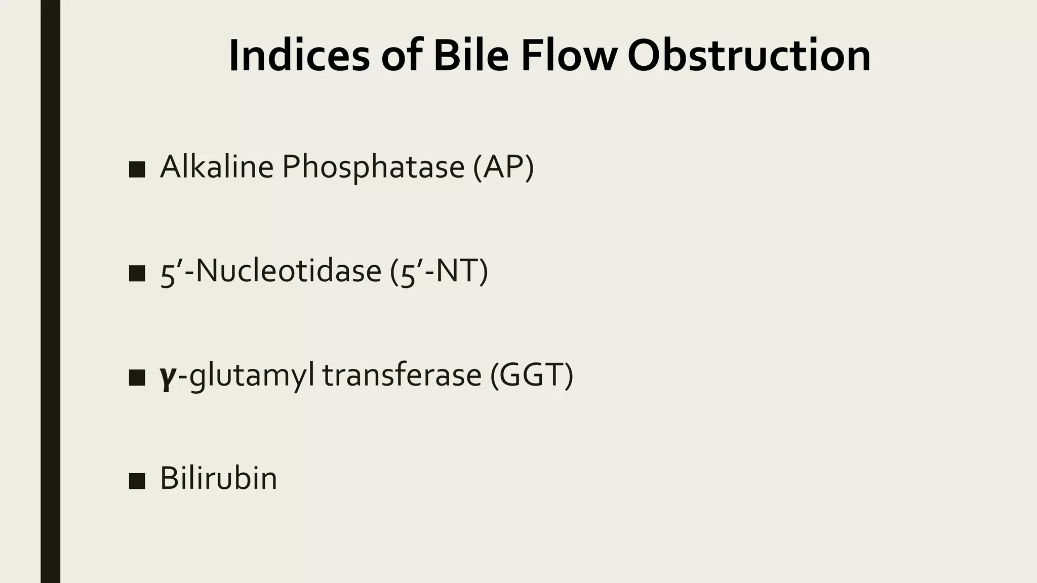
HEPATIC
PHYSIOLOGY AND
EFFECT OF
ANAESTHETIC
AGENTS
HEPATIC
PHYSIOLOGY
Presenter:
Dr.Waquas Ahmed
Junior Resident-1
Department of Anaesthesiology
& Critical Care Medicine
Moderator:
Dr. Bibha
Assistant Professor
Department of Anaesthesiology &
Critical Care Medicine
INTRODUCTION
■ Liver is the largest internal organ.
■ Weighs 1.5 kg or about 2% of the total body weight in an adult.
■ Hepatic blood flow is 1.5L/min (25% of the Cardiac Output)
■ Right lobe: Left lobe = 6:1
■ In the right mid axillary line, the liver spans from the 7th to the 11th ribs.
■ It is covered by the peritoneum (Glisson’s capsule), except for the gall bladder
bed, the inferior venacava, the bare area, and the porta hepatis.
■ Nerve plexus fibres: Sympathetic gangliaT7-T10, Right & LeftVagi & Phrenic
nerve
■ Lypmhatic vessels: glands around porta hepatis, coeliac glands, diaphragm,
mediastinal, thoracicVC
FUNCTIONAL UNIT OF LIVER: LOBULE
■ Described by Kiernan in 1833 as functional and structural unit of liver
■ Roughly 1 X 2 mm
■ Hexagonal structure
■ Human liver contains 50,000 to 100,000 lobules
■ Plates of hepatocytes located in a radial distribution about a central vein
■ Afferent blood supply from portal vein and hepatic arterioles enters at the
periphery of the lobule.
■ Bile formed in the hepatocytes flows into canaliculi located between the
plates of hepatocytes.
Fig: A sketch over the liver and
the hepatic microstructures
Hepatic Lobule
■ Central:
Tributary of hepatic vein
■ Periphery:
A triad of branches of the
 hepatic artery
 the portal vein
 the bile duct
The concept of the Liver Acinus
■ Functional microvascular unit of the liver.
■ This concept was first defined by Rappaport et al. in 1954
■ Important in describing some structural-functional relationships of the
hepatic lobule
■ There exists a gradient of oxygen and nutrition concentration along the
sinusoids
Fig: The concept of the liver
acinus
■ The activity of each hepatocyte is determined by its position
along this oxygen-nutrient gradient, which gives rise to three
zones; zone 1, zone 2 and zone 3.
■ Zone 1 is the most oxygen- and nutrient-rich zone which has a
metabolic activity different from the one in zone 3.
■ Zone 3 is exposed to the lowest concentrations of oxygen and
nutrition
TYPES OF LIVER CELLS
SINUSOIDAL ENDOTHELIAL CELLS:
 These are different form the endothelium in the rest of the body
 Large pores in between the cells are called fenestrae
 The fenestrae allow passage of relatively large particles out of the blood
like albumin, lipds and lipoproteins
 They lack the basement membrane thus have increased endothelium
permeability
TYPES OF LIVER CELLS
HEPATIC EPITHELIAL CELLS:
 Liver parenchymal cells referred to as Hepatocyte commonly.
 Highly polarized epithelial cells and serve as metabolic factories of the
liver.
 Heterogenous plasma membranes with three distinct specialized
domains.
 Ability to regenerate.
HEPATIC BLOOD FLOW
REGULATION OF HEPATIC BLOOD
FLOW
INTRINSIC MECHANISM
 HEPATIC ARTERIAL
BUFFER RESPONSE
(HABR)
 METABOLIC CONTROL
 PRESSURE-FLOW
AURTOREGULATION
EXTRINSIC MECHANISM
 NEURAL CONTROL
 HUMORAL CONTROL
INTRINSIC
MECHANISM
HEPATIC ARTERIAL BUFFER RESPONSE
■ Most important intrinsic buffer response.
■ Changes in portal venous flow cause reciprocal changes in hepatic
arterial flow.
■ When portal venous flow decreases, the HABR compensates by
increasing hepatic arterial flow and vice versa.
■ Mechanism involves the synthesis and washout of adenosine (i.e., a
vasodilator) from periportal regions.
■ HABR can nearly double the hepatic flow.
METABOLIC CONTROL
■ Decrease oxygen tension or the pH of portal venous blood increase
hepatic arterial flow.
■ Postprandial hyperosmolarity increases both hepatic and portal venous
flow.
PRESSURE-FLOW AUTOREGULATION
■ Enables tissue-specific regulators to govern organ blood flow despite
fluctuations in systemic arterial pressure.
■ Mechanism involves myogenic responses of vascular smooth muscle to
stretch.
EXTRINSIC MECHANISM
NEURAL CONTROL
■ Fibres of the vagus, phrenic and splanchnic nerves (post ganglionic
sympathetic fibers fromT5 throughT11) enter the liver at the hilum.
■ When sympathetic tone decreases, splanchnic reservoir volume
increases.
■ Sympathetic nerve stimulation may reduce hepatic blood volume by up
to 50 per cent.
HUMORAL CONTROL
■ Glucagon induces dose-dependent relaxation of hepatic arterial smooth
muscles.
■ Hence blocks the effects of physiologic vasoconstrictors on the hepatic
artery.
■ Angiotensin-II constricts the hepatic arterial and portal venous beds.
■ High blood flow of liver is due to low vascular resistance in
portal vein.
■ Average PortalVein Pressure (PVP) is 8 to 10 mm of Hg.
■ PVP exceeds hepatic venous pressure by 4 to 5 mm of Hg.
■ Hepatic venous pressure gradient > 5mm of Hg is
abnormal and called Portal Hypertension
Venous drainage of the liver:
Hepatic Sinusoids
CentralVeins
Interlobular & Supralobular veins
3 Hepatic veins
InferiorVena Cava
LYMPHATICS
■ The liver has a high blood flow and a highly permeable microcirculation.
The consequent production of interstitial fluid, intrahepatic lymph, is
formed in the perisinusoidal space of Disse between the hepatocytes
and sinusoidal lining endothelium.
■ Lymphatic vessels drain via the portal tracts, closely applied to the
hepatic arterial branches, to the hilum and thence to the thoracic duct.
■ Some interstitial fluid drains through Glisson's capsule into the
peritoneum.
■ The lymph flow rate in liver is approximately 0.5 ml/kg of liver per
minute making up 25 to 50 per cent of thoracic duct lymph flow.
Lymphatic drainage:
SUPERFICIAL DEEP
CAVAL
HEPATIC
COELIAC
PARACARDIAL
NODES AROUND
IVC & HEPATIC NODES
Liver Functions:
■ SYNTHESIS
■ METABOLISM
■ STORAGE
SYNTHESIS
■ Carrier/modulator proteins such as albumin, complement,
coagulation factors, transferrin, ceruloplasmin, thyroid-binding
globulin, haptoglobin, globulins & alpha-1-antitripsin
■ Enzymes, i.e pseudocholinesteraze
■ Bile acids
■ Lymph (50%)
■ Erythropoesis, mainly in the foetus
METABOLISM
■ Carbohydrate, protein, lipids
■ Bilirubin via conjugation
■ Hormones (cortisol, oestrogens, vasopressin, aldosteron,
thyroxine)
■ Drugs via oxidation, reduction, hydrolysis, methylation,
acetylation.
■ Old erythrocytes
■ Antigens and bacteria
STORAGE
■ Glycogen
■ Vitamins A, D, K, B12 and folate
■ Blood-approximately 10-15% of the total blood volume
LIVER STORES
■ 1 L of Blood
■ Vitamin B12 (1 year supply)
■ Vitamin D (3 month supply)
■ Vitamin A (10 month supply)
■ Iron (Ferritin)
METABOLIC FUNCTIONS
■ Carbohydrate metabolism-glucose buffer function
■ Protein metabolism
– deamination
– urea cycle
– interconversions between nonessential amino acids
■ Lipid metabolism
– β oxidation of FA→AcetylCoA→ketone bodies
– FFA esterified to formTAG.
– lipoprotein synthesis
– phospholipids & cholesterol synthesis
Glycogenesis Gloconeogenesis
EUGLYCEMIA
Glycogenolysis SURGICAL STRESS
SYNTHETIC FUNCTIONS:
■ PLASMA PROTEINS
 Albumin (12-15g/day)
 Globulins
■ CLOTTING FACTORS- all coagulation factors except factor III (tissue thromboplastin),
IV (Calcium) &VIII (vonWillebrand factor)
■ ANTITHROMBIN III, PROTEINC & S, Plasminogen activator inhibitor (PAI)
■ ENZYMES-ALP,AST (SGOT),ALT (SGPT)
■ PSEUDOCHOLINESTERASE
■ CHOLESTEROL, LIPOPROTEINS ,PHOSPHOLIPIDS
■ ACUTE PHASE REACTANTS
BILE SYNTHESIS
■ BILE EXCRETION: 600-1200ml/day
CHOLESTEROL
BILE ACIDS & BILE SALTS
ACTIVATION OF LIPASE EMULSIFICATIONOF FAT
ASSESSMENT OF HEPATIC FUNCTION
AST (Aspartate aminotransferase) &
ALT (Alanine aminotransferase)
■ Assess cellular injury & not function
■ AST is also found in nonhepatic tissues like heart, skeletal
muscle, kidney, and brain.
■ ALT is primarily localized to liver.
■ The AST/ALT ratio is helpful in differentiating aloholic liver
disease (ratio is >2) from viral hepatitis (ratio <1)
Indices of Bile Flow Obstruction
■ Alkaline Phosphatase (AP)
■ 5’-Nucleotidase (5’-NT)
■ 𝝲-glutamyl transferase (GGT)
■ Bilirubin
Tests of hepatic synthetic functioning
Serum Proteins:
Albumin, Globulin
Serum albumin is not a reliable marker
of acute liver disease as its half-life is 3
weeks.
Coagulation testing:
PT & INR
■ Sensitive indicators of hepatic
disease because of the short half-life
of factorVII.
■ The PT depends on sufficient intake
ofVitamin K, which in turn depends
on adequate biliary secretion of bile
salts.
Hepatic physiology

Hepatic physiology

  • 1.
  • 2.
    HEPATIC PHYSIOLOGY Presenter: Dr.Waquas Ahmed Junior Resident-1 Departmentof Anaesthesiology & Critical Care Medicine Moderator: Dr. Bibha Assistant Professor Department of Anaesthesiology & Critical Care Medicine
  • 3.
    INTRODUCTION ■ Liver isthe largest internal organ. ■ Weighs 1.5 kg or about 2% of the total body weight in an adult. ■ Hepatic blood flow is 1.5L/min (25% of the Cardiac Output) ■ Right lobe: Left lobe = 6:1 ■ In the right mid axillary line, the liver spans from the 7th to the 11th ribs. ■ It is covered by the peritoneum (Glisson’s capsule), except for the gall bladder bed, the inferior venacava, the bare area, and the porta hepatis. ■ Nerve plexus fibres: Sympathetic gangliaT7-T10, Right & LeftVagi & Phrenic nerve ■ Lypmhatic vessels: glands around porta hepatis, coeliac glands, diaphragm, mediastinal, thoracicVC
  • 5.
    FUNCTIONAL UNIT OFLIVER: LOBULE ■ Described by Kiernan in 1833 as functional and structural unit of liver ■ Roughly 1 X 2 mm ■ Hexagonal structure ■ Human liver contains 50,000 to 100,000 lobules ■ Plates of hepatocytes located in a radial distribution about a central vein ■ Afferent blood supply from portal vein and hepatic arterioles enters at the periphery of the lobule. ■ Bile formed in the hepatocytes flows into canaliculi located between the plates of hepatocytes.
  • 6.
    Fig: A sketchover the liver and the hepatic microstructures
  • 7.
    Hepatic Lobule ■ Central: Tributaryof hepatic vein ■ Periphery: A triad of branches of the  hepatic artery  the portal vein  the bile duct
  • 8.
    The concept ofthe Liver Acinus ■ Functional microvascular unit of the liver. ■ This concept was first defined by Rappaport et al. in 1954 ■ Important in describing some structural-functional relationships of the hepatic lobule ■ There exists a gradient of oxygen and nutrition concentration along the sinusoids
  • 10.
    Fig: The conceptof the liver acinus
  • 11.
    ■ The activityof each hepatocyte is determined by its position along this oxygen-nutrient gradient, which gives rise to three zones; zone 1, zone 2 and zone 3. ■ Zone 1 is the most oxygen- and nutrient-rich zone which has a metabolic activity different from the one in zone 3. ■ Zone 3 is exposed to the lowest concentrations of oxygen and nutrition
  • 12.
    TYPES OF LIVERCELLS SINUSOIDAL ENDOTHELIAL CELLS:  These are different form the endothelium in the rest of the body  Large pores in between the cells are called fenestrae  The fenestrae allow passage of relatively large particles out of the blood like albumin, lipds and lipoproteins  They lack the basement membrane thus have increased endothelium permeability
  • 13.
    TYPES OF LIVERCELLS HEPATIC EPITHELIAL CELLS:  Liver parenchymal cells referred to as Hepatocyte commonly.  Highly polarized epithelial cells and serve as metabolic factories of the liver.  Heterogenous plasma membranes with three distinct specialized domains.  Ability to regenerate.
  • 16.
  • 17.
    REGULATION OF HEPATICBLOOD FLOW INTRINSIC MECHANISM  HEPATIC ARTERIAL BUFFER RESPONSE (HABR)  METABOLIC CONTROL  PRESSURE-FLOW AURTOREGULATION EXTRINSIC MECHANISM  NEURAL CONTROL  HUMORAL CONTROL
  • 18.
  • 19.
    HEPATIC ARTERIAL BUFFERRESPONSE ■ Most important intrinsic buffer response. ■ Changes in portal venous flow cause reciprocal changes in hepatic arterial flow. ■ When portal venous flow decreases, the HABR compensates by increasing hepatic arterial flow and vice versa. ■ Mechanism involves the synthesis and washout of adenosine (i.e., a vasodilator) from periportal regions. ■ HABR can nearly double the hepatic flow.
  • 20.
    METABOLIC CONTROL ■ Decreaseoxygen tension or the pH of portal venous blood increase hepatic arterial flow. ■ Postprandial hyperosmolarity increases both hepatic and portal venous flow.
  • 21.
    PRESSURE-FLOW AUTOREGULATION ■ Enablestissue-specific regulators to govern organ blood flow despite fluctuations in systemic arterial pressure. ■ Mechanism involves myogenic responses of vascular smooth muscle to stretch.
  • 22.
  • 23.
    NEURAL CONTROL ■ Fibresof the vagus, phrenic and splanchnic nerves (post ganglionic sympathetic fibers fromT5 throughT11) enter the liver at the hilum. ■ When sympathetic tone decreases, splanchnic reservoir volume increases. ■ Sympathetic nerve stimulation may reduce hepatic blood volume by up to 50 per cent.
  • 24.
    HUMORAL CONTROL ■ Glucagoninduces dose-dependent relaxation of hepatic arterial smooth muscles. ■ Hence blocks the effects of physiologic vasoconstrictors on the hepatic artery. ■ Angiotensin-II constricts the hepatic arterial and portal venous beds.
  • 26.
    ■ High bloodflow of liver is due to low vascular resistance in portal vein. ■ Average PortalVein Pressure (PVP) is 8 to 10 mm of Hg. ■ PVP exceeds hepatic venous pressure by 4 to 5 mm of Hg. ■ Hepatic venous pressure gradient > 5mm of Hg is abnormal and called Portal Hypertension
  • 27.
    Venous drainage ofthe liver: Hepatic Sinusoids CentralVeins Interlobular & Supralobular veins 3 Hepatic veins InferiorVena Cava
  • 28.
    LYMPHATICS ■ The liverhas a high blood flow and a highly permeable microcirculation. The consequent production of interstitial fluid, intrahepatic lymph, is formed in the perisinusoidal space of Disse between the hepatocytes and sinusoidal lining endothelium. ■ Lymphatic vessels drain via the portal tracts, closely applied to the hepatic arterial branches, to the hilum and thence to the thoracic duct. ■ Some interstitial fluid drains through Glisson's capsule into the peritoneum. ■ The lymph flow rate in liver is approximately 0.5 ml/kg of liver per minute making up 25 to 50 per cent of thoracic duct lymph flow.
  • 29.
  • 30.
    Liver Functions: ■ SYNTHESIS ■METABOLISM ■ STORAGE
  • 31.
    SYNTHESIS ■ Carrier/modulator proteinssuch as albumin, complement, coagulation factors, transferrin, ceruloplasmin, thyroid-binding globulin, haptoglobin, globulins & alpha-1-antitripsin ■ Enzymes, i.e pseudocholinesteraze ■ Bile acids ■ Lymph (50%) ■ Erythropoesis, mainly in the foetus
  • 32.
    METABOLISM ■ Carbohydrate, protein,lipids ■ Bilirubin via conjugation ■ Hormones (cortisol, oestrogens, vasopressin, aldosteron, thyroxine) ■ Drugs via oxidation, reduction, hydrolysis, methylation, acetylation. ■ Old erythrocytes ■ Antigens and bacteria
  • 33.
    STORAGE ■ Glycogen ■ VitaminsA, D, K, B12 and folate ■ Blood-approximately 10-15% of the total blood volume
  • 34.
    LIVER STORES ■ 1L of Blood ■ Vitamin B12 (1 year supply) ■ Vitamin D (3 month supply) ■ Vitamin A (10 month supply) ■ Iron (Ferritin)
  • 35.
    METABOLIC FUNCTIONS ■ Carbohydratemetabolism-glucose buffer function ■ Protein metabolism – deamination – urea cycle – interconversions between nonessential amino acids ■ Lipid metabolism – β oxidation of FA→AcetylCoA→ketone bodies – FFA esterified to formTAG. – lipoprotein synthesis – phospholipids & cholesterol synthesis Glycogenesis Gloconeogenesis EUGLYCEMIA Glycogenolysis SURGICAL STRESS
  • 36.
    SYNTHETIC FUNCTIONS: ■ PLASMAPROTEINS  Albumin (12-15g/day)  Globulins ■ CLOTTING FACTORS- all coagulation factors except factor III (tissue thromboplastin), IV (Calcium) &VIII (vonWillebrand factor) ■ ANTITHROMBIN III, PROTEINC & S, Plasminogen activator inhibitor (PAI) ■ ENZYMES-ALP,AST (SGOT),ALT (SGPT) ■ PSEUDOCHOLINESTERASE ■ CHOLESTEROL, LIPOPROTEINS ,PHOSPHOLIPIDS ■ ACUTE PHASE REACTANTS
  • 37.
    BILE SYNTHESIS ■ BILEEXCRETION: 600-1200ml/day CHOLESTEROL BILE ACIDS & BILE SALTS ACTIVATION OF LIPASE EMULSIFICATIONOF FAT
  • 38.
  • 39.
    AST (Aspartate aminotransferase)& ALT (Alanine aminotransferase) ■ Assess cellular injury & not function ■ AST is also found in nonhepatic tissues like heart, skeletal muscle, kidney, and brain. ■ ALT is primarily localized to liver. ■ The AST/ALT ratio is helpful in differentiating aloholic liver disease (ratio is >2) from viral hepatitis (ratio <1)
  • 40.
    Indices of BileFlow Obstruction ■ Alkaline Phosphatase (AP) ■ 5’-Nucleotidase (5’-NT) ■ 𝝲-glutamyl transferase (GGT) ■ Bilirubin
  • 41.
    Tests of hepaticsynthetic functioning Serum Proteins: Albumin, Globulin Serum albumin is not a reliable marker of acute liver disease as its half-life is 3 weeks. Coagulation testing: PT & INR ■ Sensitive indicators of hepatic disease because of the short half-life of factorVII. ■ The PT depends on sufficient intake ofVitamin K, which in turn depends on adequate biliary secretion of bile salts.