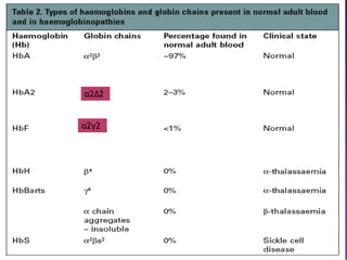
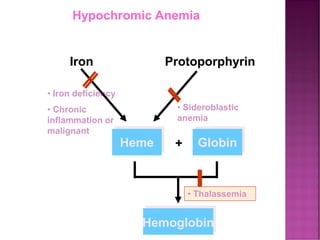
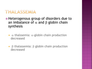
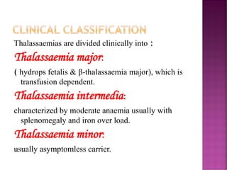
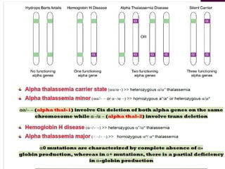
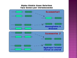
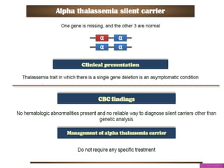
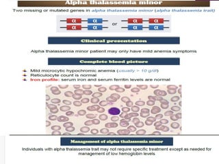
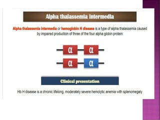
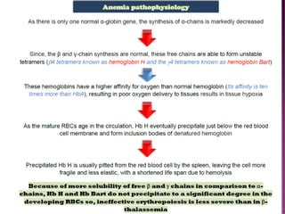
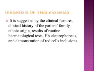
THALASSAEMIAS
Review
Haemoglobinopathies
HAEMOGLOBINOPATHIES
 Haemoglobinopathies are ‘inherited abnormalities
of globin chain synthesis’
 Encompass a clinical spectrum from asymptomatic
findings on blood film to death in utero.
 Wide spread distribution throughout the world and
increasing prevalence in multicultural Australia.
HEMOGLOBIN STRUCTURE.
 A normal hemoglobin molecule consists of:
 Four globin chains (2 alpha, 2 beta).
 Each globin chain has an iron containing
heme molecule.
 The iron in the heme molecule binds to
oxygen.
β
β
α
α
heme
Hemoglobin structure
Hb A tetramer

globin

globin
 globin  globin
α
α
α
α α
α
β γ δ
β δ
γ
HbA HbF HbA2
98% ~1% <3.5%
HEMOGLOBINS IN NORMAL
ADULTS
Copyright ©1997 BMJ Publishing Group Ltd.
α2Δ2
α2γ2
THALASSEMIAS
The thalassemias are a heterogenous
group of congenital anemias that have in
common deficient synthesis of one or
more of the globin subunits of the normal
human hemoglobins (Hbs).
CONTI.
Thalassemias are also a group of
microcytic hypochromic anaemias
(MCV & MCH are low) arise as a result of
diminished or absent production of one or
more globin chains.
It is divided on α or β thalassaemia
depending on whether there is reduced
synthesis of α or β globin
• Iron deficiency
• Chronic
inflammation or
malignant
Iron Protoporphyrin
Heme Globin
+
Hemoglobin
• Thalassemia
• Sideroblastic
anemia
Hypochromic Anemia
DEMOGRAPHICS: THALASSEMIA
• Found most
frequently in the
Mediterranean,
Africa, Western and
Southeast Asia, India
and Burma
• Distribution parallels
that of Plasmodium
falciparum
THALASSEMIA
 Heterogenous group of disorders due to
an imbalance of  and  globin chain
synthesis
  thalssemia: -globin chain production
decreased
  thalassemia:  globin chain production
decreased
*MAIN TWO TYPES OF THALASSEMIA
ARE:-
1)  thalassemia  thalassemia
2) Note:
3) *There are other types according to genetic classification
Thalassaemias are divided clinically into :
Thalassaemia major:
( hydrops fetalis & β-thalassaemia major), which is
transfusion dependent.
Thalassaemia intermedia:
characterized by moderate anaemia usually with
splenomegaly and iron over load.
Thalassaemia minor:
usually asymptomless carrier.
CLINICAL CLASSIFICATION
Genetics of Thalassemia
CON. THALASSEMIA
 The globin chains that are produced are
normal
 It is characterized by quantitative deficiency,
for example:
 o thalassemia: No -globin chain is made
 + thalassemia: decreased -globin chain is
made
 With 4  genes and 2  genes there is wide
phenotypic variation
1. ALPHA THALASSEMIA
 Normally, there are four copies of the a-globin
gene, therefore the clinical severity can be
classified according to the number of genes that
are missing or inactive.
 In case of alpha thalassemia there is inadequate
production of alpha chains
THE GENETICS OF ALPHA-
THALASSAEMIA
 Each a gene may be
deleted or (less
frequently) dysfunctional.
 The orange boxes
represent normal genes,
and the blue boxes
represent gene deletions
or dysftunctional genes.
ALPHA THALASSEMIA
There could be an excess of  globins,
leading to the formation of  globin
tetramers (4) called hemoglobin H.
Results in hemolysis, generally shortening
the life span of the red cell.
DIAGNOSIS OF THALASSEMIAS
 It is suggested by the clinical features,
clinical history of the patient´ family,
ethnic origin, results of routine
haematological tests, Hb electrophoresis,
and demonstration of red cells inclusions.
LABORATORY FINDINGS IN ALPHA
THALASSEMIA
 chains Hgb (g/dl) MCV (fl) RDW
/ Normal Normal Normal
/- 12-14 75-85 Normal
-/- or - -/ 11-13 70-75
- -/-  7-10 50-60
- -/- - - - -
RETICULOCYTOSIS IN CASE OF
HEMOLYSIS
ALPHA THALASSEMIA WITH HEMOGLOBIN H
VITAL STAIN
Hb H: Golf ball
cells
- in case of 3
alpha genes
deletion, caused
by precipitation
of aggregates of
beta globin
chains.
HB-H INCLUSIONS
CONT. ALPHA THALASSEMIA
Hemoglobin H-Constant Spring
disease.
Assignment
THANK YOU

Hemoglobinopathies/Thalassaemias- Alphaa

  • 1.
  • 2.
  • 3.
    HAEMOGLOBINOPATHIES  Haemoglobinopathies are‘inherited abnormalities of globin chain synthesis’  Encompass a clinical spectrum from asymptomatic findings on blood film to death in utero.  Wide spread distribution throughout the world and increasing prevalence in multicultural Australia.
  • 4.
    HEMOGLOBIN STRUCTURE.  Anormal hemoglobin molecule consists of:  Four globin chains (2 alpha, 2 beta).  Each globin chain has an iron containing heme molecule.  The iron in the heme molecule binds to oxygen.
  • 5.
    β β α α heme Hemoglobin structure Hb Atetramer  globin  globin  globin  globin
  • 6.
    α α α α α α β γδ β δ γ HbA HbF HbA2 98% ~1% <3.5% HEMOGLOBINS IN NORMAL ADULTS
  • 7.
    Copyright ©1997 BMJPublishing Group Ltd.
  • 8.
  • 9.
    THALASSEMIAS The thalassemias area heterogenous group of congenital anemias that have in common deficient synthesis of one or more of the globin subunits of the normal human hemoglobins (Hbs).
  • 10.
    CONTI. Thalassemias are alsoa group of microcytic hypochromic anaemias (MCV & MCH are low) arise as a result of diminished or absent production of one or more globin chains. It is divided on α or β thalassaemia depending on whether there is reduced synthesis of α or β globin
  • 11.
    • Iron deficiency •Chronic inflammation or malignant Iron Protoporphyrin Heme Globin + Hemoglobin • Thalassemia • Sideroblastic anemia Hypochromic Anemia
  • 12.
    DEMOGRAPHICS: THALASSEMIA • Foundmost frequently in the Mediterranean, Africa, Western and Southeast Asia, India and Burma • Distribution parallels that of Plasmodium falciparum
  • 14.
    THALASSEMIA  Heterogenous groupof disorders due to an imbalance of  and  globin chain synthesis   thalssemia: -globin chain production decreased   thalassemia:  globin chain production decreased
  • 15.
    *MAIN TWO TYPESOF THALASSEMIA ARE:- 1)  thalassemia  thalassemia 2) Note: 3) *There are other types according to genetic classification
  • 16.
    Thalassaemias are dividedclinically into : Thalassaemia major: ( hydrops fetalis & β-thalassaemia major), which is transfusion dependent. Thalassaemia intermedia: characterized by moderate anaemia usually with splenomegaly and iron over load. Thalassaemia minor: usually asymptomless carrier. CLINICAL CLASSIFICATION
  • 17.
  • 18.
    CON. THALASSEMIA  Theglobin chains that are produced are normal  It is characterized by quantitative deficiency, for example:  o thalassemia: No -globin chain is made  + thalassemia: decreased -globin chain is made  With 4  genes and 2  genes there is wide phenotypic variation
  • 19.
    1. ALPHA THALASSEMIA Normally, there are four copies of the a-globin gene, therefore the clinical severity can be classified according to the number of genes that are missing or inactive.  In case of alpha thalassemia there is inadequate production of alpha chains
  • 20.
    THE GENETICS OFALPHA- THALASSAEMIA  Each a gene may be deleted or (less frequently) dysfunctional.  The orange boxes represent normal genes, and the blue boxes represent gene deletions or dysftunctional genes.
  • 22.
    ALPHA THALASSEMIA There couldbe an excess of  globins, leading to the formation of  globin tetramers (4) called hemoglobin H. Results in hemolysis, generally shortening the life span of the red cell.
  • 36.
    DIAGNOSIS OF THALASSEMIAS It is suggested by the clinical features, clinical history of the patient´ family, ethnic origin, results of routine haematological tests, Hb electrophoresis, and demonstration of red cells inclusions.
  • 37.
    LABORATORY FINDINGS INALPHA THALASSEMIA  chains Hgb (g/dl) MCV (fl) RDW / Normal Normal Normal /- 12-14 75-85 Normal -/- or - -/ 11-13 70-75 - -/-  7-10 50-60 - -/- - - - -
  • 38.
  • 39.
    ALPHA THALASSEMIA WITHHEMOGLOBIN H VITAL STAIN Hb H: Golf ball cells - in case of 3 alpha genes deletion, caused by precipitation of aggregates of beta globin chains.
  • 40.
  • 41.
    CONT. ALPHA THALASSEMIA HemoglobinH-Constant Spring disease. Assignment
  • 42.