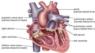
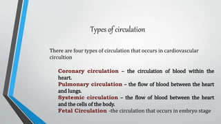
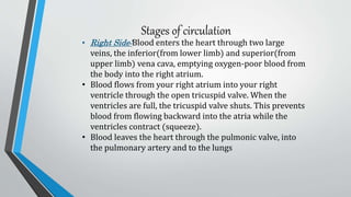
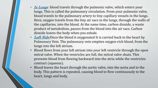
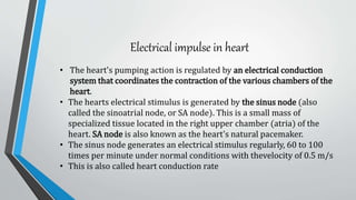
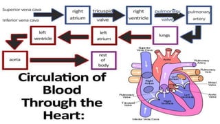
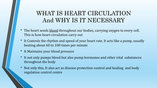
PRESENTED By: AYESHA KHAN
PRESENTED TO:DR VINOD
ROLL NO:21218
Subject:physiology
batch: IV(2026)
Circulatory Systems Consist Of?
It consist of three major units
HEART
BLOOD VESSELS
LUNGS
STRUCTURE OF HEART
It is situated between the two lungs and slightly to the left of center, behind the breastbone. It rests
on the diaphragm, the muscular partition between the chest and the abdominal cavity.
The heart consists of several layers of a tough muscular wall, the myocardium. A thin layer of tissu4e,
the pericardium, covers the outside, and another layer, the endocardium, lines the inside. The heart cavity is divided
down the middle into a right and a left heart, which in turn are subdivided into two chambers. The upper chamber
is called an atrium (or auricle), and the lower chamber is called a ventricle. The two atria(right and left) act as
receiving chambers for blood entering the heart by vena cava ; the more muscular ventricles pump the blood out of
the heart by aorta Moreover, The left (receives oxygenated blood )and right(receives deoxygenated blood) side is
devided by Mascular septumSome structures necessary for blood circulation are Aorta and Vena Cava
BLOOD VESSELS:arteries Veins and Cappillaries are major constituent of circulator system. There is
also additional structure present that is Anostomosis (network of veins and arteries)
Types of circulation
There are four types of circulation that occurs in cardiovascular
circultion
Coronary circulation – the circulation of blood within the
heart.
Pulmonary circulation – the flow of blood between the heart
and lungs.
Systemic circulation – the flow of blood between the heart
and the cells of the body.
Fetal Circulation -the circulation that occurs in embryo stage
Stages of circulation
• Right Side:Blood enters the heart through two large
veins, the inferior(from lower limb) and superior(from
upper limb) vena cava, emptying oxygen-poor blood from
the body into the right atrium.
• Blood flows from your right atrium into your right
ventricle through the open tricuspid valve. When the
ventricles are full, the tricuspid valve shuts. This prevents
blood from flowing backward into the atria while the
ventricles contract (squeeze).
• Blood leaves the heart through the pulmonic valve, into
the pulmonary artery and to the lungs
• In Lungs : blood travels through the pulmonic valve, which enters your
lungs. This is called the pulmonary circulation. From your pulmonic valve,
blood travels to the pulmonary artery to tiny capillary vessels in the lungs.
Here, oxygen travels from the tiny air sacs in the lungs, through the walls of
the capillaries, into the blood. At the same time, carbon dioxide, a waste
product of metabolism, passes from the blood into the air sacs. Carbon
dioxide leaves the body when you exhale.
• Left Side:Once the blood it oxygenated It is carried back to the heart by
Pulmonary Vein. The pulmonary vein empties oxygen-rich blood, from the
lungs into the left atrium.
• Blood flows from your left atrium into your left ventricle through the open
mitral valve. When the ventricles are full, the mitral valve shuts. This
prevents blood from flowing backward into the atria while the ventricles
contract (squeeze).
• Blood leaves the heart through the aortic valve, into the aorta and to the
body. This pattern is repeated, causing blood to flow continuously to the
heart, lungs and body.
Electrical impulse in heart
• The heart's pumping action is regulated by an electrical conduction
system that coordinates the contraction of the various chambers of the
heart.
• The hearts electrical stimulus is generated by the sinus node (also
called the sinoatrial node, or SA node). This is a small mass of
specialized tissue located in the right upper chamber (atria) of the
heart. SA node is also known as the heart's natural pacemaker.
• The sinus node generates an electrical stimulus regularly, 60 to 100
times per minute under normal conditions with thevelocity of 0.5 m/s
• This is also called heart conduction rate
WHAT IS HEART CIRCULATION
And WHY IS IT NECESSARY
• The heart sends blood throughout our bodies, carrying oxygen to every cell.
This is how heart circulation carry out
• It Controls the rhythm and speed of your heart rate. It acts like a pump, usually
beating about 60 to 100 times per minute
• It Maintains your blood pressure
• It not only pumps blood but also pump hormones and other vital substances
throughout the body
• Not only this, It also act as disease protection control and healing and body
regulation control centre
Heart and circulation

Heart and circulation

  • 2.
    PRESENTED By: AYESHAKHAN PRESENTED TO:DR VINOD ROLL NO:21218 Subject:physiology batch: IV(2026)
  • 3.
    Circulatory Systems ConsistOf? It consist of three major units HEART BLOOD VESSELS LUNGS
  • 4.
    STRUCTURE OF HEART Itis situated between the two lungs and slightly to the left of center, behind the breastbone. It rests on the diaphragm, the muscular partition between the chest and the abdominal cavity. The heart consists of several layers of a tough muscular wall, the myocardium. A thin layer of tissu4e, the pericardium, covers the outside, and another layer, the endocardium, lines the inside. The heart cavity is divided down the middle into a right and a left heart, which in turn are subdivided into two chambers. The upper chamber is called an atrium (or auricle), and the lower chamber is called a ventricle. The two atria(right and left) act as receiving chambers for blood entering the heart by vena cava ; the more muscular ventricles pump the blood out of the heart by aorta Moreover, The left (receives oxygenated blood )and right(receives deoxygenated blood) side is devided by Mascular septumSome structures necessary for blood circulation are Aorta and Vena Cava BLOOD VESSELS:arteries Veins and Cappillaries are major constituent of circulator system. There is also additional structure present that is Anostomosis (network of veins and arteries)
  • 6.
    Types of circulation Thereare four types of circulation that occurs in cardiovascular circultion Coronary circulation – the circulation of blood within the heart. Pulmonary circulation – the flow of blood between the heart and lungs. Systemic circulation – the flow of blood between the heart and the cells of the body. Fetal Circulation -the circulation that occurs in embryo stage
  • 7.
    Stages of circulation •Right Side:Blood enters the heart through two large veins, the inferior(from lower limb) and superior(from upper limb) vena cava, emptying oxygen-poor blood from the body into the right atrium. • Blood flows from your right atrium into your right ventricle through the open tricuspid valve. When the ventricles are full, the tricuspid valve shuts. This prevents blood from flowing backward into the atria while the ventricles contract (squeeze). • Blood leaves the heart through the pulmonic valve, into the pulmonary artery and to the lungs
  • 8.
    • In Lungs: blood travels through the pulmonic valve, which enters your lungs. This is called the pulmonary circulation. From your pulmonic valve, blood travels to the pulmonary artery to tiny capillary vessels in the lungs. Here, oxygen travels from the tiny air sacs in the lungs, through the walls of the capillaries, into the blood. At the same time, carbon dioxide, a waste product of metabolism, passes from the blood into the air sacs. Carbon dioxide leaves the body when you exhale. • Left Side:Once the blood it oxygenated It is carried back to the heart by Pulmonary Vein. The pulmonary vein empties oxygen-rich blood, from the lungs into the left atrium. • Blood flows from your left atrium into your left ventricle through the open mitral valve. When the ventricles are full, the mitral valve shuts. This prevents blood from flowing backward into the atria while the ventricles contract (squeeze). • Blood leaves the heart through the aortic valve, into the aorta and to the body. This pattern is repeated, causing blood to flow continuously to the heart, lungs and body.
  • 9.
    Electrical impulse inheart • The heart's pumping action is regulated by an electrical conduction system that coordinates the contraction of the various chambers of the heart. • The hearts electrical stimulus is generated by the sinus node (also called the sinoatrial node, or SA node). This is a small mass of specialized tissue located in the right upper chamber (atria) of the heart. SA node is also known as the heart's natural pacemaker. • The sinus node generates an electrical stimulus regularly, 60 to 100 times per minute under normal conditions with thevelocity of 0.5 m/s • This is also called heart conduction rate
  • 11.
    WHAT IS HEARTCIRCULATION And WHY IS IT NECESSARY • The heart sends blood throughout our bodies, carrying oxygen to every cell. This is how heart circulation carry out • It Controls the rhythm and speed of your heart rate. It acts like a pump, usually beating about 60 to 100 times per minute • It Maintains your blood pressure • It not only pumps blood but also pump hormones and other vital substances throughout the body • Not only this, It also act as disease protection control and healing and body regulation control centre