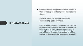
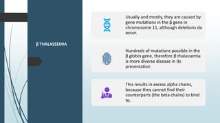
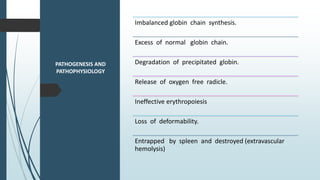
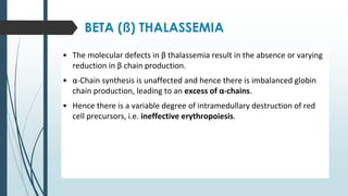
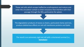
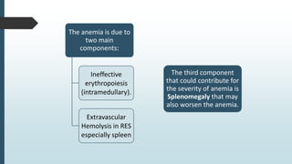
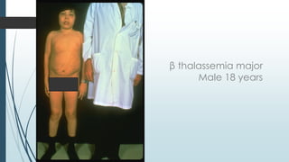
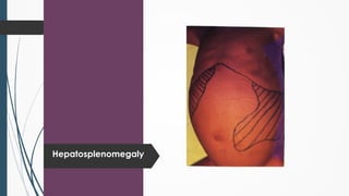
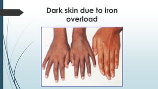
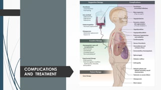
Haemoglobinopathies
Part 2
Norasidi Raffie
• Common and usually produce severe anemia in
their homozygous and compound heterozygous
states.
β Thalassemias are autosomal inherited
disorders of β-globin synthesis.
In most, globin structure is normal, but the rate
of production is reduced because of decrease in
transcription of DNA, abnormal processing of
pre-mRNA, or decreased translation of mRNA
leading to decreased HbA production (A=Adult).
β THALASSAEMIA
β THALASSEMIA
Usually and mostly, they are caused by
gene mutations in the β gene in
chromosome 11, although deletions do
occur.
Hundreds of mutations possible in the
β globin gene, therefore β thalassemia
is more diverse disease in its
presentation
This results in excess alpha chains,
because they cannot find their
counterparts (the beta chains) to bind
to.
PATHOGENESIS AND
PATHOPHYSIOLOGY
Imbalanced globin chain synthesis.
Excess of normal globin chain.
Degradation of precipitated globin.
Release of oxygen free radicle.
Ineffective erythropoiesis
Loss of deformability.
Entrapped by spleen and destroyed (extravascular
hemolysis)
BETA (ß) THALASSEMIA
• The molecular defects in β thalassemia result in the absence or varying
reduction in β chain production.
• α-Chain synthesis is unaffected and hence there is imbalanced globin
chain production, leading to an excess of α-chains.
• Hence there is a variable degree of intramedullary destruction of red
cell precursors, i.e. ineffective erythropoiesis.
The result is an extremely rigid red cell with a shortened survival (i.e.,
hemolysis).
The degradation products of excess α-chains, particularly heme and iron,
produce deleterious effects on red cell membrane proteins and lipids.
Those red cells which escape ineffective erythropoiesis and mature and
enter the circulation contain α-chain inclusions that interfere with their
passage through the RES, particularly the spleen.
The anemia is due to
two main
components:
Ineffective
erythropoiesis
(intramedullary).
Extravascular
Hemolysis in RES
especially spleen
The third component
that could contribute for
the severity of anemia is
Splenomegaly that may
also worsen the anemia.
ß THALASSEMIA
AND MALARIA
Thalassemic RBCs offers protection
against severe malaria caused by
Plasmodium falciparum.
The effect is associated with reduced
parasite multiplication within RBCs.
Among the contributing factors may be
the variable persistence of hemoglobin F,
which is relatively resistant to digestion
by malarial hemoglobinases.
Quantities Of Β Globin Chain Produced In
Different Genetic Situations Depends On The
Mutation Type
α β
β α
Homozygous Heterozygous
β
β0
β
β0
β+ β+
α β α α α β
α β
β++
α β
β++
α β
βΝ
α β
β α
x
α β
β α
x
x
Silent Carrier
State For β
Thalassemia
Are various heterozygous (from one
parent) β gene mutations that
produce only small decrease in
production of β globin chains.
Patients have nearly normal
alpha/beta chain ratio and no
hematologic abnormalities.
Have normal levels of HbA2.
β Thalassemia
Minor (Trait)
Caused by heterozygous (from one parent)
mutations that affect β globin synthesis.
Usually presents as mild, asymptomatic hemolytic
anemia unless patient in under stress such as
pregnancy, infection, or folic acid deficiency.
Have one normal β gene and one mutated β gene.
Hemoglobin level in 10-13 g/dL range with normal
or slightly elevated RBC count.
β Thalassemia
Minor (Trait)
Anemia usually hypochromic and microcytic with
slight aniso and poikilo, including target cells and
elliptocytes; also may see basophilic stippling.
Rarely see hepatomegaly or splenomegaly.
Make sure they are not diagnosed as IDA.
Mentzer index: <13
Distinguishing Thalassaemia Minor From IDA From
CBC By Applying Formulae:
Formula Thal. IDA
MCV ÷ RCC (Mentzer index) <13 >13
MCH ÷ RCC < 3.8 > 3.8
(MCV2 × MCH) ÷ 100 < 1530 > 1530
MCV – RCC – (Hb × 5) – 3.4 < 0 > 0
(MCV2 × RDW) ÷ (100xHb) < 65 > 65
RDW-CV% <14.6 >14.6
β Thalassemia
Intermediate
Patients able to maintain minimum Hb (7
g/dL or greater) without transfusion
dependence.
Expression of disorder falls between
thalassemia minor and thalassemia major.
We will see increase in both HbA2
production and HbF production.
Peripheral blood smear picture is similar to
thalassemia minor.
β Thalassemia
Intermediate
Have varying symptoms of anemia, jaundice,
splenomegaly and hepatomegaly.
Have significant increase in bilirubin levels.
Anemia usually becomes worse with infections,
pregnancy, or folic acid deficiency.
May become transfusion dependent.
Tend to develop iron overloads as result of increased
gastrointestinal absorption.
Usually survive into adulthood.
β Thalassemia
Major
Characterized by very severe microcytic,
hypochromic anemia.
Detected early in childhood:
Hb level lies between 2 and 8 g/dL.
Severe anemia causes marked bone changes due
to expansion of marrow space for increased
erythropoiesis
See characteristic changes in skull, long bones, and
hand bones.
Have protrusion upper teeth and Mongoloid facial
features.
Physical growth and development delayed.
Peripheral blood shows markedly hypochromic, microcytic
erythrocytes with extreme poikilocytosis, such as target
cells, teardrop cells and elliptocytes. See marked
basophilic stippling and numerous NRBCs.
MCV in range of 50 to 60 fl.
Retic count seen (2-8%). But it can be low relative to the
severity of anemia.
β
Thalassemia
Major
Dangers in continuous tranfusion therapy:
Development of iron
overload.
Development of
alloimmunization
(developing antibodies
to transfused RBCs).
Risk of transfusion-
transmitted diseases
(e.g., hepatitis, AIDS).
Excessive number of transfusions results in
tranfusional hemosiderosis; Without iron
chelation, patient develops cardiac disease,
liver cirrhosis, and endocrine deficiencies.
Comparison of β Thalassemias
Parameter Minor Intermedia Major
Hb 10-13 6-10 2-8
MCV (fl) 60-78 50-70 50-60
MCH (pg) 28-32 22-28 16-22
RDW Normal S. increased Increased
Micro/hypo (PBF) Mild Moderate Severe
Polychromasia V. Little Moderate Marked
Anisocytosis None Moderate Marked
Poikilocytosis None Moderate Marked
Targetting Present Present Present
β Thalassemia Major
Anisopoikilocytosis, NRBC, microcytosis, hypochromia
CLINICAL
PICTURE
Time of appearance:
 b thalassemia: 6-12 month
 a thalassemia: Incompatible with life (Hydrops fetalis)
Thalassemia minor:
 Asymptomatic or minimal pallor and mild splenomegaly
Thalassemia major:
 Severe pallor jaundice (muddy face)
 Dark urine - Mongoloid face
 Splenomegaly - Hepatomegaly
 Growth retardation - Gall stone -
Pathological fractures
LABORATORY
INVESTIGATION
◼ Peripheral blood exam:
1. Decreased RBC, hemoglobin, hematocrit
2. Microcytic hypochromic anemia (low MCV,
MCH, and MCHC)
3. Anisocytosis and poikilocytosis, target cells
4. Elevated reticulocyte count
5. Decreased serum iron, serum ferritin, and total
iron-binding capacity (TIBC)
6. Elevated serum unconjugated bilirubin and
decreased haptoglobin levels
◼ BM examination:
- Erythroid hyperplasia
◼ Alkaline denaturation test:
- Resistant
Radiological Investigations:
- X-Ray skull: wide diploic space and hair on end appearance
- Long bones: widen medullary cavities trabeculations
β Thalassemia Major
β thalassemia major
Male 18 years
Hepatosplenomegaly
Dark skin due to iron
overload
COMPLICATIONS
AND TREATMENT

α−Structural Variants
(469 var submitted, July 2002)
 Hb Anantharaj
cd11 (Lys-Glu)
 Hb Mahidol
cd74 (Asp-His)
 Hb Siam
cd15 (Gly-Arg)
 Hb Suan Dok
cd109 (Leu-Arg)
 Hb Constant spring
cd142 (stop-Gln)

β-Structural Variants
(649 var submitted, July 2002)
 Hb D-Punjab
cd121(Glu-Gln)
 Hb J-Bangkok
cd56(Gly-Asp)
 Hb S
cd6(Glu-Val)
 Hb G-Siriraj
cd7(Glu-Lys)
 Hb Tak
cd147(+AC)
 Hb E
cd26(Glu-Lys)
Sickle
Cell
Anemia
Wide spectrum of disorders
1 / 600 African Americans affected
1 / 8 African Americans - sickle trait
Hb SS ~ 60% of sickle cell disease
Hb SC and Sb-thal ~ 40%
Sickle
trait
βS/β; 8% of African-Americans
Asymptomatic
Partial protection from malaria
Sickling may occur in renal medulla → decreased urinary
concentrating ability, hematuria
Rare complications at high altitude (splenic infarction)
Sudden death following strenuous exercise (rare)
Pathophysiology of Sickle Cell Anemia
(Modified from Steinberg, M., Cecil Medicine 2007)
HbS Polymer
Vaso-occlusion
Hemolysis
Arginine NO
Sickle
Cell:
Molecular
Basis
Glutamate  Valine at 6th position b globin
Sickle Hb forms polymers when
deoxygenated
Polymerized sickle Hb injures RBC
membrane and distorts its shape
Distorted RBC is hemolyzed
Sickle Cells –
Electron
Microscopy
Sickle Cell:
Pathophysiology
Deoxygenation of mutant
Hb leads to
K+ efflux
cell density /
dehydration
polymerization
Sickled cells adhere to endothelial cells
Endothelial factors - vasoconstriction
Blood flow ¯ promotes vaso-occlusion
“Vicious cycle” with decreased blood flow, hypoxemia
/ acidosis, increased sickling
Some cells become irreversibly sickled
FACTORS THAT
INCREASE HbS
POLYMERIZATION
Decreased oxygen
Increased intracellular hemoglobin S concentration (SS
> SC, S-thal)
Increased 2,3-DPG
Decreased pH
Slowed transit time through the circulation
Endothelial adhesion
Lower concentration of HbS (compound
heterozygosity for α thalassemia)
Increased HbF levels
Genetic basis
Hydroxyurea
FACTORS THAT
INCREASE HbS
POLYMERIZATION
Clinical Features of Sickle Cell Anemia
• Painful episodes
• Pneumococcal disease
• Acute chest syndrome
• Splenic infarction
• Splenic sequestration
• Stroke
• Osteonecrosis
• Priapism
• Retinopathy
• Leg ulcers
• Gallstones
• Renal abnormalities
• Osteopenia
• Nutritional deficiencies
• Placental insufficiency
• Pulmonary hypertension
Clinical Features of Sickle Cell Anemia
Associated with higher
hemoglobin
Associated with lower
hemoglobin
Painful episodes Stroke
Acute chest syndrome Priapism
Osteonecrosis Leg Ulcers
Proliferative retinopathy
Sickle Cell – Avascular Necrosis
gait.aidi.udel.edu/.../clcsimge/sickle5 http://www.zimmer.com
Sickle Cell – Dactylitis
http://aapredbook.aappublications.org/week/116_09.jpg
Laboratory Findings
 Moderate anemia
 Reticulocytosis 3-15%
 High MCV
 Unconjugated
hyperbilirubinemia
 Elevetaed LDH
 Low haptoglobin
 Folate & iron deficit
 Peripheral smear shows
sickle cells
 Polychromasia
 Howell-jolly bodies
 Elevated WBC
 Elevated Platelets
 Low than after 18 yrs high
creatinine
Sickle Cell
Anemia -
treatment
Opiates and hydration for painful crises
Pneumococcal vaccination
Retinal surveillance
Transfusion for serious manifestations (eg stroke);
exchange transfusion
Hydroxyurea
Stem cell transplant
Hemoglobin C
Glutamate → lysine at 6th position in beta chain
Hb tends to crystallize
Prevalent in west Africa
Homozygous state – chronic hemolytic anemia
Compound heterozygosity with Hb S produces
sickle phenotype
Hemoglobin E
Mutation (glutamine → lysine at amino
acid 26)
Altered mRNA splicing, unstable mRNA
Heterozygous in 30% of SE Asians
Homozygous Hb E: microcytosis,
hypochromia, little or no anemia
Hemoglobin E / b-thal causes thalassemia-
like phenotype
Thank You

HAEMOGLOBINOPATHIES PART 2 (BETA THALASSAEMIA)

  • 1.
  • 3.
    • Common andusually produce severe anemia in their homozygous and compound heterozygous states. β Thalassemias are autosomal inherited disorders of β-globin synthesis. In most, globin structure is normal, but the rate of production is reduced because of decrease in transcription of DNA, abnormal processing of pre-mRNA, or decreased translation of mRNA leading to decreased HbA production (A=Adult). β THALASSAEMIA
  • 4.
    β THALASSEMIA Usually andmostly, they are caused by gene mutations in the β gene in chromosome 11, although deletions do occur. Hundreds of mutations possible in the β globin gene, therefore β thalassemia is more diverse disease in its presentation This results in excess alpha chains, because they cannot find their counterparts (the beta chains) to bind to.
  • 5.
    PATHOGENESIS AND PATHOPHYSIOLOGY Imbalanced globinchain synthesis. Excess of normal globin chain. Degradation of precipitated globin. Release of oxygen free radicle. Ineffective erythropoiesis Loss of deformability. Entrapped by spleen and destroyed (extravascular hemolysis)
  • 6.
    BETA (ß) THALASSEMIA •The molecular defects in β thalassemia result in the absence or varying reduction in β chain production. • α-Chain synthesis is unaffected and hence there is imbalanced globin chain production, leading to an excess of α-chains. • Hence there is a variable degree of intramedullary destruction of red cell precursors, i.e. ineffective erythropoiesis.
  • 7.
    The result isan extremely rigid red cell with a shortened survival (i.e., hemolysis). The degradation products of excess α-chains, particularly heme and iron, produce deleterious effects on red cell membrane proteins and lipids. Those red cells which escape ineffective erythropoiesis and mature and enter the circulation contain α-chain inclusions that interfere with their passage through the RES, particularly the spleen.
  • 8.
    The anemia isdue to two main components: Ineffective erythropoiesis (intramedullary). Extravascular Hemolysis in RES especially spleen The third component that could contribute for the severity of anemia is Splenomegaly that may also worsen the anemia.
  • 9.
    ß THALASSEMIA AND MALARIA ThalassemicRBCs offers protection against severe malaria caused by Plasmodium falciparum. The effect is associated with reduced parasite multiplication within RBCs. Among the contributing factors may be the variable persistence of hemoglobin F, which is relatively resistant to digestion by malarial hemoglobinases.
  • 10.
    Quantities Of ΒGlobin Chain Produced In Different Genetic Situations Depends On The Mutation Type α β β α Homozygous Heterozygous β β0 β β0 β+ β+ α β α α α β α β β++ α β β++ α β βΝ α β β α x α β β α x x
  • 11.
    Silent Carrier State Forβ Thalassemia Are various heterozygous (from one parent) β gene mutations that produce only small decrease in production of β globin chains. Patients have nearly normal alpha/beta chain ratio and no hematologic abnormalities. Have normal levels of HbA2.
  • 12.
    β Thalassemia Minor (Trait) Causedby heterozygous (from one parent) mutations that affect β globin synthesis. Usually presents as mild, asymptomatic hemolytic anemia unless patient in under stress such as pregnancy, infection, or folic acid deficiency. Have one normal β gene and one mutated β gene. Hemoglobin level in 10-13 g/dL range with normal or slightly elevated RBC count.
  • 13.
    β Thalassemia Minor (Trait) Anemiausually hypochromic and microcytic with slight aniso and poikilo, including target cells and elliptocytes; also may see basophilic stippling. Rarely see hepatomegaly or splenomegaly. Make sure they are not diagnosed as IDA. Mentzer index: <13
  • 14.
    Distinguishing Thalassaemia MinorFrom IDA From CBC By Applying Formulae: Formula Thal. IDA MCV ÷ RCC (Mentzer index) <13 >13 MCH ÷ RCC < 3.8 > 3.8 (MCV2 × MCH) ÷ 100 < 1530 > 1530 MCV – RCC – (Hb × 5) – 3.4 < 0 > 0 (MCV2 × RDW) ÷ (100xHb) < 65 > 65 RDW-CV% <14.6 >14.6
  • 15.
    β Thalassemia Intermediate Patients ableto maintain minimum Hb (7 g/dL or greater) without transfusion dependence. Expression of disorder falls between thalassemia minor and thalassemia major. We will see increase in both HbA2 production and HbF production. Peripheral blood smear picture is similar to thalassemia minor.
  • 16.
    β Thalassemia Intermediate Have varyingsymptoms of anemia, jaundice, splenomegaly and hepatomegaly. Have significant increase in bilirubin levels. Anemia usually becomes worse with infections, pregnancy, or folic acid deficiency. May become transfusion dependent. Tend to develop iron overloads as result of increased gastrointestinal absorption. Usually survive into adulthood.
  • 17.
    β Thalassemia Major Characterized byvery severe microcytic, hypochromic anemia. Detected early in childhood: Hb level lies between 2 and 8 g/dL. Severe anemia causes marked bone changes due to expansion of marrow space for increased erythropoiesis See characteristic changes in skull, long bones, and hand bones.
  • 18.
    Have protrusion upperteeth and Mongoloid facial features. Physical growth and development delayed. Peripheral blood shows markedly hypochromic, microcytic erythrocytes with extreme poikilocytosis, such as target cells, teardrop cells and elliptocytes. See marked basophilic stippling and numerous NRBCs. MCV in range of 50 to 60 fl. Retic count seen (2-8%). But it can be low relative to the severity of anemia.
  • 19.
    β Thalassemia Major Dangers in continuoustranfusion therapy: Development of iron overload. Development of alloimmunization (developing antibodies to transfused RBCs). Risk of transfusion- transmitted diseases (e.g., hepatitis, AIDS). Excessive number of transfusions results in tranfusional hemosiderosis; Without iron chelation, patient develops cardiac disease, liver cirrhosis, and endocrine deficiencies.
  • 20.
    Comparison of βThalassemias Parameter Minor Intermedia Major Hb 10-13 6-10 2-8 MCV (fl) 60-78 50-70 50-60 MCH (pg) 28-32 22-28 16-22 RDW Normal S. increased Increased Micro/hypo (PBF) Mild Moderate Severe Polychromasia V. Little Moderate Marked Anisocytosis None Moderate Marked Poikilocytosis None Moderate Marked Targetting Present Present Present
  • 21.
    β Thalassemia Major Anisopoikilocytosis,NRBC, microcytosis, hypochromia
  • 22.
    CLINICAL PICTURE Time of appearance: b thalassemia: 6-12 month  a thalassemia: Incompatible with life (Hydrops fetalis) Thalassemia minor:  Asymptomatic or minimal pallor and mild splenomegaly Thalassemia major:  Severe pallor jaundice (muddy face)  Dark urine - Mongoloid face  Splenomegaly - Hepatomegaly  Growth retardation - Gall stone - Pathological fractures
  • 23.
    LABORATORY INVESTIGATION ◼ Peripheral bloodexam: 1. Decreased RBC, hemoglobin, hematocrit 2. Microcytic hypochromic anemia (low MCV, MCH, and MCHC) 3. Anisocytosis and poikilocytosis, target cells 4. Elevated reticulocyte count 5. Decreased serum iron, serum ferritin, and total iron-binding capacity (TIBC) 6. Elevated serum unconjugated bilirubin and decreased haptoglobin levels ◼ BM examination: - Erythroid hyperplasia ◼ Alkaline denaturation test: - Resistant
  • 24.
    Radiological Investigations: - X-Rayskull: wide diploic space and hair on end appearance - Long bones: widen medullary cavities trabeculations
  • 25.
  • 26.
  • 27.
  • 28.
    Dark skin dueto iron overload
  • 29.
  • 30.
     α−Structural Variants (469 varsubmitted, July 2002)  Hb Anantharaj cd11 (Lys-Glu)  Hb Mahidol cd74 (Asp-His)  Hb Siam cd15 (Gly-Arg)  Hb Suan Dok cd109 (Leu-Arg)  Hb Constant spring cd142 (stop-Gln)
  • 31.
     β-Structural Variants (649 varsubmitted, July 2002)  Hb D-Punjab cd121(Glu-Gln)  Hb J-Bangkok cd56(Gly-Asp)  Hb S cd6(Glu-Val)  Hb G-Siriraj cd7(Glu-Lys)  Hb Tak cd147(+AC)  Hb E cd26(Glu-Lys)
  • 32.
    Sickle Cell Anemia Wide spectrum ofdisorders 1 / 600 African Americans affected 1 / 8 African Americans - sickle trait Hb SS ~ 60% of sickle cell disease Hb SC and Sb-thal ~ 40%
  • 33.
    Sickle trait βS/β; 8% ofAfrican-Americans Asymptomatic Partial protection from malaria Sickling may occur in renal medulla → decreased urinary concentrating ability, hematuria Rare complications at high altitude (splenic infarction) Sudden death following strenuous exercise (rare)
  • 34.
    Pathophysiology of SickleCell Anemia (Modified from Steinberg, M., Cecil Medicine 2007) HbS Polymer Vaso-occlusion Hemolysis Arginine NO
  • 35.
    Sickle Cell: Molecular Basis Glutamate  Valineat 6th position b globin Sickle Hb forms polymers when deoxygenated Polymerized sickle Hb injures RBC membrane and distorts its shape Distorted RBC is hemolyzed
  • 36.
  • 37.
    Sickle Cell: Pathophysiology Deoxygenation ofmutant Hb leads to K+ efflux cell density / dehydration polymerization Sickled cells adhere to endothelial cells Endothelial factors - vasoconstriction Blood flow ¯ promotes vaso-occlusion “Vicious cycle” with decreased blood flow, hypoxemia / acidosis, increased sickling Some cells become irreversibly sickled
  • 38.
    FACTORS THAT INCREASE HbS POLYMERIZATION Decreasedoxygen Increased intracellular hemoglobin S concentration (SS > SC, S-thal) Increased 2,3-DPG Decreased pH Slowed transit time through the circulation Endothelial adhesion
  • 39.
    Lower concentration ofHbS (compound heterozygosity for α thalassemia) Increased HbF levels Genetic basis Hydroxyurea FACTORS THAT INCREASE HbS POLYMERIZATION
  • 40.
    Clinical Features ofSickle Cell Anemia • Painful episodes • Pneumococcal disease • Acute chest syndrome • Splenic infarction • Splenic sequestration • Stroke • Osteonecrosis • Priapism • Retinopathy • Leg ulcers • Gallstones • Renal abnormalities • Osteopenia • Nutritional deficiencies • Placental insufficiency • Pulmonary hypertension
  • 41.
    Clinical Features ofSickle Cell Anemia Associated with higher hemoglobin Associated with lower hemoglobin Painful episodes Stroke Acute chest syndrome Priapism Osteonecrosis Leg Ulcers Proliferative retinopathy
  • 42.
    Sickle Cell –Avascular Necrosis gait.aidi.udel.edu/.../clcsimge/sickle5 http://www.zimmer.com
  • 43.
    Sickle Cell –Dactylitis http://aapredbook.aappublications.org/week/116_09.jpg
  • 44.
    Laboratory Findings  Moderateanemia  Reticulocytosis 3-15%  High MCV  Unconjugated hyperbilirubinemia  Elevetaed LDH  Low haptoglobin  Folate & iron deficit  Peripheral smear shows sickle cells  Polychromasia  Howell-jolly bodies  Elevated WBC  Elevated Platelets  Low than after 18 yrs high creatinine
  • 45.
    Sickle Cell Anemia - treatment Opiatesand hydration for painful crises Pneumococcal vaccination Retinal surveillance Transfusion for serious manifestations (eg stroke); exchange transfusion Hydroxyurea Stem cell transplant
  • 46.
    Hemoglobin C Glutamate →lysine at 6th position in beta chain Hb tends to crystallize Prevalent in west Africa Homozygous state – chronic hemolytic anemia Compound heterozygosity with Hb S produces sickle phenotype
  • 47.
    Hemoglobin E Mutation (glutamine→ lysine at amino acid 26) Altered mRNA splicing, unstable mRNA Heterozygous in 30% of SE Asians Homozygous Hb E: microcytosis, hypochromia, little or no anemia Hemoglobin E / b-thal causes thalassemia- like phenotype
  • 48.