Download to read offline
![S315
Introduction
Publication of the 2015 American Heart Association (AHA)
Guidelines Update for Cardiopulmonary Resuscitation (CPR)
and Emergency Cardiovascular Care (ECC) marks 49 years
since the first CPR guidelines were published in 1966 by an
Ad Hoc Committee on Cardiopulmonary Resuscitation estab-
lished by the National Academy of Sciences of the National
Research Council.1
Since that time, periodic revisions to the
Guidelines have been published by the AHA in 1974,2
1980,3
1986,4
1992,5
2000,6
2005,7
2010,8
and now 2015. The 2010
AHA Guidelines for CPR and ECC provided a comprehensive
review of evidence-based recommendations for resuscitation,
ECC, and first aid. The 2015 AHA Guidelines Update for CPR
and ECC focuses on topics with significant new science or
ongoing controversy, and so serves as an update to the 2010
AHA Guidelines for CPR and ECC rather than a complete
revision of the Guidelines.
The purpose of this Executive Summary is to provide an
overview of the new or revised recommendations contained in
the 2015 Guidelines Update. This document does not contain
extensive reference citations; the reader is referred to Parts 3
through 9 for more detailed review of the scientific evidence
and the recommendations on which they are based.
There have been several changes to the organization of
the 2015 Guidelines Update compared with 2010. “Part 4:
Systems of Care and Continuous Quality Improvement” is
an important new Part that focuses on the integrated struc-
tures and processes that are necessary to create systems of
care for both in-hospital and out-of-hospital resuscitation
capable of measuring and improving quality and patient out-
comes. This Part replaces the “CPR Overview” Part of the
2010 Guidelines.
Another new Part of the 2015 Guidelines Update is “Part
14: Education,” which focuses on evidence-based recommen-
dations to facilitate widespread, consistent, efficient and effec-
tive implementation of the AHA Guidelines for CPR and ECC
into practice. These recommendations will target resuscitation
education of both lay rescuers and healthcare providers. This
Part replaces the 2010 Part titled “Education, Implementation,
and Teams.” The 2015 Guidelines Update does not include a
separate Part on adult stroke because the content would rep-
licate that already offered in the most recent AHA/American
Stroke Association guidelines for the management of acute
stroke.9,10
Finally, the 2015 Guidelines Update marks the begin-
ning of a new era for the AHA Guidelines for CPR and ECC,
because the Guidelines will transition from a 5-year cycle of
periodic revisions and updates to a Web-based format that is
continuously updated. The first release of the Web-based inte-
grated Guidelines, now available online at ECCguidelines.
heart.org is based on the comprehensive 2010 Guidelines
plus the 2015 Guidelines Update. Moving forward, these
Guidelines will be updated by using a continuous evidence
evaluation process to facilitate more rapid translation of new
scientific discoveries into daily patient care.
Creation of practice guidelines is only 1 link in the chain
of knowledge translation that starts with laboratory and clini-
cal science and culminates in improved patient outcomes. The
AHA ECC Committee has set an impact goal of doubling
bystander CPR rates and doubling cardiac arrest survival by
2020. Much work will be needed across the entire spectrum of
knowledge translation to reach this important goal.
Evidence Review and Guidelines
Development Process
The process used to generate the 2015 AHA Guidelines
Update for CPR and ECC was significantly different from the
process used in prior releases of the Guidelines, and marks
the planned transition from a 5-year cycle of evidence review
to a continuous evidence evaluation process. The AHA con-
tinues to partner with the International Liaison Committee
on Resuscitation (ILCOR) in the evidence review process.
However, for 2015, ILCOR prioritized topics for systematic
review based on clinical significance and availability of new
© 2015 American Heart Association, Inc.
Circulation is available at http://circ.ahajournals.org DOI: 10.1161/CIR.0000000000000252
The American Heart Association requests that this document be cited as follows: Neumar RW, Shuster M, Callaway CW, Gent LM, Atkins DL, Bhanji
F, Brooks SC, de Caen AR, Donnino MW, Ferrer JME, Kleinman ME, Kronick SL, Lavonas EJ, Link MS, Mancini ME, Morrison LJ, O’Connor RE,
Sampson RA, Schexnayder SM, Singletary EM, Sinz EH, Travers AH, Wyckoff MH, Hazinski MF. Part 1: executive summary: 2015 American Heart
Association Guidelines Update for Cardiopulmonary Resuscitation and Emergency Cardiovascular Care. Circulation. 2015;132(suppl 2):S315–S367.
(Circulation. 2015;132[suppl 2]:S315–S367. DOI: 10.1161/CIR.0000000000000252.)
Part 1: Executive Summary
2015 American Heart Association Guidelines Update for Cardiopulmonary
Resuscitation and Emergency Cardiovascular Care
Robert W. Neumar, Chair; Michael Shuster; Clifton W. Callaway; Lana M. Gent; Dianne L. Atkins;
Farhan Bhanji; Steven C. Brooks; Allan R. de Caen; Michael W. Donnino; Jose Maria E. Ferrer;
Monica E. Kleinman; Steven L. Kronick; Eric J. Lavonas; Mark S. Link; Mary E. Mancini;
Laurie J. Morrison; Robert E. O’Connor; Ricardo A. Samson; Steven M. Schexnayder;
Eunice M. Singletary; Elizabeth H. Sinz; Andrew H. Travers; Myra H. Wyckoff; Mary Fran Hazinski
by guest on October 15, 2015http://circ.ahajournals.org/Downloaded from](https://image.slidesharecdn.com/guidelines-rcp-aha-2015-full-160515230707/85/Guidelines-rcp-aha-2015-full-4-320.jpg)
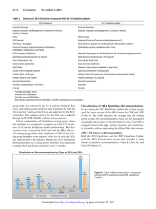
![S318 Circulation November 3, 2015
based on a report associating increased non–life-threatening
injuries with excessive compression depth.
• In adult victims of cardiac arrest, it is reasonable for
rescuers to perform chest compressions at a rate of 100
to 120/min (Class IIa, LOE C-LD). The addition of an
upper limit of compression rate is the result of 1 large
registry study associating extremely rapid compression
rates with inadequate compression depth.
• During manual CPR, rescuers should perform chest
compressions at a depth of at least 2 inches or 5 cm for
an average adult, while avoiding excessive chest com-
pression depths (greater than 2.4 inches [6 cm]) (Class
I, LOE C-LD). The addition of an upper limit of com-
pression depth followed review of 1 publication suggest-
ing potential harm from excessive chest compression
depth (greater than 6 cm, or 2.4 inches). Compression
depth may be difficult to judge without use of feedback
devices, and identification of upper limits of compres-
sion depth may be challenging.
• In adult cardiac arrest, total preshock and postshock
pauses in chest compressions should be as short as pos-
sible (Class I, LOE C-LD) because shorter pauses can
be associated with greater shock success, ROSC, and, in
some studies, higher survival to hospital discharge. The
need to reduce such pauses has received greater empha-
sis in this 2015 Guidelines Update.
• In adult cardiac arrest with an unprotected airway, it may
be reasonable to perform CPR with the goal of a chest
compression fraction as high as possible, with a target of
at least 60% (Class IIb, LOE C-LD). The addition of this
target compression fraction to the 2015 Guidelines Update
is intended to limit interruptions in compressions and to
maximize coronary perfusion and blood flow during CPR.
• For patients with known or suspected opioid addic-
tion who have a definite pulse but no normal breathing
or only gasping (ie, a respiratory arrest), in addition to
providing standard BLS care, it is reasonable for appro-
priately trained BLS providers to administer intramus-
cular or intranasal naloxone (Class IIa, LOE C-LD). It is
reasonable to provide opioid overdose response educa-
tion with or without naloxone distribution to persons at
risk for opioid overdose in any setting (Class IIa, LOE
C-LD). For more information, see “Part 10: Special
Circumstances of Resuscitation.”
• For witnessed OHCA with a shockable rhythm, it may
be reasonable for emergency medical service (EMS)
systems with priority-based, multi-tiered response to
delay positive-pressure ventilation by using a strategy
of up to 3 cycles of 200 continuous compressions with
passive oxygen insufflation and airway adjuncts (Class
IIb, LOE C-LD).
• We do not recommend the routine use of passive ven-
tilation techniques during conventional CPR for adults,
because the usefulness/effectiveness of these techniques
is unknown (Class IIb, LOE C-EO). However, in EMS
systems that use bundles of care involving continuous
chest compressions, the use of passive ventilation tech-
niques may be considered as part of that bundle (Class
IIb, LOE C-LD).
• It is recommended that emergency dispatchers deter-
mine if a patient is unconscious with abnormal breathing
after acquiring the requisite information to determine the
location of the event (Class I, LOE C-LD).
• If the patient is unconscious with abnormal or absent
breathing, it is reasonable for the emergency dispatcher
to assume that the patient is in cardiac arrest (Class IIa,
LOE C-LD).
• Dispatchers should be educated to identify unconscious-
ness with abnormal and agonal gasps across a range of clin-
ical presentations and descriptions (Class I, LOE C-LD).
• We recommend that dispatchers should provide chest
compression–only CPR instructions to callers for adults
with suspected OHCA (Class I, LOE C-LD).
• It is reasonable for healthcare providers to provide chest
compressionsandventilationforalladultpatientsincardiac
arrest, from either a cardiac or a noncardiac cause (Class
IIb, LOE C-LD). When the victim has an advanced airway
in place during CPR, rescuers no longer deliver cycles of
30 compressions and 2 breaths (ie, they no longer interrupt
compressions to deliver 2 breaths). Instead, it may be rea-
sonable for the provider to deliver 1 breath every 6 seconds
(10 breaths per minute) while continuous chest compres-
sions are being performed (Class IIb, LOE C-LD). When
the victim has an advanced airway in place during CPR,
it may be reasonable for the provider to deliver 1 breath
every 6 seconds (10 breaths per minute) while continuous
chest compressions are being performed (Class IIb, LOE
C-LD). This simple rate, rather than a range of breaths per
minute, should be easier to learn, remember, and perform.
• There is insufficient evidence to recommend the use of
artifact-filtering algorithms for analysis of electrocardio-
graphic (ECG) rhythm during CPR. Their use may be con-
sidered as part of a research program or if an EMS system
has already incorporated ECG artifact-filtering algorithms
in its resuscitation protocols (Class IIb, LOE C-EO).
• It may be reasonable to use audiovisual feedback devices
during CPR for real-time optimization of CPR perfor-
mance (Class IIb, LOE B-R).
• For victims with suspected spinal injury, rescuers should
initially use manual spinal motion restriction (eg, plac-
ing 1 hand on either side of the patient’s head to hold it
still) rather than immobilization devices, because use of
immobilization devices by lay rescuers may be harmful
(Class III: Harm, LOE C-LD).
Knowledge Gaps
• The optimal method for ensuring adequate depth of
chest compressions during manual CPR
• The duration of chest compressions after which venti-
lation should be incorporated when using Hands-Only
CPR
• The optimal chest compression fraction
• Optimal use of CPR feedback devices to increase patient
survival
Part 6: Alternative Techniques and Ancillary
Devices for Cardiopulmonary Resuscitation
High-quality conventional CPR (manual chest compressions
with rescue breaths) generates about 25% to 33% of normal
cardiac output and oxygen delivery. A variety of alternatives
by guest on October 15, 2015http://circ.ahajournals.org/Downloaded from](https://image.slidesharecdn.com/guidelines-rcp-aha-2015-full-160515230707/85/Guidelines-rcp-aha-2015-full-7-320.jpg)
![S322 Circulation November 3, 2015
acquiring a 12-lead ECG for patients with possible ACS as
early as possible in the prehospital setting. These studies reaf-
firmed previous recommendations that when STEMI is diag-
nosed in the prehospital setting, prearrival notification of the
hospital and/or prehospital activation of the catheterization
laboratory should occur without delay. These updated recom-
mendations place new emphasis on obtaining a prehospital
ECG and on both the necessity for and the timing of receiving
hospital notification.
• A prehospital 12-lead ECG should be acquired early for
patients with possible ACS (Class I, LOE B-NR).
• Prehospital notification of the hospital (if fibrinolysis is
the likely reperfusion strategy) and/or prehospital activa-
tion of the catheterization laboratory should occur for all
patients with a recognized STEMI on prehospital ECG
(Class I, LOE B-NR).
Because the rate of false-negative results of 12-lead ECGs
may be unacceptably high, a computer reading of the ECG
should not be a sole means to diagnose STEMI, but may be
used in conjunction with physician or trained provider inter-
pretation. New studies examining the accuracy of ECG inter-
pretation by trained nonphysicians have prompted a revision
of the recommendation to explicitly permit trained nonphysi-
cians to interpret ECGs for the presence of STEMI.
• We recommend that computer-assisted ECG interpreta-
tion may be used in conjunction with physician or trained
provider interpretation to recognize STEMI (Class IIb,
LOE C-LD).
• While transmission of the prehospital ECG to the ED
physician may improve the positive predictive value
(PPV) and therapeutic decision making regarding adult
patients with suspected STEMI, if transmission is not
performed, it may be reasonable for trained nonphysi-
cian ECG interpretation to be used as the basis for deci-
sion making, including activation of the catheterization
laboratory, administration of fibrinolysis, and selection
of destination hospital. (Class IIa, LOE B-NR).
High-sensitivity cardiac troponin is now widely available.
The 2015 CoSTR review examined whether a negative tropo-
nin test could reliably exclude a diagnosis of ACS in patients
who did not have signs of STEMI on ECG. For emergency
department patients with a presenting complaint consistent
with ACS, high-sensitivity cardiac troponin T (hs-cTnT) and
cardiac troponin I (cTnI) measured at 0 and 2 hours should not
be interpreted in isolation (without performing clinical risk
stratification) to exclude the diagnosis of ACS. In contrast,
high-sensitivity cardiac troponin I (hs-cTnI), cTnI, or cardiac
troponin T (cTnT) may be used in conjunction with a number
of clinical scoring systems to identify patients at low risk for
30-day major adverse cardiac events (MACE) who may be
safely discharged from the emergency department.
• We recommend that hs-cTnI measurements that are
less than the 99th percentile, measured at 0 and 2
hours, may be used together with low risk stratification
(Thrombolysis in Myocardial Infarction [TIMI] score
of 0 or 1) to predict a less-than-1% chance of 30-day
MACE (Class IIa, LOE B-NR).
• We recommend that negative cTnI or cTnT measure-
ments at 0 and between 3 and 6 hours may be used
together with very low risk stratification (Vancouver
score of 0 or North American Chest Pain score of 0 and
age less than 50 years) to predict a less-than-1% chance
of 30-day MACE (Class IIa, LOE B-NR).
New recommendations have been made regarding sev-
eral therapeutic interventions in ACS. New data from a case-
control study that compared heparin and aspirin administered
in the prehospital to the hospital setting found blood flow
rates to be higher in infarct-related arteries when heparin and
aspirin are administered in the prehospital setting. Because of
the logistical difficulties in introducing heparin to EMS sys-
tems that do not currently use this drug and the limitations in
interpreting data from a single study, initiation of adenosine
diphosphate (ADP) inhibition may be reasonable in either the
prehospital or the hospital setting in patients with suspected
STEMI who intend to undergo primary PCI.
• We recommend that EMS systems that do not cur-
rently administer heparin to suspected STEMI
patients not add this treatment, whereas those that
do administer it may continue their current practice
(Class IIb, LOE B-NR).
• In suspected STEMI patients for whom there is a planned
primary PCI reperfusion strategy, administration of
unfractionated heparin can occur either in the prehospi-
tal or the in-hospital setting (Class IIb, LOE B-NR).
Supplementary oxygen has been routinely administered to
patients with suspected ACS for years. Despite this tradition,
the usefulness of supplementary oxygen therapy has not been
established in normoxemic patients.
• The usefulness of supplementary oxygen therapy has not
been established in normoxic patients. In the prehospital,
emergency department, and hospital settings, the with-
holding of supplementary oxygen therapy in normox-
emic patients with suspected or confirmed ACS may be
considered (Class IIb, LOE C-LD).
Timely restoration of blood flow to ischemic myocar-
dium in acute STEMI remains the highest treatment priority.
While the Class of Recommendation regarding reperfu-
sion strategies remains unchanged from 2010, the choice
between fibrinolysis and PCI has been reexamined to focus
on clinical circumstances, system capabilities, and timing,
and the recommendations have been updated accordingly.
The anticipated time to PCI has been newly examined in
2015, and new time-dependent recommendations regarding
the most effective reperfusion strategy are made. In STEMI
patients, when long delays to primary PCI are anticipated
(more than 120 minutes), a strategy of immediate fibrino-
lysis followed by routine early angiography (within 3 to 24
hours) and PCI, if indicated, is reasonable. It is acknowl-
edged that fibrinolysis becomes significantly less effective
at more than 6 hours after symptom onset, and thus a longer
delay to primary PCI is acceptable in patients at more than
6 hours after symptom onset. To facilitate ideal treatment,
systems of care must factor information about hospital
by guest on October 15, 2015http://circ.ahajournals.org/Downloaded from](https://image.slidesharecdn.com/guidelines-rcp-aha-2015-full-160515230707/85/Guidelines-rcp-aha-2015-full-11-320.jpg)
![S338 Circulation November 3, 2015
2015 Devices to Support
Circulation: Mechanical Chest
Compression Devices: Piston
Device
The evidence does not demonstrate a benefit with the use of mechanical piston devices for chest
compressions versus manual chest compressions in patients with cardiac arrest. Manual chest
compressions remain the standard of care for the treatment of cardiac arrest, but mechanical
chest compressions using a piston device may be a reasonable alternative for use by properly
trained personnel (Class IIb, LOE B-R).
new for 2015
2015 Devices to Support
Circulation: Mechanical Chest
Compression Devices: Piston
Device
The use of piston devices for CPR may be considered in specific settings where the delivery of high-
quality manual compressions may be challenging or dangerous for the provider (eg, prolonged CPR
during hypothermic cardiac arrest, CPR in a moving ambulance, CPR in the angiography suite, CPR
during preparation for extracorporeal CPR [ECPR]), provided that rescuers strictly limit interruptions in
CPR during deployment and removal of the device (Class IIb, LOE C-EO).
new for 2015
2015 Devices to Support
Circulation: Load-Distributing
Band Devices
The evidence does not demonstrate a benefit with the use of LDB-CPR for chest compressions
versus manual chest compressions in patients with cardiac arrest. Manual chest compressions
remain the standard of care for the treatment of cardiac arrest, but LDB-CPR may be a reasonable
alternative for use by properly trained personnel (Class IIb, LOE B-R).
new for 2015
2015 Devices to Support
Circulation: Load-Distributing
Band Devices
The use of LDB-CPR may be considered in specific settings where the delivery of high-quality
manual compressions may be challenging or dangerous for the provider (eg, prolonged CPR
during hypothermic cardiac arrest, CPR in a moving ambulance, CPR in the angiography suite,
CPR during preparation for ECPR), provided that rescuers strictly limit interruptions in CPR during
deployment and removal of the devices (Class IIb, LOE E).
new for 2015
2015 Extracorporeal Techniques
and Invasive Perfusion
Devices: Extracorporeal CPR
There is insufficient evidence to recommend the routine use of ECPR for patients with cardiac arrest. It
may be considered for select patients for whom the suspected etiology of the cardiac arrest is potentially
reversible during a limited period of mechanical cardiorespiratory support (Class IIb, LOE C-LD).
new for 2015
The following recommendations were not reviewed in 2015. For more information, see the 2010 AHA Guidelines for CPR and ECC, “Part 7: CPR Techniques and Devices.”
2010 Open-Chest CPR Open-chest CPR can be useful if cardiac arrest develops during surgery when the chest or abdomen
is already open, or in the early postoperative period after cardiothoracic surgery (Class IIa, LOE C).
not reviewed in 2015
2010 Open-Chest CPR A resuscitative thoracotomy to facilitate open-chest CPR may be considered in very select
circumstances of adults and children with out-of-hospital cardiac arrest from penetrating trauma
with short transport times to a trauma facility (Class IIb, LOE C).
not reviewed in 2015
2010 Interposed Abdominal
Compression CPR
IAC-CPR may be considered during in-hospital resuscitation when sufficient personnel trained in
its use are available (Class IIb, LOE B).
not reviewed in 2015
2010 “Cough” CPR “Cough” CPR may be considered in settings such as the cardiac catheterization laboratory for
conscious, supine, and monitored patients if the patient can be instructed and coached to cough
forcefully every 1 to 3 seconds during the initial seconds of an arrhythmic cardiac arrest. It should
not delay definitive treatment (Class IIb, LOE C).
not reviewed in 2015
2010 Prone CPR When the patient cannot be placed in the supine position, it may be reasonable for rescuers to
provide CPR with the patient in the prone position, particularly in hospitalized patients with an
advanced airway in place (Class IIb, LOE C).
not reviewed in 2015
2010 Precordial Thump The precordial thump should not be used for unwitnessed out-of-hospital cardiac arrest (Class III,
LOE C).
not reviewed in 2015
2010 Precordial Thump The precordial thump may be considered for patients with witnessed, monitored, unstable
ventricular tachycardia including pulseless VT if a defibrillator is not immediately ready for use
(Class IIb, LOE C), but it should not delay CPR and shock delivery.
not reviewed in 2015
2010 Automatic Transport
Ventilators
During prolonged resuscitation efforts, the use of an ATV (pneumatically powered and time- or
pressure-cycled) may provide ventilation and oxygenation similar to that possible with the use
of a manual resuscitation bag, while allowing the Emergency Medical Services (EMS) team to
perform other tasks (Class IIb, LOE C).
not reviewed in 2015
2010 Manually Triggered, Oxygen-
Powered, Flow-Limited
Resuscitators
Manually triggered, oxygen-powered, flow-limited resuscitators may be considered for the
management of patients who do not have an advanced airway in place and for whom a mask is
being used for ventilation during CPR (Class IIb, LOE C).
not reviewed in 2015
2010 Manually Triggered, Oxygen-
Powered, Flow-Limited
Resuscitators
Rescuers should avoid using the automatic mode of the oxygen-powered, flow-limited resuscitator
during CPR because it may generate high positive end-expiratory pressure (PEEP) that may impede
venous return during chest compressions and compromise forward blood flow (Class III, LOE C).
not reviewed in 2015
2010 Active Compression-
Decompression CPR
There is insufficient evidence to recommend for or against the routine use of ACD-CPR. ACD-CPR
may be considered for use when providers are adequately trained and monitored (Class IIb, LOE B).
not reviewed in 2015
Part 8: Adult Advanced Cardiovascular Life Support
2015 Adjuncts to CPR When supplementary oxygen is available, it may be reasonable to use the maximal feasible
inspired oxygen concentration during CPR (Class IIb, LOE C-EO).
updated for 2015
(Continued )
2015 Guidelines Update: Master List of Recommendations, Continued
Year Last
Reviewed Topic Recommendation Comments
by guest on October 15, 2015http://circ.ahajournals.org/Downloaded from](https://image.slidesharecdn.com/guidelines-rcp-aha-2015-full-160515230707/85/Guidelines-rcp-aha-2015-full-27-320.jpg)
![Neumar et al Part 1: Executive Summary S347
2010 PCI Following ROSC After
Cardiac Arrest
It is reasonable to include cardiac catheterization and coronary angiography in standardized post–
cardiac arrest protocols as part of an overall strategy to improve neurologically intact survival in
this patient group (Class IIa, LOE B)
not reviewed in 2015
2010 PCI Following ROSC After
Cardiac Arrest
Angiography and/or PCI need not preclude or delay other therapeutic strategies including
therapeutic hypothermia (Class IIa, LOE B).
not reviewed in 2015
2010 PCI Following ROSC After
Cardiac Arrest
A 12-lead ECG should be performed as soon as possible after ROSC (Class I, LOE A). not reviewed in 2015
2010 PCI Versus Fibrinolytic
Therapy
In summary, for patients presenting within 12 hours of symptom onset and electrocardiographic
findings consistent with STEMI, reperfusion should be initiated as soon as possible – independent
of the method chosen (Class I, LOE A).
not reviewed in 2015
2010 PCI Versus Fibrinolytic
Therapy
Primary PCI performed at a high-volume center within 90 minutes of first medical contact by an
experienced operator that maintains an appropriate expert status is reasonable, as it improves
morbidity and mortality as compared with immediate fibrinolysis (30 minutes door-to-needle)
(Class I, LOE A).
not reviewed in 2015
2010 PCI Versus Fibrinolytic
Therapy
For those patients with a contraindication to fibrinolysis, PCI is recommended despite the delay,
rather than foregoing reperfusion therapy (Class I, LOE A).
not reviewed in 2015
2010 Clopidogrel On the basis of these findings, providers should administer a loading dose of clopidogrel in
addition to standard care (aspirin, anticoagulants, and reperfusion) for patients determined to
have moderate- to high-risk non-ST-segment elevation ACS and STEMl (Class I, LOE A).
not reviewed in 2015
2010 Clopidogrel It is reasonable to administer a 300-mg oral dose of clopidogrel to ED patients with suspected
ACS (without ECG or cardiac marker changes) who are unable to take aspirin because of
hypersensitivity or major gastrointestinal intolerance (Class IIa, LOE B).
not reviewed in 2015
2010 Clopidogrel Providers should administer a 300-mg oral dose of clopidogrel to ED patients up to 75 years of
age with STEMI who receive aspirin, heparin, and fibrinolysis (Class I, LOE B).
not reviewed in 2015
2010 Prasugrel Prasugrel (60 mg oral loading dose) may be substituted for clopidogrel after angiography in
patients determined to have non-ST-segment elevation ACS or STEMI who are more than 12
hours after symptom onset prior to planned PCI (Class IIa, LOE B).
not reviewed in 2015
2010 Prasugrel There is no direct evidence for the use of prasugrel in the ED or prehospital settings. In patients
who are not at high risk for bleeding, administration of prasugrel (60-mg oral loading dose) prior
to angiography in patients determined to have STEMI ≤12 hours after the initial symptoms may be
substituted for administration of clopidogrel (Class IIa, LOE B).
not reviewed in 2015
2010 Initial EMS Care Because aspirin should be administered as soon as possible after symptom onset to patients with
suspected ACS, it is reasonable for EMS dispatchers to instruct patients with no history of aspirin
allergy and without signs of active or recent gastrointestinal bleeding to chew an aspirin (160 to
325 mg) while awaiting the arrival of EMS providers (Class IIa, LOE C).
not reviewed in 2015
2010 Initial EMS Care If the patient is dyspneic, hypoxemic, or has obvious signs of heart failure, providers should titrate
therapy, based on monitoring of oxyhemoglobin saturation, to 94% (Class I, LOE C).
not reviewed in 2015
2010 Initial EMS Care EMS providers should administer nonenteric aspirin (160 [Class I, LOE B] to 325 mg [Class I, LOE C]). not reviewed in 2015
2010 Initial EMS Care Morphine is indicated in STEMI when chest discomfort is unresponsive to nitrates (Class I, LOE C); not reviewed in 2015
2010 Initial EMS Care Morphine should be used with caution in unstable angina (UA)/NSTEMI due to an association with
increased mortality in a large registry (Class IIa, LOE C).
not reviewed in 2015
2010 Interfacility Transfer These include patients who are ineligible for fibrinolytic therapy or who are in cardiogenic shock
(Class I, LOE C).
not reviewed in 2015
2010 Interfacility Transfer Transfer of high-risk patients who have received primary reperfusion with fibrinolytic therapy is
reasonable (Class IIa, LOE B).
not reviewed in 2015
2010 TIMI Risk Score These findings confirm the value of the TIMI risk score as a guide to therapeutic decisions (Class
IIa, LOE B).
not reviewed in 2015
2010 Indicators for Early Invasive
Strategies
The decision to implement an initial conservative (versus initial invasive) strategy in these patients
may be made by considering physician and patient preference (Class IIb, LOE C).
not reviewed in 2015
2010 Advanced Testing to Detect
Coronary Ischemia and CAD
For ED/CPU patients who are suspected of having ACS, have nonischemic ECG’s and negative
biomarkers, a noninvasive test for inducible myocardial ischemia or anatomic evaluation of the
coronary arteries (eg, computed tomography [CT] angiography, cardiac magnetic resonance,
myocardial perfusion imaging, stress echocardiography) can be useful in identifying patients
suitable for discharge from the ED (Class IIa, LOE B).
not reviewed in 2015
2010 Advanced Testing to Detect
Coronary Ischemia and CAD
MPS can also be used for risk stratification, especially in low- to intermediate likelihood of cardiac
events according to traditional cardiac markers (Class IIa, LOE B).
not reviewed in 2015
(Continued )
2015 Guidelines Update: Master List of Recommendations, Continued
Year Last
Reviewed Topic Recommendation Comments
by guest on October 15, 2015http://circ.ahajournals.org/Downloaded from](https://image.slidesharecdn.com/guidelines-rcp-aha-2015-full-160515230707/85/Guidelines-rcp-aha-2015-full-36-320.jpg)
![S352 Circulation November 3, 2015
2010 Cardiac Arrest Associated
With Pregnancy
Bag-mask ventilation with 100% oxygen before intubation is especially important in pregnancy
(Class IIa, LOE B).
not reviewed in 2015
2010 Cardiac Arrest Associated
With Pregnancy
If internal or external fetal monitors are attached during cardiac arrest in a pregnant woman, it is
reasonable to remove them (Class IIb, LOE C).
not reviewed in 2015
2010 Cardiac Arrest Associated
With Pregnancy
Team planning should be done in collaboration with the obstetric, neonatal, emergency,
anesthesiology, intensive care, and cardiac arrest services (Class I, LOE C).
not reviewed in 2015
2010 Cardiac Arrest Associated
With Pregnancy
During therapeutic hypothermia of the pregnant patient, it is recommended that the fetus be
continuously monitored for bradycardia as a potential complication, and obstetric and neonatal
consultation should be sought (Class I, LOE C).
not reviewed in 2015
2010 Cardiac Arrest Associated
With Pulmonary Embolism
In patients with cardiac arrest and without known PE, routine fibrinolytic treatment given during
CPR shows no benefit and is not recommended (Class III, LOE A).
not reviewed in 2015
2010 Cardiac Arrest Associated
With Life-Threatening
Electrolyte Disturbances
When cardiac arrest occurs secondary to hyperkalemia, it may be reasonable to administer
adjuvant IV therapy as outlined above for cardiotoxicity in addition to standard ACLS (Class IIb,
LOE C).
not reviewed in 2015
2010 Cardiac Arrest Associated
With Life-Threatening
Electrolyte Disturbances
The effect of bolus administration of potassium for cardiac arrest suspected to be secondary to
hypokalemia is unknown and ill advised (Class III, LOE C).
not reviewed in 2015
2010 Cardiac Arrest Associated
With Life-Threatening
Electrolyte Disturbances
Administration of calcium (calcium chloride [10%] 5 to 10 mL or calcium gluconate [10%]
15 to 30 mL IV over 2 to 5 minutes) may be considered during cardiac arrest associated with
hypermagnesemia (Class IIb, LOE C).
not reviewed in 2015
2010 Cardiac Arrest Associated
With Life-Threatening
Electrolyte Disturbances
For cardiotoxicity and cardiac arrest, IV magnesium 1 to 2 g of MgSO4 bolus IV push is
recommended (Class I, LOE C).
not reviewed in 2015
2010 Cardiac Arrest Associated
With Life-Threatening
Electrolyte Disturbances
Empirical use of calcium (calcium chloride [10%] 5 to 10 mL OR calcium gluconate [10%] 15 to
30 mL IV over 2 to 5 minutes) may be considered when hyperkalemia or hypermagnesemia is
suspected as the cause of cardiac arrest (Class IIb, LOE C).
not reviewed in 2015
2010 Cardiac Arrest Associated
With Toxic Ingestions
The administration of flumazenil to patients with undifferentiated coma confers risk and is not
recommended (Class III, LOE B).
not reviewed in 2015
2010 Cardiac Arrest Associated
With Toxic Ingestions
The recommended dose of glucagon is a bolus of 3 to 10 mg, administered slowly over 3 to 5
minutes, followed by an infusion of 3 to 5 mg/h (0.05 to 0.15 mg/kg followed by an infusion of
0.05 to 0.10 mg/kg per hour) (Class IIb, LOE C).
not reviewed in 2015
2010 Cardiac Arrest Associated
With Toxic Ingestions
Administration of high-dose insulin in patients with shock refractory to other measures may be
considered (Class IIb, LOE C).
not reviewed in 2015
2010 Cardiac Arrest Associated
With Toxic Ingestions
Administration of calcium in patients with shock refractory to other measures may be considered
(Class IIb, LOE C).
not reviewed in 2015
2010 Cardiac Arrest Associated
With Toxic Ingestions
High-dose insulin, in the doses listed in the β-blocker section above, may be effective for
restoring hemodynamic stability and improving survival in the setting of severe cardiovascular
toxicity associated with toxicity from a calcium channel blocker overdose (Class IIb, LOE B).
not reviewed in 2015
2010 Cardiac Arrest Associated
With Toxic Ingestions
Administration of calcium in patients with shock refractory to other measures may be considered
(Class IIb, LOE C).
not reviewed in 2015
2010 Cardiac Arrest Associated
With Toxic Ingestions
Antidigoxin Fab antibodies should be administered to patients with severe life-threatening cardiac
glycoside toxicity (Class I, LOE B).
not reviewed in 2015
2010 Cardiac Arrest Associated
With Toxic Ingestions
It may be reasonable to try agents that have shown efficacy in the management of acute
coronary syndrome in patients with severe cardiovascular toxicity. β-Blockers (phentolamine),
benzodiazepines (lorazepam, diazepam), calcium channel blockers (verapamil), morphine,
and sublingual nitroglycerin may be used as needed to control hypertension, tachycardia, and
agitation (Class IIb, LOE B).
not reviewed in 2015
2010 Cardiac Arrest Associated
With Toxic Ingestions
The available data do not support the use of 1 agent over another in the treatment of
cardiovascular toxicity due to cocaine (Class IIb, LOE B).
not reviewed in 2015
2010 Cardiac Arrest Associated
With Toxic Ingestions
For cocaine-induced hypertension or chest discomfort, benzodiazepines, nitroglycerin, and/or
morphine can be beneficial (Class IIa, LOE B).
not reviewed in 2015
2010 Cardiac Arrest Associated
With Toxic Ingestions
Although contradictory evidence exists, current recommendations are that pure β-blocker
medications in the setting of cocaine are not indicated (Class IIb, LOE C).
not reviewed in 2015
2010 Cardiac Arrest Associated
With Toxic Ingestions
Administration of sodium bicarbonate for cardiac arrest due to cyclic antidepressant overdose
may be considered (Class IIb, LOE C).
not reviewed in 2015
(Continued )
2015 Guidelines Update: Master List of Recommendations, Continued
Year Last
Reviewed Topic Recommendation Comments
by guest on October 15, 2015http://circ.ahajournals.org/Downloaded from](https://image.slidesharecdn.com/guidelines-rcp-aha-2015-full-160515230707/85/Guidelines-rcp-aha-2015-full-41-320.jpg)
![S368
Introduction
This Part describes the process of creating the 2015
American Heart Association (AHA) Guidelines Update
for Cardiopulmonary Resuscitation (CPR) and Emergency
Cardiovascular Care (ECC), informed by the 2015
International Consensus on CPR and ECC Science With
Treatment Recommendations (CoSTR) publication.1,2
The
process for the 2015 International Liaison Committee on
Resuscitation (ILCOR) systematic review is quite different
when compared with the process used in 2010.1–3
For the
2015 systematic review process, ILCOR used the Grading of
Recommendations Assessment, Development, and Evaluation
(GRADE) (www.gradeworkinggroup.org) approach to sys-
tematic reviews and guideline development. For the devel-
opment of this 2015 Guidelines Update, the AHA used the
ILCOR reviews as well as the AHA definition of Classes
of Recommendation (COR) and Levels of Evidence (LOE)
(Table 1). This Part summarizes the application of the ILCOR
GRADE process to inform the creation of 2015 Guidelines
Update, and the process of assigning the AHA COR and LOE.
Development of the 2015 Consensus on
Science With Treatment Recommendations
Grading of Recommendations Assessment,
Development, and Evaluation
The 2015 CoSTR summarizes the published scientific evi-
dence that was identified to answer specific resuscitation
questions. ILCOR uses the GRADE system to summarize
evidence and determine confidence in estimates of effect as
well as to formulate treatment recommendations. GRADE is
a consensus-crafted tool in wide use by many professional
societies and reference organizations, including the American
College of Physicians, the American Thoracic Society, and the
Cochrane Collaboration, as well as the Centers for Disease
Control and the World Health Organization. The choice of the
GRADE approach was based on its increasingly ubiquitous
use, practicality, and unique features. To our knowledge, the
ILCOR evidence review process represents the largest appli-
cation of the GRADE system in a healthcare-related review.
GRADE is a system to review evidence to determine the
confidence in the estimate of effect of an intervention or the
performance of a diagnostic test and to categorize the strength
of a recommendation. GRADE requires explicit documenta-
tion of the evaluation of the evidence base specific to each
outcome that was chosen and ranked as critical and important
before the evidence review. The evidence is assessed by mul-
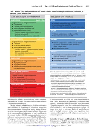
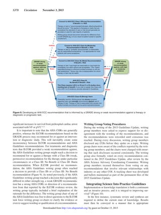
tiple criteria. Questions addressed in GRADE typically follow
a PICO (population, intervention, comparator, outcome) struc-
ture for ease of mapping to available evidence (Figure 1).
Confidence in the estimates of effect, synonymous with
and reported more succinctly as quality, is reported by a syn-
thesis of evidence informed by 1 or more studies as opposed
to studies themselves. Quality is adjudicated by a 4-part rank-
ing of our confidence in the estimate of effect (high, moderate,
low, very low) informed by study methodology and the risk of
bias. Studies start but do not necessarily end at high confidence
for randomized controlled trials (RCTs), and they start but do
not necessarily end at low confidence for observational stud-
ies. Studies may be downgraded for inconsistency, imprecision,
indirectness, and publication bias and nonrandomized observa-
tional studies may be upgraded as the result of effect size, dose-
response gradient, and plausible negative confounding; in other
words, an underestimation of the association. The direction and
strength of recommendations are driven by certainty of evidence
effect estimates, values and preferences of patients, and, to some
degree, clinicians’balance of positive and negative effects, costs
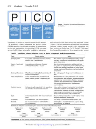
and resources, equity, acceptability, and feasibility (Table 2).
The GRADE Development Tool
The GRADE Guideline Development Tool (www.guide-
linedevelopment.org) provides a uniform interface in the form
(Circulation. 2015;132[suppl 2]:S368–S382. DOI: 10.1161/CIR.0000000000000253.)
© 2015 American Heart Association, Inc.
Circulation is available at http://circ.ahajournals.org DOI: 10.1161/CIR.0000000000000253
The American Heart Association requests that this document be cited as follows: Morrison LJ, Gent LM, Lang E, Nunnally ME, Parker MJ,
Callaway, CW, Nadkarni VM, Fernandez AR, Billi JE, Egan JR, Griffin RE, Shuster M, Hazinski MF. Part 2: evidence evaluation and management
of conflicts of interest: 2015 American Heart Association Guidelines Update for Cardiopulmonary Resuscitation and Emergency Cardiovascular Care.
Circulation. 2015;132(suppl 2):S368–S382.
*Co-chairs and equal first co-authors.
for Cardiopulmonary Resuscitation and Emergency
Cardiovascular Care
Part 2: Evidence Evaluation and
Management of Conflicts of Interest
2015 American Heart Association Guidelines Update for Cardiopulmonary
Resuscitation and Emergency Cardiovascular Care
Laurie J. Morrison, Chair; Lana M. Gent; Eddy Lang; Mark E. Nunnally; Melissa J. Parker;
Clifton W. Callaway; Vinay M. Nadkarni; Antonio R. Fernandez; John E. Billi;
Jonathan R. Egan; Russell E. Griffin; Michael Shuster; Mary Fran Hazinski
by guest on October 15, 2015http://circ.ahajournals.org/Downloaded from](https://image.slidesharecdn.com/guidelines-rcp-aha-2015-full-160515230707/85/Guidelines-rcp-aha-2015-full-58-320.jpg)
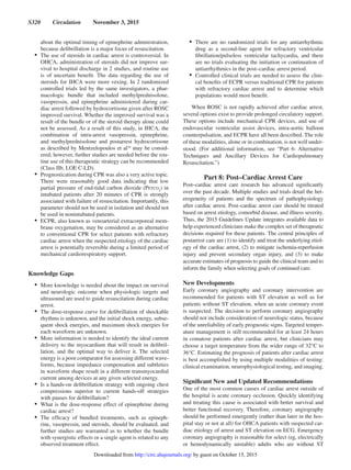
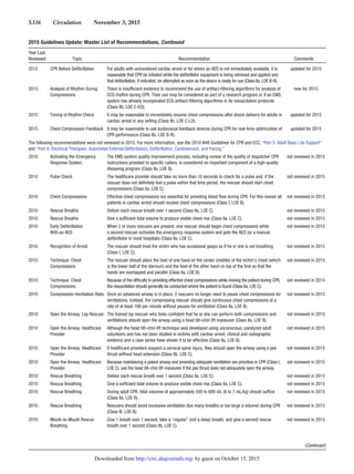
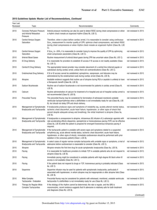
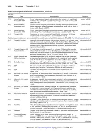
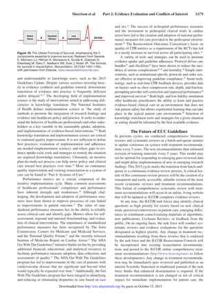
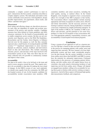
![Morrison et al Part 2: Evidence Evaluation and Conflicts of Interest S377
Identification of 2015 Guidelines Class of
Recommendation, Informed by ILCOR Consensus
Treatment Recommendation Based on GRADE
The second step in making a 2015 Guidelines Update recom-
mendation is to determine the strength of the recommenda-
tion. In many cases, after an extensive evidence review such
as that completed by ILCOR, the strength and direction
of the ILCOR treatment recommendation will be similar
to the strength and direction of the recommendation in the
2015 Guidelines Update. However, in its Clinical Practice
Guidelines Methodology Summit Report, the AHA task force
on practice guidelines12
notes that the strength of recommen-
dation and strength of evidence are each hierarchical but sepa-
rate. The classification table itself notes “COR and LOE are
determined independently, ie, any Class of Recommendation
may be paired with any Level of Evidence” (Table 1).
The writing groups for the 2015 Guidelines Update were
charged to carefully consider the 2015 ILCOR evidence review
and the ILCOR consensus treatment recommendations in light
of local training systems and the structure and resources of
out-of-hospital and in-hospital resuscitation systems. In addi-
tion, the writing groups weighed the balance between benefits
and risks and the quality of studies providing the evidence.
The writing group considered the precision, qualifications,
conditions, setting, outcomes, and limitations of the evidence
reviewed when making a final assessment. Generally, when
strong ILCOR recommendations were in favor of a treatment
or diagnostic test, the AHA Guidelines writing groups also
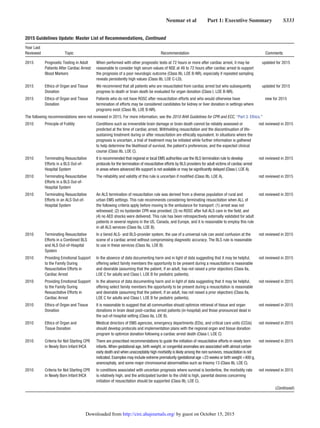
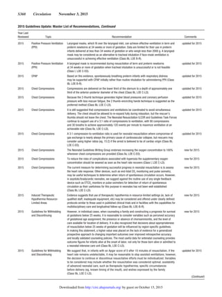
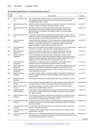
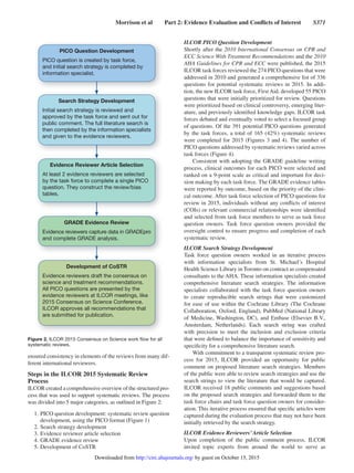
provided Class I or IIa recommendations (Figure 7). When
weak ILCOR recommendations were in favor of a treatment or
diagnostic test, the AHA Guidelines writing groups typically
provided a Class IIa, IIb, or a Class III: No Benefit recom-
mendation (Figure 8). If the AHA Guidelines writing group
reached a decision that significantly differed in either strength
(eg, a strong GRADE recommendation conversion to an AHA
Class IIb recommendation) or direction of a recommendation,
from that reported by the ILCOR evidence review, the writing
group typically included a brief explanation of the rationale
for the difference.
Ideally, strong recommendations from a scientific organi-
zation are supported by a high LOE. However, there are few
prospective RCTs and blinded clinical trials conducted in
resuscitation.As a result, it may be necessary for authors of this
2015 Guidelines Update to make recommendations to improve
survival, even in the absence of such high-quality evidence.
Such was the case in 2005, when the AHA and many other
resuscitation councils changed the treatment of pulseless arrest
associated with a shockable rhythm (ie, ventricular fibrillation
[VF] or pulseless ventricular tachycardia [pVT]) from a rec-
ommendation of 3 stacked shocks to recommending delivery
of single shocks followed by immediate CPR. Although there
were no studies documenting improved survival from VF/pVT
cardiac arrest with this approach, single shocks delivered by
biphasic defibrillators had a much higher first-shock success
than monophasic defibrillators, and experts felt strongly that
reducing interruptions in compressions would improve sur-
vival. This change in 2005, coupled with emphasis to mini-
mize interruptions in chest compressions, was associated with
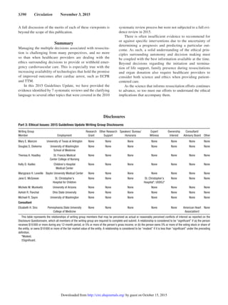
Figure 8. Developing an AHA ECC recommendation that is informed by a GRADE weak recommendation in favor of a therapy or
diagnostic or prognostic test.
by guest on October 15, 2015http://circ.ahajournals.org/Downloaded from](https://image.slidesharecdn.com/guidelines-rcp-aha-2015-full-160515230707/85/Guidelines-rcp-aha-2015-full-67-320.jpg)
![S383
The goals of resuscitation are to preserve life; restore
health; relieve suffering; limit disability; and respect indi-
viduals’ decisions, rights, and privacy. Because cardiopulmo-
nary resuscitation (CPR) efforts must be initiated immediately
at the time of arrest, a rescuer may not know who the victim is,
what that individual’s goals of care are, or if an advance direc-
tive exists. As a result, administration of CPR may be contrary
to the individual’s desires or best interests.1–3
This Part of the
2015 American Heart Association (AHA) Guidelines Update
for CPR and Emergency Cardiovascular Care provides
updates to the 2010 AHA Guidelines4
for healthcare providers
who are faced with the difficult decision to provide or with-
hold emergency cardiovascular care.
Ethical Principles
Ethical, legal, and cultural factors influence decisions about
resuscitation. Ideally, these decisions are guided by science,
patient or surrogate preferences, local policies and legal
requirements, and established ethical principles.
Principle of Respect for Autonomy
Respect for autonomy is an important social value in medical
ethics and law.5
This principle is based on society’s respect for
a competent individual’s ability to make decisions about his
or her own health care. Adults are presumed to have decision-
making capability unless they are incapacitated or declared
incompetent by a court of law. Informed decisions require that
individuals receive and understand accurate information about
their condition and prognosis as well as the nature, risks, bene-
fits, and alternatives of any proposed interventions. Individuals
must deliberate and choose among alternatives by linking their
decisions to their values and personal goals of care.
When physicians strive to understand patients’ goals
of care, decisions can be made based on the likelihood that
together they will achieve the patients’ goals of care. The fol-
lowing 3-step process may assist healthcare providers in ensur-
ing each patient understands and makes informed decisions:
(1) the patient receives and understands accurate information
about his or her condition, prognosis, nature of any proposed
interventions, alternatives, and risks and benefits; (2) the
patient is asked to paraphrase the information to give provid-
ers the opportunity to assess the patient’s understanding and
correct any misimpressions; and (3) the patient deliberates and
chooses among alternatives and justifies his or her decisions.6
When decision-making capacity is temporarily impaired
by conditions such as active illness, treatment of these condi-
tions may restore capacity. When an individual’s preferences
are unknown or uncertain, it is ethically appropriate to treat
emergency conditions until further information is available.
Pediatric Decision Making
As a general rule, minors are considered incompetent to pro-
vide legally binding consent about their health care. Parents or
guardians are generally empowered to make healthcare deci-
sions on the behalf of minors, and in most situations, parents
are given wide latitude in terms of the decisions they make
on behalf of their children. Ethically, however, a child should
be involved in decision making at a level appropriate for the
child’s maturity. Children under 14 years of age in Canada
and under 18 years of age in the United States rarely possess
the legal authority to consent to their health care except under
specific legally defined situations (eg, emancipated minors;
mature minors; minors who have specific health conditions,
such as those with sexually transmitted diseases or in need of
pregnancy-related care). However, as older children develop
the capacity to make decisions, it is ethically appropriate to
include them in discussions about their care and the treatments
using language and explanations suitable for the child’s level
of maturity and cognitive function.
Withholding and Withdrawing CPR
(Termination of Resuscitative Efforts)
Related to Out-of-Hospital Cardiac Arrest
Criteria for Not Starting CPR
While the general rule is to provide emergency treatment to
a victim of cardiac arrest, there are a few exceptions where
withholding CPR would be considered appropriate:
(Circulation. 2015;132[suppl 2]:S383–S396. DOI: 10.1161/CIR.0000000000000254.)
© 2015 American Heart Association, Inc.
Circulation is available at http://circ.ahajournals.org DOI: 10.1161/CIR.0000000000000254
The American Heart Association requests that this document be cited as follows: Mancini ME, Diekema DS, Hoadley TA, Kadlec KD, Leveille
MH, McGowan JE, Munkwitz MM, Panchal AR, Sayre MR, Sinz EH. Part 3: ethical issues: 2015 American Heart Association Guidelines Update for
Cardiopulmonary Resuscitation and Emergency Cardiovascular Care. Circulation. 2015;132(suppl 2):S383–S396.
Part 3: Ethical Issues
2015 American Heart Association Guidelines Update for Cardiopulmonary
Resuscitation and Emergency Cardiovascular Care
Mary E. Mancini, Chair; Douglas S. Diekema; Theresa A. Hoadley; Kelly D. Kadlec;
Marygrace H. Leveille; Jane E. McGowan; Michele M. Munkwitz; Ashish R. Panchal;
Michael R. Sayre; Elizabeth H. Sinz
by guest on October 15, 2015http://circ.ahajournals.org/Downloaded from](https://image.slidesharecdn.com/guidelines-rcp-aha-2015-full-160515230707/85/Guidelines-rcp-aha-2015-full-74-320.jpg)
![S388 Circulation November 3, 2015
This topic continues to be an area of active research.
The use of TTM has demonstrated the potential to improve
the neurologic outcome in certain adult patients after car-
diac arrest who might otherwise have a poor neurologic
outcome. Although the data and literature are becoming
more robust on this particular topic, there are few differ-
ences in the types of tests used in those who are and are not
treated with TTM as relates to prognosticating neurologic
outcome.
2015 Evidence Summary—New
For a full description of the evidence reviewed for each
assessment of neurologic function and prognosis for adults
who have had cardiac arrest, refer to “Part 8: Post–Cardiac
Arrest Care.”
2015 Recommendations: Clinical Examination
Findings—New
In comatose patients who are not treated with TTM, the
absence of pupillary reflex to light at 72 hours or more after
cardiac arrest is a reasonable exam finding with which to pre-
dict poor neurologic outcome (FPR [false-positive rate], 0%;
95% CI, 0%–8%; Class IIa, LOE B-NR).
In comatose patients who are treated with TTM, the
absence of pupillary reflex to light at 72 hours or more after
cardiac arrest is useful to predict poor neurologic outcome
(FPR, 0%; 95% CI, 0%–3%; Class I, LOE B-NR).
We recommend that, given their high FPRs, the findings of
either absent motor movements or extensor posturing should
not be used alone for predicting a poor neurologic outcome
(FPR, 10%; 95% CI, 7%–15% to FPR, 15%; 95% CI, 5%–
31%; Class III: Harm, LOE B-NR). The motor examination
may be a reasonable means to identify the population who
need further prognostic testing to predict poor outcome (Class
IIb, LOE B-NR).
We recommend that the presence of myoclonus, which is
distinct from status myoclonus, should not be used to predict
poor neurologic outcomes because of the high FPR (FPR, 5%;
95% CI, 3%–8% to FPR, 11%; 95% CI, 3%–26%; Class III:
Harm, LOE B-NR).
In combination with other diagnostic tests at 72 or more
hours after cardiac arrest, the presence of status myoclonus
during the first 72 hours after cardiac arrest is a reasonable
finding to help predict poor neurologic outcomes (FPR, 0%;
95% CI, 0%–4%; Class IIa, LOE B-NR).
2015 Recommendations: EEGALS 450, ALS 713
—Updated
In comatose post–cardiac arrest patients who are treated with
TTM, it may be reasonable to consider persistent absence of
EEG reactivity to external stimuli at 72 hours after cardiac
arrest, and persistent burst suppression on EEG after rewarm-
ing, to predict a poor outcome (FPR, 0%; 95% CI, 0%–3%;
Class IIb, LOE B-NR).
Intractable and persistent (more than 72 hours) status epi-
lepticus in the absence of EEG reactivity to external stimuli
may be reasonable to predict poor outcome (Class IIb, LOE
B-NR).
In comatose post–cardiac arrest patients who are not
treated with TTM, it may be reasonable to consider the pres-
ence of burst suppression on EEG at 72 hours or more after
cardiac arrest, in combination with other predictors, to pre-
dict a poor neurologic outcome (FPR, 0%; 95% CI, 0%–11%;
Class IIb, LOE B-NR).
2015 Recommendation: Evoked PotentialsALS 450
—Updated
In patients who are comatose after resuscitation from cardiac
arrest regardless of treatment with TTM, it is reasonable to
consider bilateral absence of the N20 somatosensory evoked
potentials (SSEP) wave 24 to 72 hours after cardiac arrest or
after rewarming a predictor of poor outcome (FPR, 1%; 95%
CI, 0%–3%; Class IIa, LOE B-NR).
SSEP recording requires appropriate skills and experi-
ence, and utmost care should be taken to avoid electrical inter-
ference from muscle artifacts or from the intensive care unit
environment. However, sedative drugs or temperature manip-
ulation affect SSEPs less than they affect the EEG and clinical
examination.58,59
2015 Recommendations: Imaging TestsALS 713
—New
In patients who are comatose after resuscitation from cardiac
arrest and are not treated with TTM, it may be reasonable
to use the presence of a marked reduction of the gray-white
ratio on brain computed tomography obtained within 2 hours
after cardiac arrest to predict poor outcome (Class IIb, LOE
B-NR).
It may be reasonable to consider extensive restriction of
diffusion on brain magnetic resonance imaging at 2 to 6 days
after cardiac arrest in combination with other established pre-
dictors for predicting a poor neurologic outcome (Class IIb,
LOE B-NR).
Note that acquisition and interpretation of imaging studies
have not been fully standardized and are affected by interob-
server variability.60
Therefore, brain imaging studies for prog-
nostication should be performed only in centers where specific
experience is available.
2015 Recommendations: Blood MarkersALS 713, ALS 450
Updated
Given the possibility of high FPRs, blood levels of NSE and
S-100B should not be used alone to predict a poor neurologic
outcome (Class III: Harm, LOE C-LD). When performed with
other prognostic tests at 72 hours or more after cardiac arrest,
it may be reasonable to consider high serum values of NSE at
48 to 72 hours after cardiac arrest to support the prognosis of
a poor neurologic outcome (Class IIb, LOE B-NR), especially
if repeated sampling reveals persistently high values (Class
IIb, LOE C-LD).
Laboratory standards for NSE and S-100B measurement
vary between centers, making comparison of absolute values
difficult. The kinetics of these markers have not been stud-
ied, particularly during or after TTM in cardiac arrest patients.
Finally, NSE and S-100B are not specific to neuronal damage
and can be produced by extra–central nervous system sources
(hemolysis, neuroendocrine tumors, myenteric plexus, muscle
and adipose tissue breakdown). If care is not taken when draw-
ing NSE levels and if multiple time points are not assessed,
false-positive results could occur secondary to hemolysis. All
of these limitations led the writing group to conclude that NSE
should be limited to a confirmatory test rather than a primary
method for estimating prognosis.
by guest on October 15, 2015http://circ.ahajournals.org/Downloaded from](https://image.slidesharecdn.com/guidelines-rcp-aha-2015-full-160515230707/85/Guidelines-rcp-aha-2015-full-79-320.jpg)
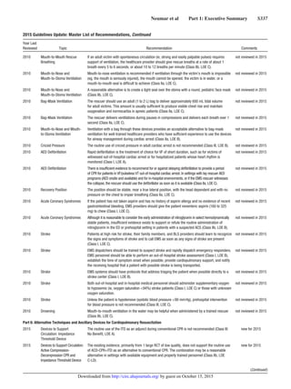
![S397
Introduction
The science and recommendations discussed in the other Parts
of the 2015 American Heart Association (AHA) Guidelines
Update for Cardiopulmonary Resuscitation (CPR) and
Emergency Cardiovascular Care (ECC) form the backbone of
resuscitation. They answer the “why”, “what,” and “when” of
performing resuscitation steps. In a perfectly controlled and
predictable environment, such as a laboratory setting, those
answers often suffice, but the “how” of actual implementa-
tion depends on knowing the “who” and “where” as well. The
ideal work flow to accomplish resuscitation successfully is
highly dependent on the system of care as a whole.
Healthcare delivery requires structure (eg, people, equip-
ment, education, prospective registry data collection) and
process (eg, policies, protocols, procedures), which, when
integrated, produce a system (eg, programs, organizations,
cultures) leading to outcomes (eg, patient safety, quality, sat-
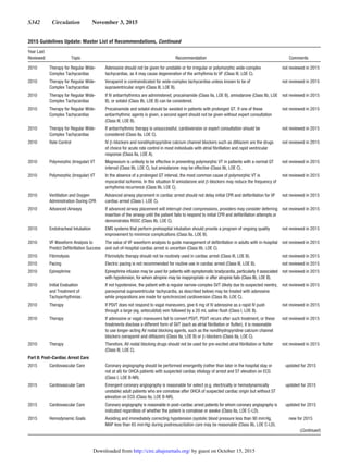
isfaction). An effective system of care (Figure 1) comprises
all of these elements—structure, process, system, and patient
outcomes—in a framework of continuous quality improve-
ment (CQI).
In this Part, we will focus on 2 distinct systems of care:
the system for patients who arrest inside the hospital and the
one for those who arrest outside it. We will set into context the
building blocks for a system of care for cardiac arrest, with
consideration of the setting, team, and available resources, as
well as CQI from the moment the patient becomes unstable
until after the patient is discharged.
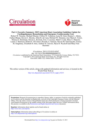
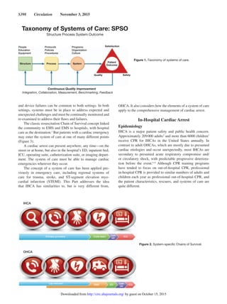
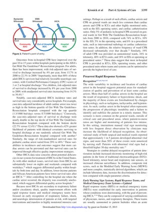
The chain of survival metaphor, first used almost 25 years
ago,1
is still very relevant. However, it may be helpful to cre-
ate 2 separate chains (Figure 2) to reflect the differences in
the steps needed for response to cardiac arrest in the hospital
(in-hospital cardiac arrest [IHCA]) and out of the hospital (out
of hospital cardiac arrest [OHCA]). Regardless of where an
arrest occurs, the care following resuscitation converges in the
hospital, generally in an emergency department (ED) or inten-
sive care unit (ICU). This post‒cardiac arrest care is depicted
as the final link in both chains, symbolized by a hospital bed
with a monitor and thermometer, which represent advanced
monitoring and targeted temperature management. As noted
above, the structure and process elements before the conver-
gence of the 2 chains, however, vary significantly.
Patients with OHCA depend on elements within the com-
munity for support. Lay rescuers must recognize the patient’s
arrest, call for help, and initiate CPR and early defibrillation
(public-access defibrillation [PAD]) until a team of profes-
sionally trained emergency medical services (EMS) provid-
ers assumes responsibility and then transports the patient to
an ED and/or cardiac catheterization lab, and then on to an
ICU for post‒cardiac arrest care. Ideally, all victims of OHCA
receive bystander CPR and defibrillation; if not, CPR and defi-
brillation won’t occur until EMS personnel arrive, and the vic-
tim’s chance of survival is then much lower.
In contrast, patients with IHCA depend on a system of
appropriate surveillance and prevention of cardiac arrest,
which is represented by a magnifying glass in the first link.
When cardiac arrest occurs, prompt notification and response
to a cardiac arrest should result in the smooth interaction of
a multidisciplinary team of professional providers, including
physicians, nurses, respiratory therapists, and others. This
team provides high-quality CPR, prompt defibrillation, and
advanced cardiovascular life support when appropriate. The
chain metaphor endures: in any resuscitation, the chain is no
stronger than its weakest link.
The level of complexity is high for both in-hospital and
out-of-hospital systems. The challenges encountered, how-
ever, are different. Teamwork and coordination among
responders is a critical determinant of patient outcomes. An
in-hospital multidisciplinary team has immediate access to
additional personnel as well as all the resources of the ED,
ICU, and laboratories, whereas in out-of-hospital settings,
2 paramedics may find themselves alone with no resources
except those they brought with them. Factors such as crowd
control, family presence, space constraints, transportation,
(Circulation. 2015;132[suppl 2]:S397–S413. DOI: 10.1161/CIR.0000000000000258.)
© 2015 American Heart Association, Inc.
Circulation is available at http://circ.ahajournals.org DOI: 10.1161/CIR.0000000000000258
The American Heart Association requests that this document be cited as follows: Kronick SL, Kurz MC, Lin S, Edelson DP, Berg RA, Billi JE, Cabanas
JG, Cone DC, Diercks DB, Foster J, Meeks RA, Travers AH, Welsford M. Part 4: systems of care and continuous quality improvement: 2015 American
HeartAssociation Guidelines Update for Cardiopulmonary Resuscitation and Emergency Cardiovascular Care. Circulation. 2015;132(suppl 2):S397–S413.
for Cardiopulmonary Resuscitation and Emergency
Cardiovascular Care
Part 4: Systems of Care and Continuous
Quality Improvement
2015 American Heart Association Guidelines Update for Cardiopulmonary
Resuscitation and Emergency Cardiovascular Care
Steven L. Kronick, Chair; Michael C. Kurz; Steve Lin; Dana P. Edelson; Robert A. Berg;
John E. Billi; Jose G. Cabanas; David C. Cone; Deborah B. Diercks; James (Jim) Foster;
Reylon A. Meeks; Andrew H. Travers; Michelle Welsford
by guest on October 15, 2015http://circ.ahajournals.org/Downloaded from](https://image.slidesharecdn.com/guidelines-rcp-aha-2015-full-160515230707/85/Guidelines-rcp-aha-2015-full-89-320.jpg)
![S410 Circulation November 3, 2015
48. Simmes FM, Schoonhoven L, Mintjes J, Fikkers BG, van der Hoeven JG.
Incidence of cardiac arrests and unexpected deaths in surgical patients
before and after implementation of a rapid response system. Ann Intensive
Care. 2012;2:20. doi: 10.1186/2110-5820-2-20.
49. Snyder CW, Patel RD, Roberson EP, Hawn MT. Unplanned intubation
after surgery: risk factors, prognosis, and medical emergency team effects.
Am Surg. 2009;75:834–838.
50. Vazquez R, Gheorghe C, Grigoriyan A, Palvinskaya T, Amoateng-
Adjepong Y, Manthous CA. Enhanced end-of-life care associated with
deploying a rapid response team: a pilot study. J Hosp Med. 2009;4:449–
452. doi: 10.1002/jhm.451.
51. Rothschild JM, Woolf S, Finn KM, Friedberg MW, Lemay C, Furbush
KA, Williams DH, Bates DW. A controlled trial of a rapid response sys-
tem in an academic medical center. Jt Comm J Qual Patient Saf. 2008;34:
417–425, 365.
52. Hunt EA, Zimmer KP, Rinke ML, Shilkofski NA, Matlin C, Garger C,
Dickson C, Miller MR. Transition from a traditional code team to a medi-
cal emergency team and categorization of cardiopulmonary arrests in a
children’s center. Arch Pediatr Adolesc Med. 2008;162:117–122. doi:
10.1001/archpediatrics.2007.33.
53. Sharek PJ, Parast LM, Leong K, Coombs J, Earnest K, Sullivan J,
Frankel LR, Roth SJ. Effect of a rapid response team on hospital-wide
mortality and code rates outside the ICU in a Children’s Hospital. JAMA.
2007;298:2267–2274. doi: 10.1001/jama.298.19.2267.
54. Anwar-ul-Haque, Saleem AF, Zaidi S, Haider SR. Experience of pediat-
ric rapid response team in a tertiary care hospital in Pakistan. Indian J
Pediatr. 2010;77:273–276. doi: 10.1007/s12098-010-0032-2.
55. Kotsakis A, Lobos AT, Parshuram C, Gilleland J, Gaiteiro R, Mohseni-
Bod H, Singh R, Bohn D; Ontario Pediatric Critical Care Response Team
Collaborative. Implementation of a multicenter rapid response system in
pediatric academic hospitals is effective. Pediatrics. 2011;128:72–78. doi:
10.1542/peds.2010-0756.
56. Hanson CC, Randolph GD, Erickson JA, Mayer CM, Bruckel JT, Harris
BD, Willis TS. A reduction in cardiac arrests and duration of clinical insta-
bility after implementation of a paediatric rapid response system. Postgrad
Med J. 2010;86:314–318. doi: 10.1136/qshc.2007.026054.
57. Zenker P, Schlesinger A, Hauck M, Spencer S, Hellmich T, Finkelstein
M, Thygeson MV, Billman G. Implementation and impact of a rapid
response team in a children’s hospital. Jt Comm J Qual Patient Saf.
2007;33:418–425.
58. Brilli RJ, Gibson R, Luria JW, Wheeler TA, Shaw J, Linam M, Kheir J,
McLain P, Lingsch T, Hall-Haering A, McBride M. Implementation of a
medical emergency team in a large pediatric teaching hospital prevents
respiratory and cardiopulmonary arrests outside the intensive care unit.
Pediatr Crit Care Med. 2007;8:236–246; quiz 247. doi: 10.1097/01.
PCC.0000262947.72442.EA.
59. Tibballs J, Kinney S. Reduction of hospital mortality and of preventable
cardiac arrest and death on introduction of a pediatric medical emer-
gency team. Pediatr Crit Care Med. 2009;10:306–312. doi: 10.1097/
PCC.0b013e318198b02c.
60. Randhawa S, Roberts-Turner R, Woronick K, DuVal J. Implementing
and sustaining evidence-based nursing practice to reduce pediat-
ric cardiopulmonary arrest. West J Nurs Res. 2011;33:443–456. doi:
10.1177/0193945910379585.
61. Cardoso LT, Grion CM, Matsuo T, Anami EH, Kauss IA, Seko L,
Bonametti AM. Impact of delayed admission to intensive care units on
mortality of critically ill patients: a cohort study. Crit Care. 2011;15:R28.
doi: 10.1186/cc9975.
62. Meaney PA, Bobrow BJ, Mancini ME, Christenson J, de Caen AR,
Bhanji F, Abella BS, Kleinman ME, Edelson DP, Berg RA, Aufderheide
TP, Menon V, Leary M; CPR Quality Summit Investigators, the
American Heart Association Emergency Cardiovascular Care
Committee, and the Council on Cardiopulmonary, Critical Care,
Perioperative and Resuscitation. Cardiopulmonary resuscitation qual-
ity: [corrected] improving cardiac resuscitation outcomes both inside
and outside the hospital: a consensus statement from the American
Heart Association. Circulation. 2013;128:417–435. doi: 10.1161/
CIR.0b013e31829d8654.
63. Edelson DP,Yuen TC, Mancini ME, Davis DP, Hunt EA, Miller JA, Abella
BS. Hospital cardiac arrest resuscitation practice in the United States: a
nationally representative survey. J Hosp Med. 2014;9:353–357. doi:
10.1002/jhm.2174.
64. Mancini ME, Soar J, Bhanji F, Billi JE, Dennett J, Finn J, Ma
MH, Perkins GD, Rodgers DL, Hazinski MF, Jacobs I, Morley PT;
on behalf of the Education, Implementation, and Teams Chapter
Collaborators. Part 12: education, implementation, and teams:
2010 International Consensus on Cardiopulmonary Resuscitation
and Emergency Cardiovascular Care Science With Treatment
Recommendations. Circulation. 2010;122(suppl 2):S539–S581. doi:
10.1161/CIRCULATIONAHA.110.971143.
65. Soar J, Mancini ME, Bhanji F, Billi JE, Dennett J, Finn J, Ma MH,
Perkins GD, Rodgers DL, Hazinski MF, Jacobs I, Morley PT; Education,
Implementation, and Teams Chapter Collaborators. Part 12: educa-
tion, implementation, and teams: 2010 International Consensus on
Cardiopulmonary Resuscitation and Emergency Cardiovascular Care
Science with Treatment Recommendations. Resuscitation. 2010;81
suppl 1:e288–e330. doi: 10.1016/j.resuscitation.2010.08.030.
66. Hunziker S, Johansson AC, Tschan F, Semmer NK, Rock L, Howell
MD, Marsch S. Teamwork and leadership in cardiopulmonary resus-
citation. J Am Coll Cardiol. 2011;57:2381–2388. doi: 10.1016/j.
jacc.2011.03.017.
67. Hunziker S, Bühlmann C, Tschan F, Balestra G, Legeret C, Schumacher
C, Semmer NK, Hunziker P, Marsch S. Brief leadership instructions
improve cardiopulmonary resuscitation in a high-fidelity simulation: a
randomized controlled trial. Crit Care Med. 2010;38:1086–1091. doi:
10.1097/CCM.0b013e3181cf7383.
68. Ten Eyck RP, Tews M, Ballester JM, Hamilton GC. Improved fourth-
year medical student clinical decision-making performance as a resus-
citation team leader after a simulation-based curriculum. Simul Healthc.
2010;5:139–145. doi: 10.1097/SIH.0b013e3181cca544.
69. Fernandez Castelao E, Russo SG, Cremer S, Strack M, Kaminski L,
Eich C, Timmermann A, Boos M. Positive impact of crisis resource
management training on no-flow time and team member verbalisa-
tions during simulated cardiopulmonary resuscitation: a randomised
controlled trial. Resuscitation. 2011;82:1338–1343. doi: 10.1016/j.
resuscitation.2011.05.009.
70. Ornato JP, Peberdy MA. Applying lessons from commercial aviation
safety and operations to resuscitation. Resuscitation. 2014;85:173–176.
doi: 10.1016/j.resuscitation.2013.10.029.
71. Neumar RW, Otto CW, Link MS, Kronick SL, Shuster M, Callaway
CW, Kudenchuk PJ, Ornato JP, McNally B, Silvers SM, Passman RS,
White RD, Hess EP, Tang W, Davis D, Sinz E, Morrison LJ. Part 8: adult
advanced cardiovascular life support: 2010 American Heart Association
Guidelines for Cardiopulmonary Resuscitation and Emergency
Cardiovascular Care. Circulation. 2010;122(suppl 3):S729–S767. doi:
10.1161/CIRCULATIONAHA.110.970988.
72. Kleinman ME, Chameides L, Schexnayder SM, Samson RA, Hazinski
MF, Atkins DL, Berg MD, de Caen AR, Fink EL, Freid EB, Hickey
RW, Marino BS, Nadkarni VM, Proctor LT, Qureshi FA, Sartorelli
K, Topjian A, van der Jagt EW, Zaritsky AL. Part 14: pediatric
advanced life support: 2010 American Heart Association Guidelines
for Cardiopulmonary Resuscitation and Emergency Cardiovascular
Care. Circulation. 2010;122(suppl 3):S876–S908. doi: 10.1161/
CIRCULATIONAHA.110.971101.
73. Cooper S. Developing leaders for advanced life support: evaluation of a
training programme. Resuscitation. 2001;49:33–38.
74. DePriest J, Fee-Mulhearn AL, Teleron A. A novel ACLS team leader
checklist implemented to improve resuscitation efforts. Resuscitation.
2013;84:e115. doi: 10.1016/j.resuscitation.2013.03.002.
75. Bhanji F, Mancini ME, Sinz E, Rodgers DL, McNeil MA, Hoadley TA,
Meeks RA, Hamilton MF, Meaney PA, Hunt EA, Nadkarni VM, Hazinski
MF. Part 16: education, implementation, and teams: 2010 American
Heart Association Guidelines for Cardiopulmonary Resuscitation and
Emergency Cardiovascular Care. Circulation. 2010;122(suppl 3):
S920–S933. doi: 10.1161/CIRCULATIONAHA.110.971135.
76. Weidman EK, Bell G, Walsh D, Small S, Edelson DP. Assessing the
impact of immersive simulation on clinical performance during actual
in-hospital cardiac arrest with CPR-sensing technology: a randomized
feasibility study. Resuscitation. 2010;81:1556–1561. doi: 10.1016/j.
resuscitation.2010.05.021.
77. Yeung J, Meeks R, Edelson D, Gao F, Soar J, Perkins GD. The use of
CPR feedback/prompt devices during training and CPR performance:
A systematic review. Resuscitation. 2009;80:743–751. doi: 10.1016/j.
resuscitation.2009.04.012.
78. Niles D, Sutton RM, DonoghueA, Kalsi MS, Roberts K, Boyle L, Nishisaki
A, Arbogast KB, Helfaer M, Nadkarni V. “Rolling Refreshers”: a novel
approach to maintain CPR psychomotor skill competence. Resuscitation.
2009;80:909–912. doi: 10.1016/j.resuscitation.2009.04.021.
79. Sutton RM, Niles D, Meaney PA,Aplenc R, French B,Abella BS, Lengetti
EL, Berg RA, Helfaer MA, Nadkarni V. Low-dose, high-frequency
by guest on October 15, 2015http://circ.ahajournals.org/Downloaded from](https://image.slidesharecdn.com/guidelines-rcp-aha-2015-full-160515230707/85/Guidelines-rcp-aha-2015-full-102-320.jpg)
![S412 Circulation November 3, 2015
108. Hallstrom AP. Dispatcher-assisted “phone” cardiopulmonary resuscita-
tion by chest compression alone or with mouth-to-mouth ventilation.
Crit Care Med. 2000;28(11 suppl):N190–N192.
109. Hüpfl M, Selig HF, Nagele P. Chest-compression-only versus stan-
dard cardiopulmonary resuscitation: a meta-analysis. Lancet.
2010;376:1552–1557. doi: 10.1016/S0140-6736(10)61454-7.
110. Bray JE, Deasy C, Walsh J, Bacon A, Currell A, Smith K. Changing
EMS dispatcher CPR instructions to 400 compressions before mouth-
to-mouth improved bystander CPR rates. Resuscitation. 2011;82:1393–
1398. doi: 10.1016/j.resuscitation.2011.06.018.
111. Culley LL, Clark JJ, Eisenberg MS, Larsen MP. Dispatcher-assisted
telephone CPR: common delays and time standards for delivery. Ann
Emerg Med. 1991;20:362–366.
112. Rea TD, Eisenberg MS, Culley LL, Becker L. Dispatcher-assisted car-
diopulmonary resuscitation and survival in cardiac arrest. Circulation.
2001;104:2513–2516.
113. Ringh M, Fredman D, Nordberg P, Stark T, Hollenberg J. Mobile
phone technology identifies and recruits trained citizens to per-
form CPR on out-of-hospital cardiac arrest victims prior to ambu-
lance arrival. Resuscitation. 2011;82:1514–1518. doi: 10.1016/j.
resuscitation.2011.07.033.
114. Zijlstra JA, Stieglis R, Riedijk F, Smeekes M, van der Worp WE, Koster
RW. Local lay rescuers with AEDs, alerted by text messages, contrib-
ute to early defibrillation in a Dutch out-of-hospital cardiac arrest
dispatch system. Resuscitation. 2014;85:1444–1449. doi: 10.1016/j.
resuscitation.2014.07.020.
114a. Ringh M, Rosenqvist M, Hollenberg J, Jonsson M, Fredman D, Nordberg
P, Järnbert-Pettersson H, Hasselqvist-Ax I, Riva G, Svensson L. Mobile-
phone dispatch of laypersons for CPR in out-of-hospital cardiac arrest.
N Engl J Med. 2015;24:2316–2325. doi: 10.1056/NEJMoa1406038.
115. Perkins GD, Jacobs IG, Nadkarni VM, Berg RA, Bhanji F, Biarent D,
Bossaert LL, Brett SJ, Chamberlain D, de Caen AR, Deakin CD, Finn JC,
GräsnerJ-T,HazinskiMF,IwamiT,KosterRW,LimSH,Huei-MingMaM,
McNally BF, Morley PT, Morrison LJ, Monsieurs KG, Montgomery W,
Nichol G, Okada K, Eng Hock Ong M, Travers AH, Nolan JP; for the
Utstein Collaborators. Cardiac arrest and cardiopulmonary resuscitation
outcome reports: update of the Utstein Resuscitation Registry Templates
for Out-of-Hospital CardiacArrest: a statement for healthcare professionals
from a task force of the International Liaison Committee on Resuscitation
(American Heart Association, European Resuscitation Council, Australian
and New Zealand Council on Resuscitation, Heart and Stroke Foundation
of Canada, InterAmerican Heart Foundation, Resuscitation Council of
Southern Africa, Resuscitation Council of Asia); and the American Heart
Association Emergency Cardiovascular Care Committee and the Council
on Cardiopulmonary, Critical Care, Perioperative and Resuscitation.
Circulation. 2015;132:1286–1300. doi: 10.1161/CIR.0000000000000144.
116. Le May MR, So DY, Dionne R, Glover CA, Froeschl MP, Wells GA,
Davies RF, Sherrard HL, Maloney J, Marquis JF, O’Brien ER, Trickett
J, Poirier P, Ryan SC, Ha A, Joseph PG, Labinaz M. A citywide proto-
col for primary PCI in ST-segment elevation myocardial infarction. N
Engl J Med. 2008;358:231–240. doi: 10.1056/NEJMoa073102.
117. Mercuri M, Velianou JL, Welsford M, Gauthier L, Natarajan MK.
Improving the timeliness of care for patients with acute ST-elevation
myocardial infarction: implications of “self-transport” versus use of
EMS. Healthc Q. 2010;13:105–109.
118. Kajino K, Iwami T, Daya M, Nishiuchi T, Hayashi Y, Kitamura T,
Irisawa T, Sakai T, Kuwagata Y, Hiraide A, Kishi M, Yamayoshi S.
Impact of transport to critical care medical centers on outcomes after
out-of-hospital cardiac arrest. Resuscitation. 2010;81:549–554. doi:
10.1016/j.resuscitation.2010.02.008.
119. Callaway CW, Schmicker R, Kampmeyer M, Powell J, Rea TD, Daya
MR, Aufderheide TP, Davis DP, Rittenberger JC, Idris AH, Nichol G;
Resuscitation Outcomes Consortium (ROC) Investigators. Receiving
hospital characteristics associated with survival after out-of-hospital
cardiac arrest. Resuscitation. 2010;81:524–529. doi: 10.1016/j.
resuscitation.2009.12.006.
120. Carr BG, Goyal M, Band RA, Gaieski DF, Abella BS, Merchant RM,
Branas CC, Becker LB, Neumar RW.A national analysis of the relation-
ship between hospital factors and post-cardiac arrest mortality. Intensive
Care Med. 2009;35:505–511. doi: 10.1007/s00134-008-1335-x.
121. Cudnik MT, Sasson C, Rea TD, Sayre MR, Zhang J, Bobrow BJ, Spaite
DW, McNally B, Denninghoff K, Stolz U. Increasing hospital volume
is not associated with improved survival in out of hospital cardiac arrest
of cardiac etiology. Resuscitation. 2012;83:862–868. doi: 10.1016/j.
resuscitation.2012.02.006.
122. Engdahl J, Abrahamsson P, Bång A, Lindqvist J, Karlsson T, Herlitz J.
Is hospital care of major importance for outcome after out-of-hospital
cardiac arrest? Experience acquired from patients with out-of-hospital
cardiac arrest resuscitated by the same Emergency Medical Service and
admitted to one of two hospitals over a 16-year period in the municipal-
ity of Göteborg. Resuscitation. 2000;43:201–211.
123. Hansen M, Fleischman R, Meckler G, Newgard CD. The associa-
tion between hospital type and mortality among critically ill chil-
dren in US EDs. Resuscitation. 2013;84:488–491. doi: 10.1016/j.
resuscitation.2012.07.032.
124. Herlitz J, Engdahl J, Svensson L, Angquist KA, Silfverstolpe J,
Holmberg S. Major differences in 1-month survival between hos-
pitals in Sweden among initial survivors of out-of-hospital car-
diac arrest. Resuscitation. 2006;70:404–409. doi: 10.1016/j.
resuscitation.2006.01.014.
125. Kjaergaard J, Bro-Jeppesen J, Rasmussen LS, Nielsen SL, Folke F,
Lippert F, Wanscher MC, Hassager C. [Differences between hospitals
in prognosis after resuscitated out-of-hospital cardiac arrest patients].
Ugeskr Laeger. 2009;171:2169–2173.
126. Stub D, Smith K, Bray JE, Bernard S, Duffy SJ, Kaye DM. Hospital
characteristics are associated with patient outcomes following out-
of-hospital cardiac arrest. Heart. 2011;97:1489–1494. doi: 10.1136/
hrt.2011.226431.
127. Davis DP, Fisher R, Aguilar S, Metz M, Ochs G, McCallum-Brown L,
Ramanujam P, Buono C, Vilke GM, Chan TC, Dunford JV. The fea-
sibility of a regional cardiac arrest receiving system. Resuscitation.
2007;74:44–51. doi: 10.1016/j.resuscitation.2006.11.009.
128. Heffner AC, Pearson DA, Nussbaum ML, Jones AE. Regionalization
of post-cardiac arrest care: implementation of a cardiac resusci-
tation center. Am Heart J. 2012;164:493–501.e2. doi: 10.1016/j.
ahj.2012.06.014.
129. Mooney MR, Unger BT, Boland LL, Burke MN, Kebed KY, Graham
KJ, Henry TD, Katsiyiannis WT, Satterlee PA, Sendelbach S,
Hodges JS, Parham WM. Therapeutic hypothermia after out-of-
hospital cardiac arrest: evaluation of a regional system to increase
access to cooling. Circulation. 2011;124:206–214. doi: 10.1161/
CIRCULATIONAHA.110.986257.
130. Spaite DW, Bobrow BJ, Vadeboncoeur TF, Chikani V, Clark L, Mullins
T, Sanders AB. The impact of prehospital transport interval on survival
in out-of-hospital cardiac arrest: implications for regionalization of
post-resuscitation care. Resuscitation. 2008;79:61–66. doi: 10.1016/j.
resuscitation.2008.05.006.
131. Spaite DW, Stiell IG, Bobrow BJ, de Boer M, Maloney J, Denninghoff
K, Vadeboncoeur TF, Dreyer J, Wells GA. Effect of transport
interval on out-of-hospital cardiac arrest survival in the OPALS
study: implications for triaging patients to specialized cardiac
arrest centers. Ann Emerg Med. 2009;54:248–255. doi: 10.1016/j.
annemergmed.2008.11.020.
132. Bosson N, Kaji AH, Niemann JT, Eckstein M, Rashi P, Tadeo R,
Gorospe D, Sung G, French WJ, Shavelle D, Thomas JL, Koenig W.
Survival and neurologic outcome after out-of-hospital cardiac arrest:
results one year after regionalization of post-cardiac arrest care in a
large metropolitan area. Prehosp Emerg Care. 2014;18:217–223. doi:
10.3109/10903127.2013.856507.
133. Fothergill RT, Watson LR, Virdi GK, Moore FP, Whitbread M. Survival
of resuscitated cardiac arrest patients with ST-elevation myocardial
infarction (STEMI) conveyed directly to a Heart Attack Centre by
ambulance clinicians. Resuscitation. 2014;85:96–98. doi: 10.1016/j.
resuscitation.2013.09.010.
134. Lick CJ, Aufderheide TP, Niskanen RA, Steinkamp JE, Davis SP,
Nygaard SD, Bemenderfer KK, Gonzales L, Kalla JA, Wald SK,
Gillquist DL, Sayre MR, Osaki Holm SY, Oski Holm SY, Oakes DA,
Provo TA, Racht EM, Olsen JD, Yannopoulos D, Lurie KG. Take Heart
America: A comprehensive, community-wide, systems-based approach
to the treatment of cardiac arrest. Crit Care Med. 2011;39:26–33. doi:
10.1097/CCM.0b013e3181fa7ce4.
135. Lund-Kordahl I, Olasveengen TM, Lorem T, Samdal M, Wik L, Sunde
K. Improving outcome after out-of-hospital cardiac arrest by strengthen-
ing weak links of the local Chain of Survival; quality of advanced life
support and post-resuscitation care. Resuscitation. 2010;81:422–426.
doi: 10.1016/j.resuscitation.2009.12.020.
136. Spaite DW, Bobrow BJ, Stolz U, Berg RA, Sanders AB, Kern KB,
Chikani V, Humble W, Mullins T, Stapczynski JS, Ewy GA; Arizona
Cardiac Receiving Center Consortium. Statewide regionalization of
postarrest care for out-of-hospital cardiac arrest: association with
by guest on October 15, 2015http://circ.ahajournals.org/Downloaded from](https://image.slidesharecdn.com/guidelines-rcp-aha-2015-full-160515230707/85/Guidelines-rcp-aha-2015-full-104-320.jpg)
![S414
Introduction
As with other Parts of the 2015 American Heart Association
(AHA) Guidelines Update for Cardiopulmonary Resuscitation
(CPR) and Emergency Cardiovascular Care (ECC), Part 5 is
based on the International Liaison Committee on Resuscitation
(ILCOR) 2015 international evidence review process. ILCOR
Basic Life Support (BLS) Task Force members identified
and prioritized topics and questions with the newest or most
controversial evidence, or those that were thought to be most
important for resuscitation. This 2015 Guidelines Update is
based on the systematic reviews and recommendations of
the 2015 International Consensus on CPR and ECC Science
With Treatment Recommendations, “Part 3: Adult Basic Life
Support and Automated External Defibrillation.”1,2
In the
online version of this document, live links are provided so the
reader can connect directly to the systematic reviews on the
ILCOR Scientific Evidence Evaluation and Review System
(SEERS) website. These links are indicated by a combination
of letters and numbers (eg, BLS 740). We encourage readers
to use the links and review the evidence and appendix.
As with all AHA Guidelines, each 2015 recommenda-
tion is labeled with a Class of Recommendation (COR) and a
Level of Evidence (LOE). The 2015 Guidelines Update uses
the newest AHA COR and LOE classification system, which
contains modifications of the Class III recommendation and
introduces LOE B-R (randomized studies) and B-NR (non-
randomized studies) as well as LOE C-LD (based on limited
data) and LOE C-EO (consensus of expert opinion).
The AHA process for identification and management of
potential conflicts of interest was used, and potential conflicts
for writing group members are listed at the end of each Part
of the 2015 Guidelines Update. For additional information
about the systematic review process or management of poten-
tial conflicts of interest, see “Part 2: Evidence Evaluation and
Management of Conflicts of Interest” in this 2015 Guidelines
Update and the related publication, “Part 2: Evidence
Evaluation and Management of Conflicts of Interest” in the
ILCOR 2015 International Consensus on CPR and ECC
Science With Treatment Recommendations.2a
Because this 2015 publication represents the first
Guidelines Update, it includes an appendix with all the 2015
recommendations for adult BLS as well as the recommenda-
tions from the 2010 Guidelines. If the 2015 ILCOR review
resulted in a new or significantly revised Guidelines recom-
mendation, that recommendation will be labeled New or
Updated.
It is important to note that the 2010 recommendations used
a previous version of the AHA COR and LOE classification
system that was current in 2010. Any of the 2010 algorithms
that have been revised as a result of recommendations in the
2015 Guidelines Update are contained in this publication. To
emphasize that the algorithm has been modified, the words
2015 Update will appear in the title of the algorithm.
Adult BLS and CPR Quality Overview
Sudden cardiac arrest remains a leading cause of death in
the United States. Seventy percent of out-of-hospital cardiac
arrests (OHCAs) occur in the home, and approximately 50%
are unwitnessed. Outcome from OHCA remains poor: only
10.8% of adult patients with nontraumatic cardiac arrest who
have received resuscitative efforts from emergency medical
services (EMS) survive to hospital discharge.3
In-hospital car-
diac arrest (IHCA) has a better outcome, with 22.3% to 25.5%
of adults surviving to discharge.4
BLS is the foundation for saving lives after cardiac arrest.
Fundamental aspects of adult BLS include immediate recog-
nition of sudden cardiac arrest and activation of the emergency
response system, early CPR, and rapid defibrillation with an
automated external defibrillator (AED). Initial recognition
and response to heart attack and stroke are also considered
part of BLS. This section presents the updated recommenda-
tions for adult BLS guidelines for lay rescuers and healthcare
providers. Key changes and continued points of emphasis in
this 2015 Guidelines Update include the following:
• The crucial links in the adult out-of-hospital Chain of
Survival are unchanged from 2010; however, there
is increased emphasis on the rapid identification of
(Circulation. 2015;132[suppl 2]:S414–S435. DOI: 10.1161/CIR.0000000000000259.)
© 2015 American Heart Association, Inc.
Circulation is available at http://circ.ahajournals.org DOI: 10.1161/CIR.0000000000000259
The American Heart Association requests that this document be cited as follows: Kleinman ME, Brennan EE, Goldberger ZD, Swor RA, Terry M,
Bobrow BJ, Gazmuri RJ, Travers AH, Rea T. Part 5: adult basic life support and cardiopulmonary resuscitation quality: 2015 American Heart Association
Guidelines Update for Cardiopulmonary Resuscitation and Emergency Cardiovascular Care. Circulation. 2015;132(suppl 2):S414–S435.
for Cardiopulmonary Resuscitation and Emergency
Cardiovascular Care
Part 5: Adult Basic Life Support and
Cardiopulmonary Resuscitation Quality
2015 American Heart Association Guidelines Update for Cardiopulmonary
Resuscitation and Emergency Cardiovascular Care
Monica E. Kleinman, Chair; Erin E. Brennan; Zachary D. Goldberger; Robert A. Swor;
Mark Terry; Bentley J. Bobrow; Raúl J. Gazmuri; Andrew H. Travers; Thomas Rea
by guest on October 15, 2015http://circ.ahajournals.org/Downloaded from](https://image.slidesharecdn.com/guidelines-rcp-aha-2015-full-160515230707/85/Guidelines-rcp-aha-2015-full-107-320.jpg)
![S416 Circulation November 3, 2015
of chest compressions. The 2015 ILCOR BLS Task Force
reviewed the most recent evidence evaluating the impact of
this change in sequence on resuscitation.
2015 Evidence Review
Additional evidence published since 2010 showed that begin-
ning the CPR sequence with compressions minimized time to
first chest compression.30–32
2015 Recommendation—Updated
Similar to the 2010 Guidelines, it may be reasonable for res-
cuers to initiate CPR with chest compressions (Class IIb, LOE
C-LD). The characteristics of effective chest compressions are
described in the following section on BLS skills. As in the
2010 sequence, once chest compressions have been started,
a trained rescuer delivers rescue breaths by mouth-to-mask
or bag-mask device to provide oxygenation and ventilation.
Recommendations regarding the duration of each breath and
the need to make the chest rise were not updated in 2015.
Early Defibrillation With an AED
After activating the emergency response system, the lone res-
cuer retrieves anAED (if nearby and easily accessible) and then
returns to the victim to attach and use the AED and provide
CPR. When 2 or more trained rescuers are present, 1 rescuer
begins CPR, starting with chest compressions, while a second
rescuer activates the emergency response system and gets the
AED (or a manual defibrillator in most hospitals) and other
emergency equipment. The AED or manual defibrillator is used
as rapidly as possible, and both rescuers are expected to provide
CPR with chest compressions and ventilation. The sequence for
using an AED has not been updated from the 2010 Guidelines.
Rescuer-Specific CPR Strategies: Putting It All
TogetherBLS 359, BLS 372
This section summarizes the sequence of CPR interventions
to be performed by 3 types of prototypical rescuers after they
activate the emergency response system. The specific steps for
rescuers and healthcare providers (compression-only [Hands-
Only™] CPR, conventional CPR with rescue breaths, and CPR
withAED use) are determined by the rescuer’s level of training.
Untrained Lay Rescuer—Updated
Bystander CPR may prevent VF from deteriorating to asys-
tole, and it also increases the chance of defibrillation, contrib-
utes to preservation of heart and brain function, and improves
survival from OHCA.33
Bystander CPR rates remain unaccept-
ably low in many communities. Because compression-only
CPR is easier to teach, remember, and perform, it is preferred
for “just-in-time” teaching for untrained lay rescuers.
2015 Evidence Review
When telephone guidance is needed, survival is improved when
compression-only CPR is provided as compared with conven-
tional CPR for adult victims of cardiac arrest.34
Multiple studies
haveshownnodifferenceinsurvivalwhenadultvictimsofOHCA
receive compression-only CPR versus conventional CPR.27,29,35–42
2015 Recommendations—Updated
Untrained lay rescuers should provide compression-only
CPR, with or without dispatcher assistance (Class I, LOE
C-LD). The rescuer should continue compression-only CPR
until the arrival of an AED or rescuers with additional training
(Class I, LOE C-LD).
Trained Lay Rescuer
The 2010 Guidelines recommended that trained rescuers
should provide rescue breaths in addition to chest compres-
sions because they may encounter victims with asphyxial
causes of cardiac arrest or they may be providing CPR for
prolonged periods of time before additional help arrives.
2015 Recommendations—Updated
All lay rescuers should, at a minimum, provide chest compres-
sions for victims of cardiac arrest (Class I, LOE C-LD). In
Table 1. Basic Life Support Sequence
Step Lay Rescuer Not Trained Lay Rescuer Trained Healthcare Provider
1 Ensure scene safety. Ensure scene safety. Ensure scene safety.
2 Check for response. Check for response. Check for response.
3 Shout for nearby help. Phone or ask someone
to phone 9-1-1 (the phone or caller with the
phone remains at the victim’s side, with the
phone on speaker).
Shout for nearby help and activate the
emergency response system (9-1-1, emergency
response). If someone responds, ensure that
the phone is at the side of the victim if at all
possible.
Shout for nearby help/activate the resuscitation
team; can activate the resuscitation team at this
time or after checking breathing and pulse.
4 Follow the dispatcher’s instructions. Check for no breathing or only gasping; if none,
begin CPR with compressions.
Check for no breathing or only gasping and
check pulse (ideally simultaneously). Activation
and retrieval of the AED/emergency equipment
by either the lone healthcare provider or by the
second person sent by the rescuer must occur
no later than immediately after the check for
no normal breathing and no pulse identifies
cardiac arrest.
5 Look for no breathing or only gasping, at the
direction of the dispatcher.
Answer the dispatcher’s questions, and follow
the dispatcher’s instructions.
Immediately begin CPR, and use the AED/
defibrillator when available.
6 Follow the dispatcher’s instructions. Send the second person to retrieve an AED, if
one is available.
When the second rescuer arrives, provide
2-person CPR and use AED/defibrillator.
AED indicates automated external defibrillator; and CPR, cardiopulmonary resuscitation.
by guest on October 15, 2015http://circ.ahajournals.org/Downloaded from](https://image.slidesharecdn.com/guidelines-rcp-aha-2015-full-160515230707/85/Guidelines-rcp-aha-2015-full-109-320.jpg)
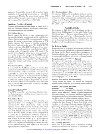
![S418 Circulation November 3, 2015
is superior to conventional CPR alone for the management
of adults and children with suspected opioid-associated
cardiac or respiratory arrest in the prehospital setting.
However, the additional search for available evidence
regarding overdose education and naloxone distribution
programs yielded 3 observational before-and-after stud-
ies. One study observed a dose-response effect with 0.73
(95% confidence interval [CI], 0.57–0.91) and 0.54 (95%
CI, 0.39–0.76) adjusted rate ratios for lethal overdose in
communities with low and high implementation, respec-
tively.49
The remaining 2 observational studies reported
reductions in rate ratios for lethal overdose of 0.62 (95%
CI, 0.54–0.72)50
and 0.70 (95% CI, 0.65–0.74) in individual
communities that implemented programs to address opioid
overdose.51
2015 Recommendations—New
For a patient with known or suspected opioid overdose who
has a definite pulse but no normal breathing or only gasp-
ing (ie, a respiratory arrest), in addition to providing stan-
dard BLS care, it is reasonable for appropriately trained BLS
healthcare providers to administer intramuscular or intrana-
sal naloxone (Class IIa, LOE C-LD). For patients in cardiac
arrest, medication administration is ineffective without con-
comitant chest compressions for drug delivery to the tissues,
so naloxone administration may be considered after initiation
of CPR if there is high suspicion for opiate overdose (Class
IIb, LOE C-EO). It is reasonable to provide opioid overdose
response education with or without naloxone distribution
to persons at risk for opioid overdose (or those living with
or in frequent contact with such persons) (Class IIa, LOE
C-LD). Information regarding lay rescuer education and the
use of naloxone for known or suspected victims of opioid
overdose is discussed in “Part 10: Special Circumstances of
Resuscitation.”
Scenario: Pulse Absent, No Breathing or Only Gasping
As in the 2010 Guidelines, rescuers should initiate CPR and
use an AED as soon as possible. By this point in all poten-
tial scenarios, the emergency response system is activated,
and a defibrillator and emergency equipment are retrieved or
requested.
No,
nonshockable
Yes,
shockable
No normal
breathing,
has pulse
© 2015 American Heart Association
AED arrives.
Check rhythm.
Shockable rhythm?
Give 1 shock. Resume CPR
immediately for about 2 minutes
(until prompted by AED to allow
rhythm check).
Continue until ALS providers take
over or victim starts to move.
Provide rescue breathing:
1 breath every 5-6 seconds, or
about 10-12 breaths/min.
• Activate emergency response
system (if not already done)
after 2 minutes.
• Continue rescue breathing;
check pulse about every
2 minutes. If no pulse, begin
CPR (go to “CPR” box).
• If possible opioid overdose,
administer naloxone if
available per protocol.
Resume CPR immediately for
about 2 minutes (until prompted
by AED to allow rhythm check).
Continue until ALS providers take
over or victim starts to move.
CPR
Begin cycles of
30 compressions and 2 breaths.
Use AED as soon as it is available.
Monitor until
emergency
responders arrive.
Verify scene safety.
Victim is unresponsive.
Shout for nearby help.
Activate emergency response system
via mobile device (if appropriate).
Get AED and emergency equipment
(or send someone to do so).
Look for no breathing
or only gasping and check
pulse (simultaneously).
Is pulse definitely felt
within 10 seconds?
Normal
breathing,
has pulse
No breathing
or only gasping,
no pulse
By this time in all scenarios, emergency
response system or backup is activated,
and AED and emergency equipment are
retrieved or someone is retrieving them.
BLS Healthcare Provider
Adult Cardiac Arrest Algorithm—2015 Update
Figure 1. BLS Healthcare Provider Adult Cardiac Arrest Algorithm—2015 Update.
by guest on October 15, 2015http://circ.ahajournals.org/Downloaded from](https://image.slidesharecdn.com/guidelines-rcp-aha-2015-full-160515230707/85/Guidelines-rcp-aha-2015-full-111-320.jpg)
![Kleinman et al Part 5: Adult BLS and CPR S419
Technique: Chest Compressions—Updated
Chest compressions are the key component of effective CPR.
Characteristics of chest compressions include their depth, rate,
and degree of recoil. The quality of CPR can also be charac-
terized by the frequency and duration of interruptions in chest
compressions—when such interruptions are minimized, the chest
compression fraction (percent of total resuscitation time that
compressions are performed) is higher. Finally, with high-quality
CPR, the rescuer avoids excessive ventilation. These CPR perfor-
mance elements affect intrathoracic pressure, coronary perfusion
pressure, cardiac output, and, in turn, clinical outcomes.
Hand Position During CompressionsBLS 357
—Updated
The 2015 ILCOR systematic review addressed whether hand
position placement for chest compressions affected resusci-
tation outcomes. Different rescuer hand positions alter the
mechanics of chest compressions and may, in turn, influence
their quality and effectiveness.
2015 Summary of Evidence
Only a few human studies involving a total of fewer than
100 cardiac arrest patients have evaluated hand position dur-
ing CPR.52–54
These investigations assessed hand placement
on the lower third of the sternum compared with the center
of the chest in a crossover design, and they measured physi-
ologic endpoints, such as blood pressure and end-tidal carbon
dioxide (ETCO2
). The studies have not provided conclusive
or consistent results about the effects of hand placement on
resuscitation outcomes.
2015 Recommendation—Unchanged
Consistent with the 2010 Guidelines, it is reasonable to
position hands for chest compressions on the lower half of
the sternum in adults with cardiac arrest (Class IIa, LOE
C-LD).
Chest Compression RateBLS 343
—Updated
In the 2010 Guidelines, the recommended compression
rate was at least 100 compressions per minute. The 2015
Guidelines Update incorporates new evidence about the
potential for an upper threshold of rate beyond which outcome
may be adversely affected.
The 2015 ILCOR systematic review addressed whether
compression rates different from 100/min influence physi-
ologic or clinical outcomes. Chest compression rate is defined
as the actual rate used during each continuous period of chest
compressions. This rate differs from the number of chest
compressions delivered per unit of time, which takes into
account any interruptions in chest compressions.
2015 Summary of Evidence
Evidence involving compression rate is derived from obser-
vational human studies that evaluate the relationship between
compression rate and outcomes including survival to hos-
pital discharge, return of spontaneous circulation (ROSC),
and various physiologic measures, such as blood pressure
and end-tidal CO2
. These investigations suggest that there
may be an optimal zone for the rate of manual chest com-
pressions—between 100/min and 120/min—that on average
is associated with improved survival.55,56
Importantly, there
is an interdependent relationship between compression rate
and compression depth during manual chest compressions:
as rate increases to greater than 120/min, depth decreases in
a dose-dependent manner.55
For example, the proportion of
compressions less than 38 mm (less than 3.8 cm or 1.5 inches)
was about 35% for a compression rate of 100 to 119/min but
increased to 50% for a compression rate of 120 to 139/min
and 70% for a compression rate of greater than 140/min.
2015 Recommendation—Updated
In adult victims of cardiac arrest, it is reasonable for rescuers
to perform chest compressions at a rate of 100/min to 120/min
(Class IIa, LOE C-LD).
Chest Compression DepthBLS 366
—Updated
The 2015 ILCOR systematic review addressed whether a
chest compression depth different from 2 inches (5 cm) influ-
ences physiologic or clinical outcomes. The depth of chest
compression can affect the relative increase in intrathoracic
pressure and, in turn, influence forward blood flow from the
heart and great vessels to the systemic circulation. In the 2010
Guidelines, the recommended compression depth was at least
2 inches (5 cm). The 2015 Guidelines Update incorporates
new evidence about the potential for an upper threshold of
compression depth beyond which outcomes may be adversely
affected.
2015 Summary of Evidence
Evidence involving compression depth is derived from obser-
vational human studies that evaluate the relationship between
compression depth and outcomes including survival with
favorable neurologic outcome, survival to hospital discharge,
and ROSC. Studies often classify compression depth differ-
ently, using distinct categories of depth or using an average
depth for a given portion of the resuscitation.
Even with this heterogeneity, there is consistent evidence
that achieving compression depth of approximately 5 cm is
associated with greater likelihood of favorable outcomes com-
pared with shallower compressions.57–65
In the largest study
to date (n=9136), the optimal compression depth with regard
to survival occurred within the range of 41 to 55 mm (4.1 to
5.5 cm, or 1.61 to 2.2 inches).60
Less evidence is available
about whether there is an upper threshold beyond which com-
pressions may be too deep. During manual CPR, injuries are
more common when compression depth is greater than 6 cm
(2.4 inches) than when it is between 5 and 6 cm (2 and 2.4
inches).66
Importantly, chest compressions performed by pro-
fessional rescuers are more likely to be too shallow (ie, less
than 40 mm [4 cm] or 1.6 inches) and less likely to exceed
55 mm (5.5 cm or 2.2 inches).60
2015 Recommendation—Updated
During manual CPR, rescuers should perform chest compres-
sions to a depth of at least 2 inches or 5 cm for an average
adult, while avoiding excessive chest compression depths
(greater than 2.4 inches or 6 cm) (Class I, LOE C-LD).
Chest Wall RecoilBLS 367
The 2015 ILCOR systematic reviews addressed whether full
chest wall recoil compared with incomplete recoil influenced
by guest on October 15, 2015http://circ.ahajournals.org/Downloaded from](https://image.slidesharecdn.com/guidelines-rcp-aha-2015-full-160515230707/85/Guidelines-rcp-aha-2015-full-112-320.jpg)
![S432 Circulation November 3, 2015
51. Maxwell S, Bigg D, Stanczykiewicz K, Carlberg-Racich S. Prescribing nal-
oxone to actively injecting heroin users: a program to reduce heroin over-
dose deaths. J Addict Dis. 2006;25:89–96. doi: 10.1300/J069v25n03_11.
52. Cha KC, Kim HJ, Shin HJ, Kim H, Lee KH, Hwang SO. Hemodynamic
effect of external chest compressions at the lower end of the sternum in
cardiac arrest patients. J Emerg Med. 2013;44:691–697. doi: 10.1016/j.
jemermed.2012.09.026.
53. Qvigstad E, Kramer-Johansen J, Tømte Ø, Skålhegg T, Sørensen Ø, Sunde
K, Olasveengen TM. Clinical pilot study of different hand positions during
manual chest compressions monitored with capnography. Resuscitation.
2013;84:1203–1207. doi: 10.1016/j.resuscitation.2013.03.010.
54. Orlowski JP. Optimum position for external cardiac compression in infants
and young children. Ann Emerg Med. 1986;15:667–673.
55. Idris AH, Guffey D, Pepe PE, Brown SP, Brooks SC, Callaway CW,
Christenson J, Davis DP, Daya MR, Gray R, Kudenchuk PJ, Larsen J, Lin
S, Menegazzi JJ, Sheehan K, Sopko G, Stiell I, Nichol G, Aufderheide TP;
Resuscitation Outcomes Consortium Investigators. Chest compression
rates and survival following out-of-hospital cardiac arrest. Crit Care Med.
2015;43:840–848. doi: 10.1097/CCM.0000000000000824.
56. Idris AH, Guffey D, Aufderheide TP, Brown S, Morrison LJ, Nichols
P, Powell J, Daya M, Bigham BL, Atkins DL, Berg R, Davis D, Stiell
I, Sopko G, Nichol G; Resuscitation Outcomes Consortium (ROC)
Investigators. Relationship between chest compression rates and outcomes
from cardiac arrest. Circulation. 2012;125:3004–3012. doi: 10.1161/
CIRCULATIONAHA.111.059535.
57. Vadeboncoeur T, Stolz U, Panchal A, Silver A, Venuti M, Tobin J, Smith
G, Nunez M, Karamooz M, Spaite D, Bobrow B. Chest compression depth
and survival in out-of-hospital cardiac arrest. Resuscitation. 2014;85:182–
188. doi: 10.1016/j.resuscitation.2013.10.002.
58. Hostler D, Everson-Stewart S, Rea TD, Stiell IG, Callaway CW,
Kudenchuk PJ, Sears GK, Emerson SS, Nichol G; Resuscitation Outcomes
Consortium Investigators. Effect of real-time feedback during cardiopul-
monary resuscitation outside hospital: prospective, cluster-randomised
trial. BMJ. 2011;342:d512.
59. Stiell IG, Brown SP, Christenson J, Cheskes S, Nichol G, Powell J,
Bigham B, Morrison LJ, Larsen J, Hess E, Vaillancourt C, Davis DP,
Callaway CW; Resuscitation Outcomes Consortium (ROC) Investigators.
What is the role of chest compression depth during out-of-hospital cardiac
arrest resuscitation? Crit Care Med. 2012;40:1192–1198. doi: 10.1097/
CCM.0b013e31823bc8bb.
60. Stiell IG, Brown SP, Nichol G, Cheskes S, Vaillancourt C, Callaway CW,
Morrison LJ, Christenson J, Aufderheide TP, Davis DP, Free C, Hostler D,
Stouffer JA, Idris AH; Resuscitation Outcomes Consortium Investigators.
What is the optimal chest compression depth during out-of-hospital car-
diac arrest resuscitation of adult patients? Circulation. 2014;130:1962–
1970. doi: 10.1161/CIRCULATIONAHA.114.008671.
61. Kramer-Johansen J, Myklebust H, Wik L, Fellows B, Svensson L,
Sørebø H, Steen PA. Quality of out-of-hospital cardiopulmonary
resuscitation with real time automated feedback: a prospective inter-
ventional study. Resuscitation. 2006;71:283–292. doi: 10.1016/j.
resuscitation.2006.05.011.
62. Edelson DP, Abella BS, Kramer-Johansen J, Wik L, Myklebust H, Barry
AM, Merchant RM, Hoek TL, Steen PA, Becker LB. Effects of com-
pression depth and pre-shock pauses predict defibrillation failure dur-
ing cardiac arrest. Resuscitation. 2006;71:137–145. doi: 10.1016/j.
resuscitation.2006.04.008.
63. Edelson DP, Litzinger B, Arora V, Walsh D, Kim S, Lauderdale DS,
Vanden Hoek TL, Becker LB, Abella BS. Improving in-hospital cardiac
arrest process and outcomes with performance debriefing. Arch Intern
Med. 2008;168:1063–1069. doi: 10.1001/archinte.168.10.1063.
64. Babbs CF, Kemeny AE, Quan W, Freeman G. A new paradigm for
human resuscitation research using intelligent devices. Resuscitation.
2008;77:306–315. doi: 10.1016/j.resuscitation.2007.12.018.
65. Bohn A, Weber TP, Wecker S, Harding U, Osada N, Van Aken H, Lukas
RP. The addition of voice prompts to audiovisual feedback and debrief-
ing does not modify CPR quality or outcomes in out of hospital cardiac
arrest–a prospective, randomized trial. Resuscitation. 2011;82:257–262.
doi: 10.1016/j.resuscitation.2010.11.006.
66. Hellevuo H, Sainio M, Nevalainen R, Huhtala H, Olkkola KT, Tenhunen
J, Hoppu S. Deeper chest compression - more complications for car-
diac arrest patients? Resuscitation. 2013;84:760–765. doi: 10.1016/j.
resuscitation.2013.02.015.
67. Zuercher M, Hilwig RW, Ranger-Moore J, Nysaether J, Nadkarni VM,
Berg MD, Kern KB, Sutton R, Berg RA. Leaning during chest compres-
sions impairs cardiac output and left ventricular myocardial blood flow in
piglet cardiac arrest. Crit Care Med. 2010;38:1141–1146. doi: 10.1097/
CCM.0b013e3181ce1fe2.
68. Niles DE, Sutton RM, Nadkarni VM, Glatz A, Zuercher M, Maltese MR,
Eilevstjønn J, Abella BS, Becker LB, Berg RA. Prevalence and hemo-
dynamic effects of leaning during CPR. Resuscitation. 2011;82 suppl
2:S23–S26. doi: 10.1016/S0300-9572(11)70147-2.
69. Fried DA, Leary M, Smith DA, Sutton RM, Niles D, Herzberg DL, Becker
LB, Abella BS. The prevalence of chest compression leaning during in-
hospital cardiopulmonary resuscitation. Resuscitation. 2011;82:1019–
1024. doi: 10.1016/j.resuscitation.2011.02.032.
70. Niles D, Nysaether J, Sutton R, Nishisaki A, Abella BS, Arbogast K,
Maltese MR, Berg RA, Helfaer M, Nadkarni V. Leaning is common
during in-hospital pediatric CPR, and decreased with automated cor-
rective feedback. Resuscitation. 2009;80:553–557. doi: 10.1016/j.
resuscitation.2009.02.012.
71. Yannopoulos D, McKnite S, Aufderheide TP, Sigurdsson G, Pirrallo
RG, Benditt D, Lurie KG. Effects of incomplete chest wall decompres-
sion during cardiopulmonary resuscitation on coronary and cerebral
perfusion pressures in a porcine model of cardiac arrest. Resuscitation.
2005;64:363–372. doi: 10.1016/j.resuscitation.2004.10.009.
72. Glatz AC, Nishisaki A, Niles DE, Hanna BD, Eilevstjonn J, Diaz LK,
Gillespie MJ, Rome JJ, Sutton RM, Berg RA, Nadkarni VM. Sternal
wall pressure comparable to leaning during CPR impacts intrathoracic
pressure and haemodynamics in anaesthetized children during cardiac
catheterization. Resuscitation. 2013;84:1674–1679. doi: 10.1016/j.
resuscitation.2013.07.010.
73. Meaney PA, Bobrow BJ, Mancini ME, Christenson J, de Caen AR, Bhanji
F, Abella BS, Kleinman ME, Edelson DP, Berg RA, Aufderheide TP,
Menon V, Leary M; CPR Quality Summit Investigators, the American
Heart Association Emergency Cardiovascular Care Committee, and
the Council on Cardiopulmonary, Critical Care, Perioperative and
Resuscitation. Cardiopulmonary resuscitation quality: [corrected] improv-
ing cardiac resuscitation outcomes both inside and outside the hospital: a
consensus statement from the American Heart Association. Circulation.
2013;128:417–435. doi: 10.1161/CIR.0b013e31829d8654.
74. Sell RE, Sarno R, Lawrence B, Castillo EM, Fisher R, Brainard C, Dunford
JV, Davis DP. Minimizing pre- and post-defibrillation pauses increases the
likelihood of return of spontaneous circulation (ROSC). Resuscitation.
2010;81:822–825. doi: 10.1016/j.resuscitation.2010.03.013.
75. Cheskes S, Schmicker RH, Christenson J, Salcido DD, Rea T, Powell
J, Edelson DP, Sell R, May S, Menegazzi JJ, Van Ottingham L, Olsufka
M, Pennington S, Simonini J, Berg RA, Stiell I, Idris A, Bigham B,
Morrison L; Resuscitation Outcomes Consortium (ROC) Investigators.
Perishock pause: an independent predictor of survival from out-of-hospi-
tal shockable cardiac arrest. Circulation. 2011;124:58–66. doi: 10.1161/
CIRCULATIONAHA.110.010736.
76. Cheskes S, Schmicker RH, Verbeek PR, Salcido DD, Brown SP, Brooks S,
Menegazzi JJ, Vaillancourt C, Powell J, May S, Berg RA, Sell R, Idris A,
Kampp M, Schmidt T, Christenson J; Resuscitation Outcomes Consortium
(ROC) investigators. The impact of peri-shock pause on survival from out-
of-hospital shockable cardiac arrest during the Resuscitation Outcomes
Consortium PRIMED trial. Resuscitation. 2014;85:336–342. doi:
10.1016/j.resuscitation.2013.10.014.
77. Christenson J, Andrusiek D, Everson-Stewart S, Kudenchuk P, Hostler D,
Powell J, Callaway CW, Bishop D, Vaillancourt C, Davis D, Aufderheide
TP, Idris A, Stouffer JA, Stiell I, Berg R; Resuscitation Outcomes
Consortium Investigators. Chest compression fraction determines sur-
vival in patients with out-of-hospital ventricular fibrillation. Circulation.
2009;120:1241–1247. doi: 10.1161/CIRCULATIONAHA.109.852202.
78. Vaillancourt C, Everson-Stewart S, Christenson J, Andrusiek D, Powell
J, Nichol G, Cheskes S, Aufderheide TP, Berg R, Stiell IG; Resuscitation
Outcomes Consortium Investigators. The impact of increased chest com-
pression fraction on return of spontaneous circulation for out-of-hospi-
tal cardiac arrest patients not in ventricular fibrillation. Resuscitation.
2011;82:1501–1507. doi: 10.1016/j.resuscitation.2011.07.011.
79. Jost D, Degrange H, Verret C, Hersan O, Banville IL, Chapman FW, Lank
P, Petit JL, Fuilla C, Migliani R, Carpentier JP; DEFI 2005 Work Group.
DEFI 2005: a randomized controlled trial of the effect of automated exter-
nal defibrillator cardiopulmonary resuscitation protocol on outcome from
out-of-hospital cardiac arrest. Circulation. 2010;121:1614–1622. doi:
10.1161/CIRCULATIONAHA.109.878389.
80. Beesems SG, Wijmans L, Tijssen JG, Koster RW. Duration of ven-
tilations during cardiopulmonary resuscitation by lay rescuers and
first responders: relationship between delivering chest compressions
by guest on October 15, 2015http://circ.ahajournals.org/Downloaded from](https://image.slidesharecdn.com/guidelines-rcp-aha-2015-full-160515230707/85/Guidelines-rcp-aha-2015-full-125-320.jpg)
![S436
Introduction
Conventional cardiopulmonary resuscitation (CPR) consist-
ing of manual chest compressions with rescue breaths is inher-
ently inefficient with respect to generating cardiac output. A
variety of alternatives and adjuncts to conventional CPR have
been developed, with the aim of enhancing perfusion during
resuscitation from cardiac arrest. Since the publication of the
2010 American Heart Association (AHA) Guidelines for CPR
and Emergency Cardiovascular Care (ECC),1
a number of
clinical trials have provided additional data on the effective-
ness of these alternatives and adjuncts. Compared with con-
ventional CPR, many of these techniques and devices require
specialized equipment and training. Some have only been
tested in highly selected subgroups of cardiac arrest patients;
this context must be considered when rescuers or healthcare
systems are considering implementation.
Methodology
The recommendations in this 2015 AHA Guidelines Update
for CPR and ECC are based on an extensive evidence review
process that was begun by the International Liaison Committee
on Resuscitation (ILCOR) after the publication of the
ILCOR 2010 International Consensus on Cardiopulmonary
Resuscitation and Emergency Cardiovascular Care Science
With Treatment Recommendations2,3
and was completed in
February 2015.4,5
In this in-depth evidence review process, the ILCOR
Advanced Life Support (ALS) Task Force examined topics
and then generated a prioritized list of questions for systematic
review. Questions were first formulated in PICO (population,
intervention, comparator, outcome) format,6
search strate-
gies and criteria for inclusion and exclusion of articles were
defined, and then a search for relevant articles was performed.
The evidence was evaluated by the ILCOR ALS Task Force by
using the standardized methodological approach proposed by
the Grading of Recommendations Assessment, Development,
and Evaluation (GRADE) Working Group.7
The quality of the evidence was categorized based on the
study methodologies and the 5 core GRADE domains of risk
of bias, inconsistency, indirectness, imprecision, and other
considerations (including publication bias). Then, where
possible, consensus-based treatment recommendations were
created.
To create this 2015 AHA Guidelines Update for CPR
and ECC, the AHA formed 15 writing groups, with careful
attention to manage conflicts of interest, to assess the ILCOR
treatment recommendations, and to write AHA Guidelines
and treatment recommendations by using the AHA Class of
Recommendation and Level of Evidence (LOE) system. The
recommendations made in the 2015 AHA Guidelines Update
for CPR and ECC are informed by the ILCOR recommenda-
tions and GRADE classification, in the context of the delivery
of medical care in North America. Throughout the online ver-
sion of this publication, live links are provided so the reader
can connect directly to the systematic reviews on the ILCOR
Scientific Evidence Evaluation and Review System (SEERS)
website. These links are indicated by a superscript combi-
nation of letters and numbers (eg, ALS 579). We encourage
readers to use the links and review the evidence and appen-
dixes, such as the GRADE tables. For further information,
please see Part 2 of this supplement, “Evidence Evaluation
and Management of Conflicts of Interest.”
The following CPR techniques and devices were last
reviewed in 20102,3
: open-chest CPR, interposed abdominal
compression, “cough” CPR, prone CPR, precordial thump,
percussion pacing, and devices to assist ventilation. The
reader is referred to the 2010 Guidelines for details of those
recommendations.1
A listing of all of the recommendations in
this 2015 Guidelines Update and the recommendations from
“Part 7: CPR Techniques and Devices” of the 2010 Guidelines
can be found in the Appendix.
Devices to Support Circulation
Impedance Threshold DeviceALS 579
The impedance threshold device (ITD) is a pressure-sensitive
valve that is attached to an endotracheal tube (ETT), supraglot-
tic airway, or face mask. The ITD limits air entry into the lungs
(Circulation. 2015;132[suppl 2]:S436–S443. DOI: 10.1161/CIR.0000000000000260.)
© 2015 American Heart Association, Inc.
Circulation is available at http://circ.ahajournals.org DOI: 10.1161/CIR.0000000000000260
The American Heart Association requests that this document be cited as follows: Brooks SC, Anderson ML, Bruder E, Daya MR, Gaffney A, Otto CW,
Singer AJ, Thiagarajan RR, Travers AH. Part 6: alternative techniques and ancillary devices for cardiopulmonary resuscitation: 2015 American Heart
Association Guidelines Update for Cardiopulmonary Resuscitation and Emergency Cardiovascular Care. Circulation. 2015;132(suppl 2):S436–S443.
for Cardiopulmonary Resuscitation and Emergency
Cardiovascular Care
Part 6: Alternative Techniques and Ancillary
Devices for Cardiopulmonary Resuscitation
2015 American Heart Association Guidelines Update for Cardiopulmonary
Resuscitation and Emergency Cardiovascular Care
Steven C. Brooks, Chair; Monique L. Anderson; Eric Bruder; Mohamud R. Daya;
Alan Gaffney; Charles W. Otto; Adam J. Singer; Ravi R. Thiagarajan; Andrew H. Travers
by guest on October 15, 2015http://circ.ahajournals.org/Downloaded from](https://image.slidesharecdn.com/guidelines-rcp-aha-2015-full-160515230707/85/Guidelines-rcp-aha-2015-full-130-320.jpg)
![S438 Circulation November 3, 2015
out to 1 year. For survival to hospital discharge with favorable
neurologic function, this translates into a number needed to
treat of 45 with very wide confidence limits (95% confidence
interval, 25–333), making interpretation of the true clinical
effect challenging. There was no difference in the overall inci-
dence of adverse events, although pulmonary edema was more
common withACD-CPR+ITD as compared with conventional
CPR (11.3% versus 7.9%; P=0.002). The ResQ Trial had a
number of important limitations, including lack of blinding,
different CPR feedback elements between the study arms (ie,
co-intervention), lack of CPR quality assessment, and early
termination. Although improved neurologic function was
noted with the use of the ACD-CPR+ITD combination at both
hospital discharge and 1-year follow-up, additional trials are
needed to confirm these findings.
2015 Recommendation—New
The existing evidence, primarily from 1 large RCT of low
quality, does not support the routine use of ACD-CPR+ITD as
an alternative to conventional CPR. The combination may be
a reasonable alternative in settings with available equipment
and properly trained personnel (Class IIb, LOE C-LD).
Mechanical Chest Compression Devices:
Piston DeviceALS 782
A mechanical piston device consists of an automated com-
pressed gas- or electric-powered plunger positioned over
the sternum, which compresses the chest at a set rate. Some
devices incorporate a suction cup at the end of the piston that
is designed to actively decompress the chest after each com-
pression, whereas others do not.
2015 Evidence Review
The Lund University Cardiac Arrest System (LUCAS) is a
gas- (oxygen or air) or electric-powered piston device that
produces a consistent chest compression rate and depth.
It incorporates a suction cup on the end of the piston that
attaches to the sternum and returns the sternum to the start-
ing position when it retracts. A small pilot RCT found similar
survival in patients randomly assigned to mechanical versus
manual chest compressions.17
Subsequently, 2 large RCTs,
the Prehospital Randomised Assessment of a Mechanical
Compression Device in Cardiac Arrest (PARAMEDIC)18
and
LUCAS in Cardiac Arrest (LINC)19
trials, have compared
the use of LUCAS against manual compressions for patients
with OHCA. Together, these studies enrolled 7060 patients,
and neither demonstrated a benefit for mechanical CPR over
manual CPR with respect to early (4-hour) and late (1- and
6-month) survival.18,19
The PARAMEDIC study demonstrated
a negative association between mechanical chest compres-
sions and survival with good neurologic outcome (Cerebral
Performance Category 1–2) at 3 months as compared with
manual compressions.
A number of other mechanical piston devices have been
compared with manual chest compressions in studies of
OHCA. There are no large-scale RCTs with these devices.
Three small (largest sample size of 50 patients) RCTs found
no differences in early survival20–22
despite improvements in
end-tidal CO2
in patients randomly assigned to mechanical
piston devices in 2 of these 3 studies.21,22
However, in neither
of these studies did any patient survive to hospital discharge.
Time-motion analysis of manual versus mechanical chest
compressions showed that it took considerable time to deploy
the mechanical piston device, prolonging the no-chest com-
pression interval during CPR.23
2015 Recommendations—New
The evidence does not demonstrate a benefit with the use of
mechanical piston devices for chest compressions versus man-
ual chest compressions in patients with cardiac arrest. Manual
chest compressions remain the standard of care for the treat-
ment of cardiac arrest, but mechanical piston devices may be
a reasonable alternative for use by properly trained personnel
(Class IIb, LOE B-R). The use of mechanical piston devices
may be considered in specific settings where the delivery of
high-quality manual compressions may be challenging or dan-
gerous for the provider (eg, limited rescuers available, pro-
longed CPR, during hypothermic cardiac arrest, in a moving
ambulance, in the angiography suite, during preparation for
extracorporeal CPR [ECPR]), provided that rescuers strictly
limit interruptions in CPR during deployment and removal of
the devices (Class IIb, LOE C-EO).
Load-Distributing Band DevicesALS 782
The load-distributing band (LDB) is a circumferential chest
compression device composed of a pneumatically or electri-
cally actuated constricting band and backboard.
2015 Evidence Summary
While early case series24–26
of patients treated with LDB-
CPR were encouraging, an observational study exploring a
number of treatments related to new guideline implementa-
tion suggested that the use of LDB-CPR was associated with
lower odds of 30-day survival when compared with concur-
rent patients receiving only manual CPR.27
One multicenter
prospective RCT28
comparing LDB-CPR (Autopulse device)
with manual CPR for OHCA demonstrated no improvement
in 4-hour survival and worse neurologic outcome when the
device was compared with manual CPR. Site-specific factors29
and experience with deployment of the device30
may have
influenced the outcomes in this study. In a high-quality mul-
ticenter RCT of 4753 OHCA patients, LDB-CPR (Autopulse
device) and manual chest compressions were shown to be
equivalent with respect to the outcome of survival to hospital
discharge. Both approaches in this study were carefully moni-
tored to minimize hands-off time and to optimize compression
technique.31
2015 Recommendations—New
The evidence does not demonstrate a benefit with the use
of LDB-CPR for chest compressions versus manual chest
compressions in patients with cardiac arrest. Manual chest
compressions remain the standard of care for the treatment
of cardiac arrest, but LDB-CPR may be a reasonable alter-
native for use by properly trained personnel (Class IIb, LOE
B-R). The use of LDB-CPR may be considered in specific set-
tings where the delivery of high-quality manual compressions
may be challenging or dangerous for the provider (eg, limited
rescuers available, prolonged CPR, during hypothermic car-
diac arrest, in a moving ambulance, in the angiography suite,
by guest on October 15, 2015http://circ.ahajournals.org/Downloaded from](https://image.slidesharecdn.com/guidelines-rcp-aha-2015-full-160515230707/85/Guidelines-rcp-aha-2015-full-132-320.jpg)
![Brooks et al Part 6: Alternative Techniques and Ancillary Devices for CPR S441
Appendix
2015 Guidelines Update: Part 6 Recommendations
Year Last
Reviewed Topic Recommendation Comments
2015 Devices to Support Circulation: Impedance
Threshold Device
The routine use of the ITD as an adjunct during conventional CPR is not
recommended (Class III: No Benefit, LOE A).
new for 2015
2015 Devices to Support Circulation: Active
Compression-Decompression CPR and
Impedance Threshold Device
The existing evidence, primarily from 1 large RCT of low quality, does not
support the routine use of ACD-CPR+ITD as an alternative to conventional
CPR. The combination may be a reasonable alternative in settings with
available equipment and properly trained personnel (Class IIb, LOE C-LD).
new for 2015
2015 Devices to Support Circulation: Mechanical
Chest Compression Devices: Piston Device
The evidence does not demonstrate a benefit with the use of mechanical
piston devices for chest compressions versus manual chest compressions
in patients with cardiac arrest. Manual chest compressions remain the
standard of care for the treatment of cardiac arrest, but mechanical chest
compressions using a piston device may be a reasonable alternative for use
by properly trained personnel (Class IIb, LOE B-R).
new for 2015
2015 Devices to Support Circulation: Mechanical
Chest Compression Devices: Piston Device
The use of piston devices for CPR may be considered in specific settings
where the delivery of high-quality manual compressions may be challenging
or dangerous for the provider (eg, limited rescuers available, prolonged
CPR, during hypothermic cardiac arrest, in a moving ambulance, in the
angiography suite, during preparation for extracorporeal CPR [ECPR]),
provided that rescuers strictly limit interruptions in CPR during deployment
and removal of the device (Class IIb, LOE C-EO).
new for 2015
2015 Devices to Support Circulation: Load-
Distributing Band Devices
The evidence does not demonstrate a benefit with the use of LDB-CPR for
chest compressions versus manual chest compressions in patients with
cardiac arrest. Manual chest compressions remain the standard of care for
the treatment of cardiac arrest, but LDB-CPR may be a reasonable alternative
for use by properly trained personnel (Class IIb, LOE B-R).
new for 2015
2015 Devices to Support Circulation: Load-
Distributing Band Devices
The use of LDB-CPR may be considered in specific settings where the
delivery of high-quality manual compressions may be challenging or
dangerous for the provider (eg, limited rescuers available, prolonged
CPR, during hypothermic cardiac arrest, in a moving ambulance, in the
angiography suite, during preparation for extracorporeal CPR [ECPR]),
provided that rescuers strictly limit interruptions in CPR during deployment
and removal of the devices (Class IIb, LOE E).
new for 2015
2015 Extracorporeal Techniques and Invasive
Perfusion Devices: Extracorporeal CPR
There is insufficient evidence to recommend the routine use of ECPR for
patients with cardiac arrest. It may be considered for select patients for
whom the suspected etiology of the cardiac arrest is potentially reversible
during a limited period of mechanical cardiorespiratory support (Class IIb,
LOE C-LD).
new for 2015
The following recommendations were not reviewed in 2015. For more information, see the 2010 AHA Guidelines for CPR and ECC, “Part 7: CPR Techniques and Devices”
2010 Open-Chest CPR Open-chest CPR can be useful if cardiac arrest develops during surgery when
the chest or abdomen is already open, or in the early postoperative period
after cardiothoracic surgery (Class IIa, LOE C).
not reviewed in 2015
2010 Open-Chest CPR A resuscitative thoracotomy to facilitate open-chest CPR may be considered
in very select circumstances of adults and children with out-of-hospital
cardiac arrest from penetrating trauma with short transport times to a trauma
facility (Class IIb, LOE C).
not reviewed in 2015
2010 Interposed Abdominal Compression-CPR IAC-CPR may be considered during in-hospital resuscitation when sufficient
personnel trained in its use are available (Class IIb, LOE B).
not reviewed in 2015
2010 “Cough” CPR “Cough” CPR may be considered in settings such as the cardiac
catheterization laboratory for conscious, supine, and monitored patients if
the patient can be instructed and coached to cough forcefully every 1 to 3
seconds during the initial seconds of an arrhythmic cardiac arrest. It should
not delay definitive treatment (Class IIb, LOE C).
not reviewed in 2015
2010 Prone CPR When the patient cannot be placed in the supine position, it may be
reasonable for rescuers to provide CPR with the patient in the prone position,
particularly in hospitalized patients with an advanced airway in place (Class
IIb, LOE C).
not reviewed in 2015
2010 Precordial Thump The precordial thump should not be used for unwitnessed out-of-hospital
cardiac arrest (Class III, LOE C).
not reviewed in 2015
(Continued )
by guest on October 15, 2015http://circ.ahajournals.org/Downloaded from](https://image.slidesharecdn.com/guidelines-rcp-aha-2015-full-160515230707/85/Guidelines-rcp-aha-2015-full-135-320.jpg)
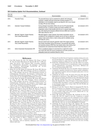

![S444
Introduction
Basic life support (BLS), advanced cardiovascular life sup-
port (ACLS), and post–cardiac arrest care are labels of con-
venience that each describe a set of skills and knowledge that
are applied sequentially during the treatment of patients who
have a cardiac arrest. There is overlap as each stage of care
progresses to the next, but generallyACLS comprises the level
of care between BLS and post–cardiac arrest care.
ACLS training is recommended for advanced providers
of both prehospital and in-hospital medical care. In the past,
much of the data regarding resuscitation was gathered from
out-of-hospital arrests, but in recent years, data have also been
collected from in-hospital arrests, allowing for a comparison
of cardiac arrest and resuscitation in these 2 settings. While
there are many similarities, there are also some differences
between in- and out-of-hospital cardiac arrest etiology, which
may lead to changes in recommended resuscitation treatment
or in sequencing of care. The consideration of steroid admin-
istration for in-hospital cardiac arrest (IHCA) versus out-of-
hospital cardiac arrest (OHCA) is one such example discussed
in this Part.
The recommendations in this 2015 American Heart
Association (AHA) Guidelines Update for Cardiopulmonary
Resuscitation (CPR) and Emergency Cardiovascular Care
(ECC) are based on an extensive evidence review process
that was begun by the International Liaison Committee
on Resuscitation (ILCOR) after the publication of the
ILCOR 2010 International Consensus on Cardiopulmonary
Resuscitation and Emergency Cardiovascular Care Science
With Treatment Recommendations1
and was completed in
February 2015.2
In this in-depth evidence review process, the ILCOR
task forces examined topics and then generated prioritized
lists of questions for systematic review. Questions were first
formulated in PICO (population, intervention, comparator,
outcome) format,3
and then a search strategy and inclusion
and exclusion criteria were defined and a search for relevant
articles was performed. The evidence was evaluated by using
the standardized methodological approach proposed by the
Grading of Recommendations Assessment, Development, and
Evaluation (GRADE) Working Group.4
The quality of the evidence was categorized based on the
study methodologies and the 5 core GRADE domains of risk
of bias, inconsistency, indirectness, imprecision, and other
considerations (including publication bias). Then, where
possible, consensus-based treatment recommendations were
created.
To create this 2015 Guidelines Update, the AHA formed
15 writing groups, with careful attention to avoid or manage
conflicts of interest, to assess the ILCOR treatment recom-
mendations and to write AHA treatment recommendations
by using the AHA Class of Recommendation and Level of
Evidence (LOE) system.
The recommendations made in this 2015 Guidelines
Update are informed by the ILCOR recommendations and
GRADE classification, in the context of the delivery of medical
care in North America. The AHA ACLS writing group made
new recommendations only on topics specifically reviewed by
ILCOR in 2015. This chapter delineates any instances where
the AHA writing group developed recommendations that are
substantially different than the ILCOR statements. In the
online version of this document, live links are provided so the
reader can connect directly to the systematic reviews on the
Scientific Evidence Evaluation and Review System (SEERS)
website. These links are indicated by a superscript combina-
tion of letters and numbers (eg, ALS 433).
This update uses the newest AHA COR and LOE classi-
fication system, which contains modifications of the Class III
recommendation and introduces LOE B-R (randomized stud-
ies) and B-NR (nonrandomized studies) as well as LOE C-LD
(limited data) and LOE C-EO (consensus of expert opinion).
All recommendations made in this 2015 Guidelines Update,
as well as in the 2010 Guidelines, are listed in the Appendix.
For further information, see “Part 2: Evidence Evaluation and
Management of Conflicts of Interest.”
The ILCOR ACLS Task Force addressed 37 PICO
questions related to ACLS care (presented in this Part) in
2015. These questions included oxygen dose during CPR,
(Circulation. 2015;132[suppl 2]:S444–S464. DOI: 10.1161/CIR.0000000000000261.)
© 2015 American Heart Association, Inc.
Circulation is available at http://circ.ahajournals.org DOI: 10.1161/CIR.0000000000000261
The American Heart Association requests that this document be cited as follows: Link MS, Berkow LC, Kudenchuk PJ, Halperin HR, Hess EP, Moitra
VK, Neumar RW, O’Neil BJ, Paxton JH, Silvers SM, White RD, Yannopoulos D, Donnino MW. Part 7: adult advanced cardiovascular life support:
2015 American Heart Association Guidelines Update for Cardiopulmonary Resuscitation and Emergency Cardiovascular Care. Circulation. 2015;
132(suppl 2):S444–S464.
for Cardiopulmonary Resuscitation and Emergency
Cardiovascular Care
Part 7: Adult Advanced Cardiovascular Life Support
2015 American Heart Association Guidelines Update for Cardiopulmonary
Resuscitation and Emergency Cardiovascular Care
Mark S. Link, Chair; Lauren C. Berkow; Peter J. Kudenchuk; Henry R. Halperin; Erik P. Hess;
Vivek K. Moitra; Robert W. Neumar; Brian J. O’Neil; James H. Paxton; Scott M. Silvers;
Roger D. White; Demetris Yannopoulos; Michael W. Donnino
by guest on October 15, 2015http://circ.ahajournals.org/Downloaded from](https://image.slidesharecdn.com/guidelines-rcp-aha-2015-full-160515230707/85/Guidelines-rcp-aha-2015-full-139-320.jpg)
![S446 Circulation November 3, 2015
Ultrasound During Cardiac ArrestALS 658
Bedside cardiac and noncardiac ultrasound are frequently used
as diagnostic and prognostic tools for critically ill patients.44
Ultrasound may be applied to patients receiving CPR to help
assess myocardial contractility and to help identify potentially
treatable causes of cardiac arrest such as hypovolemia, pneu-
mothorax, pulmonary thromboembolism, or pericardial tam-
ponade.45
However, it is unclear whether important clinical
outcomes are affected by the routine use of ultrasound among
patients experiencing cardiac arrest.
2015 Evidence Summary
One limited study with a small sample size was identified that
specifically addressed the utility of ultrasound during car-
diac arrest. This study evaluated bedside cardiac ultrasound
use during ACLS among adult patients in pulseless electri-
cal activity arrest and found no difference in the incidence of
ROSC when ultrasound was used.46
2015 Recommendations—Updated
Ultrasound (cardiac or noncardiac) may be considered during
the management of cardiac arrest, although its usefulness has
not been well established (Class IIb, LOE C-EO).
If a qualified sonographer is present and use of ultrasound
does not interfere with the standard cardiac arrest treatment
protocol, then ultrasound may be considered as an adjunct to
standard patient evaluation (Class IIb, LOE C-EO).
Adjuncts for Airway Control and Ventilation
This portion of the 2015 Guidelines Update focuses on recom-
mendations for airway management based on rate of survival
and favorable neurologic outcome.
Bag-Mask Ventilation Compared With Any
Advanced Airway During CPRALS 783
Bag-mask ventilation is a commonly used method for provid-
ing oxygenation and ventilation in patients with respiratory
insufficiency or arrest. When cardiac arrest occurs, provid-
ers must determine the best way to support ventilation and
oxygenation. Options include standard bag-mask ventilation
versus the placement of an advanced airway (ie, endotracheal
tube [ETT], supraglottic airway device [SGA]). Previous
guidelines recommended that prolonged interruptions in
chest compressions should be avoided during transitions from
bag-mask ventilation to an advanced airway device. In 2015,
ILCOR evaluated the evidence comparing the effect of bag-
mask ventilation versus advanced airway placement on over-
all survival and neurologic outcome from cardiac arrest.
2015 Evidence Summary
There is inadequate evidence to show a difference in survival
or favorable neurologic outcome with the use of bag-mask
ventilation compared with endotracheal intubation47–53
or other
advanced airway devices.47,49–51,54
The majority of these retro-
spective observational studies demonstrated slightly worse
survival with the use of an advanced airway when compared
with bag-mask ventilation. However, interpretation of these
results is limited by significant concerns of selection bias. Two
additional observational studies54,55
showed no difference in
survival.
Advanced Airway Placement Choice
Advanced airway devices are frequently placed by experi-
enced providers during CPR if bag-mask ventilation is inad-
equate or as a stepwise approach to airway management.
Placement of an advanced airway may result in interruption
of chest compressions, and the ideal timing of placement to
maximize outcome has not been adequately studied. The use
of an advanced airway device such as an ETT or SGA and the
effect of ventilation technique on overall survival and neuro-
logic outcome was evaluated in 2015.
2015 Evidence Summary
Endotracheal Intubation Versus Bag-Mask Ventilation
There is no high-quality evidence favoring the use of endo-
tracheal intubation compared with bag-mask ventilation or an
advanced airway device in relation to overall survival or favor-
able neurologic outcome.47–53
Evaluating retrospective studies
that compare bag-mask ventilation to endotracheal intubation
is challenging because patients with more severe physiologic
compromise will typically receive more invasive care (includ-
ing endotracheal intubation) than patients who are less com-
promised and more likely to survive. Within that context, a
number of retrospective studies show an association of worse
outcome in those who were intubated as compared with those
receiving bag-mask ventilation. While the studies did attempt
to control for confounders, bias still may have been pres-
ent, limiting the interpretation of these investigations. These
studies illustrate that endotracheal intubation can be associ-
ated with a number of complications and that the procedure
requires skill and experience. Risks of endotracheal intubation
during resuscitation include unrecognized esophageal intuba-
tion and increased hands-off time.
Supraglottic Airway Devices
Several retrospective studies compared a variety of supraglot-
tic devices (laryngeal mask airway, laryngeal tube, Combitube,
esophageal obturator airway) to both bag-mask ventilation
and endotracheal intubation. There is no high-quality evi-
dence demonstrating a difference in survival rate or favorable
neurologic outcome from use of an SGA compared with bag-
mask ventilation47,49–51
or endotracheal intubation.47,49,50,54,56–61
Three observational studies demonstrated a lower rate of both
overall survival and favorable neurologic outcome when SGA
use was compared with bag-mask ventilation,47,49,51
whereas
another observational study demonstrated similar survival
rates.50
In studies comparing SGA insertion to endotracheal intu-
bation, no high-quality studies have demonstrated a difference
in overall survival or favorable neurologic outcome.50,54,56–58,61
Several retrospective observational studies show more favor-
able outcome with the use of an SGA device, whereas other
studies favor the use of endotracheal intubation.47,49,50,59–61
2015 Recommendations—Updated
Either a bag-mask device or an advanced airway may be used
for oxygenation and ventilation during CPR in both the in-
hospital and out-of-hospital setting (Class IIb, LOE C-LD).
For healthcare providers trained in their use, either an
SGA device or an ETT may be used as the initial advanced
airway during CPR (Class IIb, LOE C-LD).
by guest on October 15, 2015http://circ.ahajournals.org/Downloaded from](https://image.slidesharecdn.com/guidelines-rcp-aha-2015-full-160515230707/85/Guidelines-rcp-aha-2015-full-141-320.jpg)
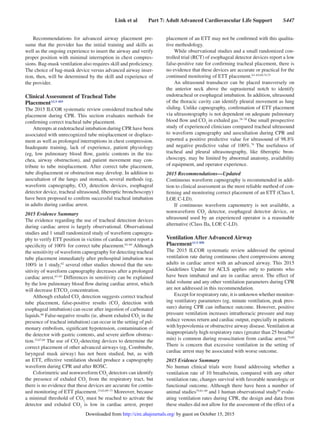
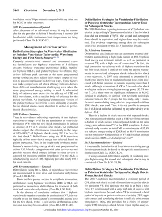
![S452 Circulation November 3, 2015
No
2
9
Yes No
4
6
8
Yes
Yes
10
No
12
Yes
No
No Yes
Shock
Shock
Shock
11
5
7
1
3
Rhythm
shockable?
Rhythm
shockable?
Rhythm
shockable?
Rhythm
shockable?
Rhythm
shockable?
• If no signs of return of
spontaneous circulation
(ROSC), go to 10 or 11
• If ROSC, go to
Post–Cardiac Arrest Care
Go to 5 or 7
VF/pVT Asystole/PEA
CPR Quality
• Push hard (at least 2 inches
[5 cm]) and fast (100-120/min)
and allow complete chest recoil.
• Minimize interruptions in
compressions.
• Avoid excessive ventilation.
• Rotate compressor every
2 minutes, or sooner if fatigued.
• If no advanced airway,
30:2 compression-ventilation
ratio.
• Quantitative waveform
capnography
– If PETCO2
10 mm Hg, attempt
to improve CPR quality.
• Intra-arterial pressure
– If relaxation phase (dia-
stolic) pressure 20 mm Hg,
attempt to improve CPR
quality.
Shock Energy for Defibrillation
• Biphasic: Manufacturer
recommendation (eg, initial
dose of 120-200 J); if unknown,
use maximum available.
Second and subsequent doses
should be equivalent, and higher
doses may be considered.
• Monophasic: 360 J
Drug Therapy
• Epinephrine IV/IO dose:
1 mg every 3-5 minutes
• Amiodarone IV/IO dose: First
dose: 300 mg bolus. Second
dose: 150 mg.
Advanced Airway
• Endotracheal intubation or
supraglottic advanced airway
• Waveform capnography or
capnometry to confirm and
monitor ET tube placement
• Once advanced airway in place,
give 1 breath every 6 seconds
(10 breaths/min) with continuous
chest compressions
Return of Spontaneous
Circulation (ROSC)
• Pulse and blood pressure
• Abrupt sustained increase in
PETCO2
(typically ≥40 mm Hg)
• Spontaneous arterial pressure
waves with intra-arterial
monitoring
Reversible Causes
• Hypovolemia
• Hypoxia
• Hydrogen ion (acidosis)
• Hypo-/hyperkalemia
• Hypothermia
• Tension pneumothorax
• Tamponade, cardiac
• Toxins
• Thrombosis, pulmonary
• Thrombosis, coronary
© 2015 American Heart Association
Start CPR
• Give oxygen
• Attach monitor/defibrillator
CPR 2 min
• IV/IO access
• Epinephrine every 3-5 min
• Consider advanced airway,
capnography
CPR 2 min
• Epinephrine every 3-5 min
• Consider advanced airway,
capnography
CPR 2 min
• Amiodarone
• Treat reversible causes
CPR 2 min
• Treat reversible causes
CPR 2 min
• IV/IO access
Adult Cardiac Arrest Algorithm—2015 Update
Figure 1. Adult Cardiac Arrest Algorithm―2015 Update.
with late administration. When neurologically intact survival
to discharge was assessed,133,134,136
however, there was vari-
able benefit with early administration of epinephrine for both
shockable and nonshockable rhythms. Later administration of
epinephrine was associated with a worse outcome. ROSC was
generally improved with early administration of epinephrine in
studies of more than 210 000 patients.120,133,135,137
Design flaws
in the majority of these observational OHCA studies, how-
ever, included the use of a “no epinephrine” control arm as the
comparator (thus not allowing for estimates on the effect of
timing), and the lack of known timing of epinephrine adminis-
tration upon arrival in the emergency department. In addition,
by guest on October 15, 2015http://circ.ahajournals.org/Downloaded from](https://image.slidesharecdn.com/guidelines-rcp-aha-2015-full-160515230707/85/Guidelines-rcp-aha-2015-full-147-320.jpg)
![Link et al Part 7: Adult Advanced Cardiovascular Life Support S461
References
1. Morrison LJ, Deakin CD, Morley PT, Callaway CW, Kerber RE, Kronick
SL, Lavonas EJ, Link MS, Neumar RW, Otto CW, Parr M, Shuster
M, Sunde K, Peberdy MA, Tang W, Hoek TL, Böttiger BW, Drajer S,
Lim SH, Nolan JP; Advanced Life Support Chapter Collaborators.
Part 8: advanced life support: 2010 International Consensus on
Cardiopulmonary Resuscitation and Emergency Cardiovascular Care
Science With Treatment Recommendations. Circulation. 2010;122
(suppl 2):S345–S421. doi: 10.1161/CIRCULATIONAHA.110.971051.
2. Callaway CW, Soar J, Aibiki M, Böttiger BW, Brooks SC, Deakin CD,
Donnino MW, Drajer S, Kloeck W, Morley PT, Morrison LJ, Neumar
RW, Nicholson TC, Nolan JP, Okada K, O’Neil BJ, Paiva EF, Parr
MJ, Wang TL, Witt J; on behalf of the Advanced Life Support Chapter
Collaborators. Part 4: advanced life support: 2015 International Consensus
on Cardiopulmonary Resuscitation and Emergency Cardiovascular Care
Science With Treatment Recommendations. Circulation. 2015;132(suppl
1):S84–S145. doi: 10.1161/CIR.0000000000000273.
3. O’Connor D, Green S, Higgins J, eds. Chapter 5: defining the review
questions and developing criteria for including studies. In: The Cochrane
Collaboration. Higgans JPT, Green S, eds. Cochrane Handbook for
Systematic Reviews of Interventions. Version 5.1.0. 2011. http://handbook.
cochrane.org/. Accessed May 6, 2015.
4. Schünemann H, Brożek J, Guyatt G, Oxman A. GRADE Handbook.
2013. http://www.guidelinedevelopment.org/handbook/. Accessed May
6, 2015.
5. Spindelboeck W, Schindler O, Moser A, Hausler F, Wallner S, Strasser C,
Haas J, Gemes G, Prause G. Increasing arterial oxygen partial pressure
during cardiopulmonary resuscitation is associated with improved rates
of hospital admission. Resuscitation. 2013;84:770–775. doi: 10.1016/j.
resuscitation.2013.01.012.
6. Meaney PA, Bobrow BJ, Mancini ME, Christenson J, de Caen AR, Bhanji
F, Abella BS, Kleinman ME, Edelson DP, Berg RA, Aufderheide TP,
Menon V, Leary M; CPR Quality Summit Investigators, the American
Heart Association Emergency Cardiovascular Care Committee, and
the Council on Cardiopulmonary, Critical Care, Perioperative and
Resuscitation. Cardiopulmonary resuscitation quality: [corrected] improv-
ing cardiac resuscitation outcomes both inside and outside the hospital: a
consensus statement from the American Heart Association. Circulation.
2013;128:417–435. doi: 10.1161/CIR.0b013e31829d8654.
7. Halperin HR, Tsitlik JE, Gelfand M, Weisfeldt ML, Gruben KG, Levin
HR, Rayburn BK, Chandra NC, Scott CJ, Kreps BJ. A preliminary study
of cardiopulmonary resuscitation by circumferential compression of the
chest with use of a pneumatic vest. N Engl J Med. 1993;329:762–768. doi:
10.1056/NEJM199309093291104.
8. Kern KB, Hilwig RW, Berg RA, Ewy GA. Efficacy of chest compres-
sion-only BLS CPR in the presence of an occluded airway. Resuscitation.
1998;39:179–188.
9. Levine RL, Wayne MA, Miller CC. End-tidal carbon dioxide and outcome
of out-of-hospital cardiac arrest. N Engl J Med. 1997;337:301–306. doi:
10.1056/NEJM199707313370503.
10. Lindner KH, Prengel AW, Pfenninger EG, Lindner IM, Strohmenger HU,
Georgieff M, Lurie KG. Vasopressin improves vital organ blood flow
during closed-chest cardiopulmonary resuscitation in pigs. Circulation.
1995;91:215–221.
11. Little CM, Angelos MG, Paradis NA. Compared to angiotensin II, epi-
nephrine is associated with high myocardial blood flow following
return of spontaneous circulation after cardiac arrest. Resuscitation.
2003;59:353–359.
12. Paradis NA, Martin GB, Rivers EP, Goetting MG, Appleton TJ, Feingold
M, Nowak RM. Coronary perfusion pressure and the return of sponta-
neous circulation in human cardiopulmonary resuscitation. JAMA.
1990;263:1106–1113.
13. Wayne MA, Levine RL, Miller CC. Use of end-tidal carbon diox-
ide to predict outcome in prehospital cardiac arrest. Ann Emerg Med.
1995;25:762–767.
14. Bhende MS, Thompson AE. Evaluation of an end-tidal CO2 detector dur-
ing pediatric cardiopulmonary resuscitation. Pediatrics. 1995;95:395–399.
15. Ornato JP, Shipley JB, Racht EM, Slovis CM, Wrenn KD, Pepe PE,
Almeida SL, Ginger VF, Fotre TV. Multicenter study of a portable, hand-
size, colorimetric end-tidal carbon dioxide detection device. Ann Emerg
Med. 1992;21:518–523.
16. Callaham M, Barton C. Prediction of outcome of cardiopulmonary resus-
citation from end-tidal carbon dioxide concentration. Crit Care Med.
1990;18:358–362.
17. Sanders AB, Ogle M, Ewy GA. Coronary perfusion pressure during cardio-
pulmonary resuscitation. Am J Emerg Med. 1985;3:11–14.
18. Rivers EP, Martin GB, Smithline H, Rady MY, Schultz CH, Goetting
MG, Appleton TJ, Nowak RM. The clinical implications of continuous
central venous oxygen saturation during human CPR. Ann Emerg Med.
1992;21:1094–1101.
19. Cantineau JP, Lambert Y, Merckx P, Reynaud P, Porte F, Bertrand C,
Duvaldestin P. End-tidal carbon dioxide during cardiopulmonary resusci-
tation in humans presenting mostly with asystole: a predictor of outcome.
Crit Care Med. 1996;24:791–796.
20. Grmec S, Kupnik D. Does the Mainz Emergency Evaluation Scoring
(MEES) in combination with capnometry (MEESc) help in the prognosis
of outcome from cardiopulmonary resuscitation in a prehospital setting?
Resuscitation. 2003;58:89–96.
21. Grmec S, Lah K, Tusek-Bunc K. Difference in end-tidal CO2 between
asphyxia cardiac arrest and ventricular fibrillation/pulseless ventricu-
lar tachycardia cardiac arrest in the prehospital setting. Crit Care.
2003;7:R139–R144. doi: 10.1186/cc2369.
22. Grmec S, Klemen P. Does the end-tidal carbon dioxide (EtCO2) concen-
tration have prognostic value during out-of-hospital cardiac arrest? Eur J
Emerg Med. 2001;8:263–269.
23. Kolar M, Krizmaric M, Klemen P, Grmec S. Partial pressure of end-tidal
carbon dioxide successful predicts cardiopulmonary resuscitation in the
field: a prospective observational study. Crit Care. 2008;12:R115. doi:
10.1186/cc7009.
24. Steedman DJ, Robertson CE. Measurement of end-tidal carbon dioxide
concentration during cardiopulmonary resuscitation. Arch Emerg Med.
1990;7:129–134.
25. Pokorná M, Necas E, Kratochvíl J, Skripský R, Andrlík M, Franek O.
A sudden increase in partial pressure end-tidal carbon dioxide (P(ET)
CO(2)) at the moment of return of spontaneous circulation. J Emerg Med.
2010;38:614–621. doi: 10.1016/j.jemermed.2009.04.064.
26. Sehra R, Underwood K, Checchia P. End tidal CO2 is a quantitative mea-
sure of cardiac arrest. Pacing Clin Electrophysiol. 2003;26(1 Pt 2):515–517.
27. Grmec S, Krizmaric M, Mally S, Kozelj A, Spindler M, Lesnik B. Utstein
style analysis of out-of-hospital cardiac arrest–bystander CPR and end
expired carbon dioxide. Resuscitation. 2007;72:404–414. doi: 10.1016/j.
resuscitation.2006.07.012.
28. Entholzner E, Felber A, Mielke L, Hargasser S, Breinbauer B,
Hundelshausen VB, Hipp R. Assessment of end-tidal CO2 measure-
ment in reanimation. Anasthesiol Intensivmed Notfallmed Schmerzther.
1992;27:473–476.
29. Garnett AR, Ornato JP, Gonzalez ER, Johnson EB. End-tidal car-
bon dioxide monitoring during cardiopulmonary resuscitation. JAMA.
1987;257:512–515.
30. Bhende MS, Karasic DG, Karasic RB. End-tidal carbon dioxide changes
during cardiopulmonary resuscitation after experimental asphyxial
cardiac arrest. Am J Emerg Med. 1996;14:349–350. doi: 10.1016/
S0735-6757(96)90046-7.
31. Falk JL, Rackow EC, Weil MH. End-tidal carbon dioxide concentration
during cardiopulmonary resuscitation. N Engl J Med. 1988;318:607–611.
doi: 10.1056/NEJM198803103181005.
32. Axelsson C, Karlsson T, Axelsson AB, Herlitz J. Mechanical active com-
pression-decompression cardiopulmonary resuscitation (ACD-CPR) ver-
sus manual CPR according to pressure of end tidal carbon dioxide (P(ET)
CO2) during CPR in out-of-hospital cardiac arrest (OHCA). Resuscitation.
2009;80:1099–1103. doi: 10.1016/j.resuscitation.2009.08.006.
33. Berryman CR, Phillips GM. Interposed abdominal compression-CPR in
human subjects. Ann Emerg Med. 1984;13:226–229.
34. Cha KC, Kim HJ, Shin HJ, Kim H, Lee KH, Hwang SO. Hemodynamic
effect of external chest compressions at the lower end of the sternum in
cardiac arrest patients. J Emerg Med. 2013;44:691–697. doi: 10.1016/j.
jemermed.2012.09.026.
35. Duchateau FX, Gueye P, Curac S, Tubach F, Broche C, Plaisance P,
Payen D, Mantz J, Ricard-Hibon A. Effect of the AutoPulse automated
band chest compression device on hemodynamics in out-of-hospital car-
diac arrest resuscitation. Intensive Care Med. 2010;36:1256–1260. doi:
10.1007/s00134-010-1784-x.
36. Kern KB, Sanders AB, Raife J, Milander MM, Otto CW, Ewy GA. A
study of chest compression rates during cardiopulmonary resuscitation in
humans. The importance of rate-directed chest compressions. Arch Intern
Med. 1992;152:145–149.
37. Manning JE. Feasibility of blind aortic catheter placement in the pre-
hospital environment to guide resuscitation in cardiac arrest. J Trauma
by guest on October 15, 2015http://circ.ahajournals.org/Downloaded from](https://image.slidesharecdn.com/guidelines-rcp-aha-2015-full-160515230707/85/Guidelines-rcp-aha-2015-full-156-320.jpg)
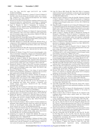
![S465
Introduction
The recommendations in this 2015 American Heart
Association (AHA) Guidelines Update for Cardiopulmonary
Resuscitation and Emergency Cardiovascular Care are based
on an extensive evidence review process that was begun
by the International Liaison Committee on Resuscitation
(ILCOR) after the publication of the 2010 International
Consensus on Cardiopulmonary Resuscitation and
Emergency Cardiovascular Care Science With Treatment
Recommendations1,2
and was completed in February 2015.3,4
In this in-depth evidence review process, ILCOR exam-
ined topics and then generated a prioritized list of questions
for systematic review. Questions were first formulated in
PICO (population, intervention, comparator, outcome) for-
mat,5
and then search strategies and inclusion and exclusion
criteria were defined and a search for relevant articles was
performed. The evidence was evaluated by the ILCOR task
forces by using the standardized methodological approach
proposed by the Grading of Recommendations Assessment,
Development and Evaluation (GRADE) Working Group.6
The quality of the evidence was categorized based on the
study methodologies and the 5 core GRADE domains of risk
of bias, inconsistency, indirectness, imprecision, and other con-
siderations (including publication bias). Then, where possible,
consensus-based treatment recommendations were created.
To create this 2015 Guidelines Update, the AHA formed
15 writing groups, with careful attention to manage conflicts
of interest, to assess the ILCOR treatment recommendations
and to write AHA treatment recommendations by using the
AHA Class of Recommendation (COR) and Level of Evidence
(LOE) system. The recommendations made in the Guidelines
are informed by the ILCOR recommendations and GRADE
classification, in the context of the delivery of medical care
in North America. The AHA writing group made new recom-
mendations only on topics specifically reviewed by ILCOR in
2015. This chapter delineates instances where the AHA writ-
ing group developed recommendations that are significantly
stronger or weaker than the ILCOR statements. In the online
version of this publication, live links are provided so the
reader can connect directly to the systematic reviews on the
Scientific Evidence Evaluation and Review System (SEERS)
website. These links are indicated by a combination of letters
and numbers (eg, ALS 790). We encourage readers to use the
links and review the evidence and appendixes, including the
GRADE tables.
This update uses the newest AHA COR and LOE classi-
fication system, which contains modifications of the Class III
recommendation and introduces LOE B-R (randomized stud-
ies) and B-NR (nonrandomized studies) as well as LOE C-LD
(limited data) and LOE C-EO (consensus of expert opinion).
All recommendations made in this 2015 Guidelines Update,
as well as in the 2010 Guidelines for post‒cardiac arrest
care, are listed in the Appendix. For further information, see
“Part 2: Evidence Evaluation and Management of Conflicts of
Interest” in this 2015 Guidelines Update.
Overview of Post–Cardiac Arrest Care
The 2010 Guidelines emphasized that cardiac arrest can
result from many different diseases. Regardless of cause, the
hypoxemia, ischemia, and reperfusion that occur during car-
diac arrest and resuscitation may cause damage to multiple
organ systems.7
The severity of damage can vary widely
among patients and among organ systems within individual
patients. Therefore, effective post–cardiac arrest care consists
of identification and treatment of the precipitating cause of
cardiac arrest combined with the assessment and mitigation of
ischemia-reperfusion injury to multiple organ systems. Care
must be tailored to the particular disease and dysfunction that
affect each patient. Therefore, individual patients may require
few, many, or all of the specific interventions discussed in the
remainder of this Part.
Cardiovascular Care
Acute Cardiovascular InterventionsACS 340, ACS 885
The 2010 Guidelines recommended obtaining a 12-lead elec-
trocardiogram (ECG) as soon as possible after return of spon-
taneous circulation (ROSC) to identify if acute ST elevation
is present, and to perform urgent coronary angiography with
prompt recanalization of any infarct-related artery in select
(Circulation. 2015;132[suppl 1]:S465–S482. DOI: 10.1161/CIR.0000000000000262.)
© 2015 American Heart Association, Inc.
Circulation is available at http://circ.ahajournals.org DOI: 10.1161/CIR.0000000000000262
The American Heart Association requests that this document be cited as follows: Callaway CW, Donnino MW, Fink EL, Geocadin RG, Golan E, Kern
KB, Leary M, Meurer WJ, Peberdy MA, Thompson TM, Zimmerman JL. Part 8: post–cardiac arrest care: 2015 American Heart Association Guidelines
Update for Cardiopulmonary Resuscitation and Emergency Cardiovascular Care. Circulation. 2015;132(suppl 2):S465–S482.
Part 8: Post–Cardiac Arrest Care
2015 American Heart Association Guidelines Update for Cardiopulmonary
Resuscitation and Emergency Cardiovascular Care
Clifton W. Callaway, Chair; Michael W. Donnino; Ericka L. Fink; Romergryko G. Geocadin;
Eyal Golan; Karl B. Kern; Marion Leary; William J. Meurer; Mary Ann Peberdy;
Trevonne M. Thompson; Janice L. Zimmerman
by guest on October 15, 2015http://circ.ahajournals.org/Downloaded from](https://image.slidesharecdn.com/guidelines-rcp-aha-2015-full-160515230707/85/Guidelines-rcp-aha-2015-full-161-320.jpg)
![S466 Circulation November 3, 2015
post–cardiac arrest patients in whom ST-segment elevation
was identified. Acute coronary syndromes are a common
etiology for out-of-hospital cardiac arrest (OHCA) in adults
with no obvious extracardiac cause of arrest8–10
and also can
precipitate some in-hospital cardiac arrest. In series in which
consecutive post–cardiac arrest patients with suspected car-
diovascular cause were taken to coronary angiography, a
coronary artery lesion amenable to emergency treatment was
found in 96% of patients with ST elevation and in 58% of
patients without ST elevation.10
The 2015 ILCOR systematic review examined immediate
coronary angiography for patients after cardiac arrest.
2015 Evidence Summary
Numerous observational studies evaluate the relationship
between coronary angiography, survival, and functional out-
come in post–cardiac arrest patients, but there are no prospec-
tive randomized trials evaluating an interventional strategy in
postarrest patients. The timing of immediate coronary angi-
ography was defined in various ways in different studies, but
all studies considered immediate angiography as a procedure
performed on the same day as the cardiac arrest, as opposed
to later in the hospital stay. Fifteen observational studies
reported improved survival to hospital discharge associated
with emergency coronary angiography in patients with ST
elevation after cardiac arrest.11–25
Nine observational studies
showed improved neurologically favorable outcome associ-
ated with emergency coronary angiography in patients with
ST elevation after cardiac arrest.11–13,16,18–21,23
Fewer data are available to evaluate coronary angiogra-
phy in patients without ST elevation on the initial ECG. Two
observational studies reported improved survival to hospital
discharge and improved neurologically favorable outcome
associated with emergency coronary angiography in patients
without ST elevation on initial ECG.11,16
2015 Recommendations—Updated
Coronary angiography should be performed emergently
(rather than later in the hospital stay or not at all) for OHCA
patients with suspected cardiac etiology of arrest and ST ele-
vation on ECG (Class I, LOE B-NR).
Emergency coronary angiography is reasonable for select
(eg, electrically or hemodynamically unstable) adult patients
who are comatose after OHCA of suspected cardiac origin but
without ST elevation on ECG (Class IIa, LOE B-NR).
Coronary angiography is reasonable in post–cardiac arrest
patients for whom coronary angiography is indicated regard-
less of whether the patient is comatose or awake (Class IIa,
LOE C-LD).
Early invasive approaches are preferred for patients with
ST-segment elevation myocardial infarction (STEMI), mak-
ing these recommendations for post–cardiac arrest patients
consistent with global recommendations for all patients with
STEMI.26
Early invasive approaches also are suggested for
treatment of select post–cardiac arrest patients with acute
coronary syndromes without ST elevation. Considerations for
selecting patients are complex and may consider factors such
as hemodynamic or electrical instability as well as comorbidi-
ties, evidence of ongoing ischemia, and other patient charac-
teristics.27
Knowledge of coronary anatomy and opportunity
for placement of temporary support devices are other potential
benefits derived from early catheterization. Therefore, these
recommendations for post–cardiac arrest care are consis-
tent with recommendations for all patients with non-STEMI
acute coronary syndromes. Both the European Society of
Cardiology and the combined entity of the American College
of Cardiology Foundation and the AHA have published
STEMI guidelines recommending immediate coronary angi-
ography, and percutaneous coronary intervention when indi-
cated, for resuscitated OHCA patients whose ECGs show
STEMI.26,28
None of these guidelines recommended different
treatment of patients based on the initial cardiac arrest rhythm
(ventricular fibrillation [VF] or non-VF).
Previous consensus statements have discussed how public
reporting of postprocedure death creates an incentive to avoid
emergency coronary angiography in comatose patients who
are at higher risk of death as a consequence of poor neurologic
recovery.29
However, the probability of neurologic recovery
cannot be determined reliably at the time that emergency car-
diovascular interventions are performed (see Prognostication
of Outcome section in this Part). Therefore, the best care for
the patient requires separation of decisions about cardiovas-
cular intervention from assessment of neurologic prognosis.
Hemodynamic GoalsALS 570
Post–cardiac arrest patients are often hemodynamically unsta-
ble, which can occur for multiple reasons that include the
underlying etiology of the arrest as well as the ischemia-reper-
fusion injury from the arrest. Management of these patients
can be challenging, and optimal hemodynamic goals remain
undefined. In 2015, ILCOR evaluated the optimal hemody-
namic targets in post–cardiac arrest patients, primarily consid-
ering blood pressure goals.
2015 Evidence Summary
There are several observational studies evaluating the rela-
tionship between blood pressure and outcome in post–cardiac
arrest patients, but there are no interventional studies targeting
blood pressure in isolation and no trials evaluating one spe-
cific strategy for improving blood pressure over another (ie,
fluids, vasopressors). Observational studies found that post–
cardiac arrest systolic blood pressure less than 90 mm Hg30,31
or greater than 100 mm Hg32
was associated with higher mor-
tality and diminished functional recovery. One observational
study found that mean arterial pressure (MAP) greater than
100 mm Hg during 2 hours after ROSC was associated with
better neurologic recovery at hospital discharge.33
Another
observational study found that survivors, compared with non-
survivors, had higher MAP at 1 hour (96 versus 84 mm Hg)
and at 6 hours (96 versus 90 mm Hg).34
While no studies evaluated blood pressure in isolation,
several before-and-after studies implemented bundles of care
that included blood pressure goals. In these studies, the indi-
vidual effect of blood pressure was impossible to separate
from the effects of the remainder of the bundle. One bundle
with a MAP target of greater than 80 mm Hg improved mor-
tality and neurologic outcome at hospital discharge.35
One
bundle with a goal of MAP over 75 mm Hg found no change
in functional recovery at hospital discharge.36
One bundle with
by guest on October 15, 2015http://circ.ahajournals.org/Downloaded from](https://image.slidesharecdn.com/guidelines-rcp-aha-2015-full-160515230707/85/Guidelines-rcp-aha-2015-full-162-320.jpg)
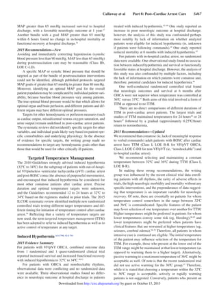
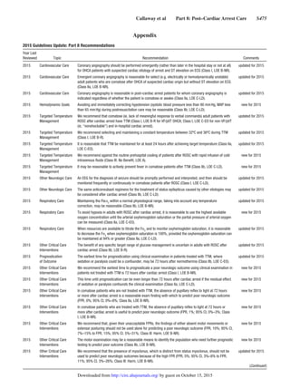

![S483
Introduction
Clinicians often struggle with uncertainty and complexity in
deciding which course of treatment will likely lead to an opti-
mal outcome for an individual patient. Scientific research pro-
vides information on how patient populations have responded
to treatment regimens, and this information, combined with a
knowledge of the individual patient, can help guide the clini-
cian’s decisions.
The recommendations in this 2015 American Heart
Association (AHA) Guidelines Update for Cardiopulmonary
Resuscitation (CPR) and Emergency Cardiovascular Care
(ECC) are based on an extensive evidence review process
that was begun by the International Liaison Committee
on Resuscitation (ILCOR) after the publication of the
ILCOR 2010 International Consensus on Cardiopulmonary
Resuscitation and Emergency Cardiovascular Care Science
With Treatment Recommendations1,2
and was completed in
February 2015.3,4
In this in-depth evidence review process, ILCOR exam-
ined topics and then generated a prioritized list of questions
for systematic review. Questions were first formulated in
PICO (population, intervention, comparator, outcome) for-
mat,5
and then a search for relevant articles was performed.
The evidence was evaluated by the ILCOR task forces by
using the standardized methodologic approach proposed by
the Grading of Recommendations Assessment, Development
and Evaluation (GRADE) Working Group.6
The quality of the evidence was categorized based on the
study methodologies and the 5 core GRADE domains of risk
of bias, inconsistency, indirectness, imprecision, and other
considerations (including publication bias). Then, where
possible, consensus-based treatment recommendations were
created.
To create this 2015 AHA Guidelines Update for CPR and
ECC, the AHA formed 15 writing groups, with careful atten-
tion to avoid conflicts of interest, to assess the ILCOR treatment
recommendations, and to write AHA treatment recommenda-
tions by using the AHA Class of Recommendation and Level
of Evidence (LOE) system. The recommendations made in the
2015 Guidelines Update for CPR and ECC are informed by the
ILCOR recommendations and GRADE classification, in the
context of the delivery of medical care in North America. In
the online version of this publication, live links are provided so
the reader can connect directly to the systematic reviews on the
Scientific Evidence Evaluation and Review System (SEERS)
website. These links are indicated by a superscript combina-
tion of letters and numbers (eg, ACS 873).
This 2015 Guidelines Update offers recommendations for
the care of patients with acute coronary syndromes (ACS).
The recommendations made here update those made in the
2010 Guidelines and address only those issues that were
reviewed in 2015. The ILCOR ACS Task Force did not review
areas in which it found a paucity of new evidence between
2010 and 2015; therefore, the 2010 Guidelines for these unre-
viewed areas remain current. For example, acetylsalicylic acid
administration has been shown to be of benefit in ACS and
was recommended by the 2010 Guidelines.7
Acetylsalicylic
acid was not reviewed by the ACS Task Force in 2015, so the
recommendations from 2010 should be used. (Note: The First
Aid section of this 2015 Guidelines Update makes recommen-
dations on acetylsalicylic acid administration by nonmedical
personnel—see “Part 15: First Aid”). The recommendations
that were not reviewed in 2015 will either be reviewed and
included in future AHA Guidelines for CPR and ECC or will
be in the most recent ACC/AHA Guidelines.8–10
A table of recommendations made in this update, as well
as the recommendations made in “Part 10: Acute Coronary
Syndromes” of the 2010 Guidelines,7
can be found in the
Appendix.
The 2015 Guidelines for ACS are directed toward prac-
titioners who provide care for patients with suspected ACS
from the time of first medical contact until disposition from
the emergency department (ED). Care providers during this
time may include emergency medical service (EMS) dispatch-
ers, first responders, EMT-Bs, paramedics, nurses, physicians,
and other independent practitioners.
Methodology
ILCOR performed 18 systematic reviews (14 based on
meta-analyses) on more than 110 relevant studies that span
(Circulation. 2015;132[suppl 2]:S483–S500. DOI: 10.1161/CIR.0000000000000263.)
© 2015 American Heart Association, Inc.
Circulation is available at http://circ.ahajournals.org DOI: 10.1161/CIR.0000000000000263
The American Heart Association requests that this document be cited as follows: O’Connor RE, Al Ali AS, Brady WJ, Ghaemmaghami CA, Menon V,
Welsford M, Shuster M. Part 9: acute coronary syndromes: 2015 American Heart Association Guidelines Update for Cardiopulmonary Resuscitation and
Emergency Cardiovascular Care. Circulation. 2015;132(suppl 2):S483–S500.
Part 9: Acute Coronary Syndromes
2015 American Heart Association Guidelines Update for Cardiopulmonary
Resuscitation and Emergency Cardiovascular Care
Robert E. O’Connor, Chair; Abdulaziz S. Al Ali; William J. Brady; Chris A. Ghaemmaghami;
Venu Menon; Michelle Welsford; Michael Shuster
by guest on October 15, 2015http://circ.ahajournals.org/Downloaded from](https://image.slidesharecdn.com/guidelines-rcp-aha-2015-full-160515230707/85/Guidelines-rcp-aha-2015-full-180-320.jpg)
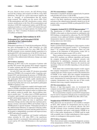
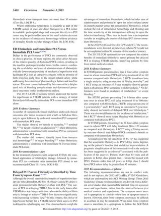
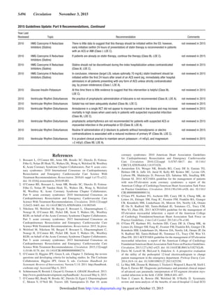

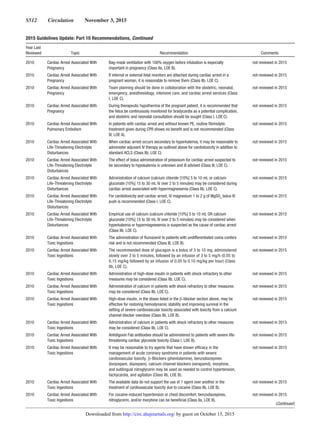

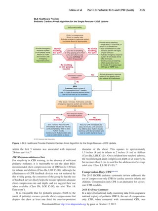
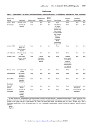
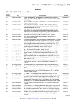
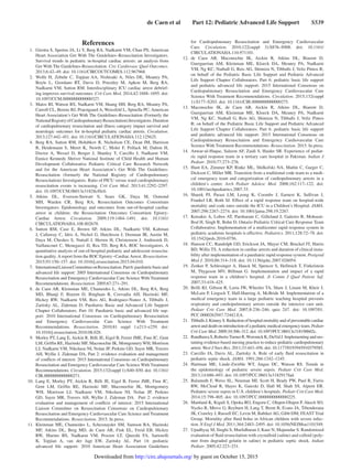


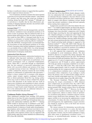


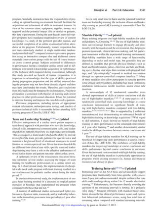

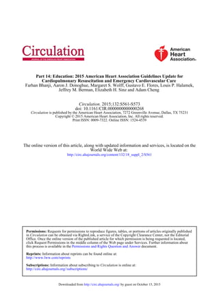
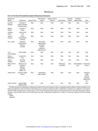
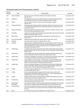


The 2015 AHA Guidelines update for CPR and ECC consists of 15 parts that were published to update recommendations based on new evidence or ongoing controversies since the 2010 guidelines. Key changes include a new Part 4 on systems of care and quality improvement, and a new Part 14 on education. The guidelines transitioned to a web-based format for continuous updating going forward. The evidence review process was also changed from a 5-year cycle to continuous evaluation to more rapidly translate new science into practice. The overall goal is to double bystander CPR rates and cardiac arrest survival by 2020.


![谷歌留痕技术 [ 𝙩𝙤𝙥 𝟮𝟯𝟯. 𝙘 𝙤𝙢 ]](https://cdn.slidesharecdn.com/ss_thumbnails/top233-260130174328-3833018c-thumbnail.jpg?width=640&height=640&fit=bounds)

