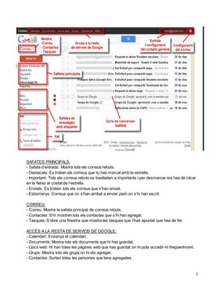
SAFATES PRINCIPALS:
-Safata d’entrada: Mostra tots els correus rebuts.
- Destacats: Es troben els correus que tu has marcat amb la estrella.
- Important: Tots els correus rebuts es traslladen a importants i per desmarcar-los has de clicar
en la fletxa al costat de l’estrella.
- Enviats: Es troben tots els correus que s’han enviat.
- Esborranys: Correus que no s’han arribat a enviar però on s’hi han escrit.
1
CORREU:
- Correu: Mostra la safata principal de correus rebuts.
- Contactes: S’hi mostren tots els contactes que s’hi han agregat.
- Tasques: S’obre una finestra que mostra les tasques que t’has apuntat que has de fer.
ACCÉS A LA RESTA DE SERVEIS DE GOOGLE:
- Calendari: Ensenya el calendari.
- Documents: Mostra tots els documents que hi has guardat.
- Llocs web: Hi han totes les pàgines web que has guardat on hi pots accedir-hi freqüentment.
- Grups: Mostra tots els grups on hi ets agregat.
- Contactes: Surten totes les persones que tens agregades.
2.
2
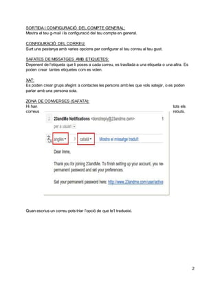
SORTIDA ICONFIGURACIÓ DEL COMPTE GENERAL:
Mostra el teu g-mail i la configuració del teu compte en general.
CONFIGURACIÓ DEL CORREU:
Surt una pestanya amb varies opcions per configurar el teu correu al teu gust.
SAFATES DE MISSATGES AMB ETIQUETES:
Depenent de l’etiqueta que li poses a cada correu, es trasllada a una etiqueta o una altra. Es
poden crear tantes etiquetes com es volen.
XAT:
Es poden crear grups afegint a contactes les persons amb les que vols xatejar, o es poden
parlar amb una persona sola.
ZONA DE CONVERSES (SAFATA):
Hi han tots els
correus rebuts.
Quan escrius un correu pots triar l’opció de que te’l tradueixi.
3.
3
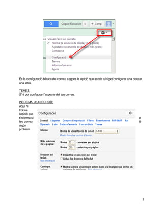
És laconfiguració bàsica del correu, segons la opició que es tria s’hi pot configurar una cosa o
una altra.
TEMES:
S’hi pot configurar l’aspecte del teu correu.
INFORMA D’UN ERROR:
Aquí hi
trobes
l’opció que
t’informa si el
teu correu té
algún
problem.
4.
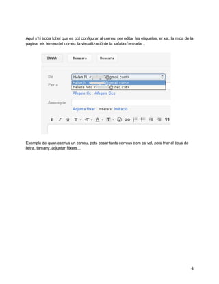
Aquí s’hi trobatot el que es pot configurar al correu, per editar les etiquetes, el xat, la mida de la
pàgina, els temes del correu, la visualització de la safata d’entrada…
4
Exemple de quan escrius un correu, pots posar tants correus com es vol, pots triar el tipus de
lletra, tamany, adjuntar fitxers...