Cytogenetics Overview
Cytogenetics isthe branch of genetics that studies the structure and
behavior of chromosomes. It includes karyotyping, analysis of
chromosome number, structure, and abnormalities.
Clinical Importance
• Detection of genetic disorders (e.g., Down syndrome, Turner
syndrome)
• Prenatal diagnostics
• Cancer cytogenetics
• Reproductive failure and infertility evaluation
3.
Abnormalities of ChromosomeNumber
(Aneuploidies)
Mechanism: Nondisjunction, the failure of homologous chromosomes
or sister chromatids to separate properly during meiosis or mitosis.
Leads to trisomy (extra chromosome) or monosomy (missing
chromosome).
4.
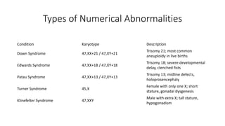
Types of NumericalAbnormalities
Condition Karyotype Description
Down Syndrome 47,XX+21 / 47,XY+21
Trisomy 21; most common
aneuploidy in live births
Edwards Syndrome 47,XX+18 / 47,XY+18
Trisomy 18; severe developmental
delay, clenched fists
Patau Syndrome 47,XX+13 / 47,XY+13
Trisomy 13; midline defects,
holoprosencephaly
Turner Syndrome 45,X
Female with only one X; short
stature, gonadal dysgenesis
Klinefelter Syndrome 47,XXY
Male with extra X; tall stature,
hypogonadism
5.
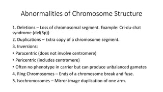
Abnormalities of ChromosomeStructure
1. Deletions – Loss of chromosomal segment. Example: Cri-du-chat
syndrome (del(5p))
2. Duplications – Extra copy of a chromosome segment.
3. Inversions:
• Paracentric (does not involve centromere)
• Pericentric (includes centromere)
• Often no phenotype in carrier but can produce unbalanced gametes
4. Ring Chromosomes – Ends of a chromosome break and fuse.
5. Isochromosomes – Mirror image duplication of one arm.
6.
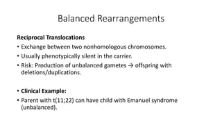
Balanced Rearrangements
Reciprocal Translocations
•Exchange between two nonhomologous chromosomes.
• Usually phenotypically silent in the carrier.
• Risk: Production of unbalanced gametes → offspring with
deletions/duplications.
• Clinical Example:
• Parent with t(11;22) can have child with Emanuel syndrome
(unbalanced).
7.
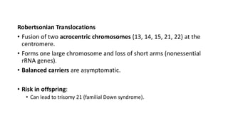
Robertsonian Translocations
• Fusionof two acrocentric chromosomes (13, 14, 15, 21, 22) at the
centromere.
• Forms one large chromosome and loss of short arms (nonessential
rRNA genes).
• Balanced carriers are asymptomatic.
• Risk in offspring:
• Can lead to trisomy 21 (familial Down syndrome).
8.
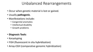
Unbalanced Rearrangements
• Occurwhen genetic material is lost or gained.
• Usually pathogenic.
• Manifestations include:
• Congenital anomalies
• Intellectual disability
• Growth problems
• Diagnosis Tools:
• Karyotyping
• FISH (fluorescent in situ hybridization)
• Array-CGH (comparative genomic hybridization)
9.
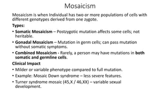
Mosaicism
Mosaicism is whenIndividual has two or more populations of cells with
different genotypes derived from one zygote.
Types:
• Somatic Mosaicism – Postzygotic mutation affects some cells; not
heritable.
• Gonadal Mosaicism – Mutation in germ cells; can pass mutation
without somatic symptoms.
• Combined Mosaicism - Rarely, a person may have mutations in both
somatic and germline cells.
Clinical Impact
• Milder or variable phenotype compared to full mutation.
• Example: Mosaic Down syndrome – less severe features.
• Turner syndrome mosaic (45,X / 46,XX) – variable sexual
development.
10.
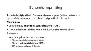
Genomic Imprinting
Parent-of-origin effect:Only one allele of a gene (either maternal or
paternal) is expressed; the other is epigenetically silenced.
Mechanism
• Controlled by imprinting control regions (ICRs).
• DNA methylation and histone modification silence one allele.
Relevance
• Imprinting disorders occur when:
• The active allele is deleted/mutated.
• There is uniparental disomy (UPD).
• ICR is abnormally methylated.
11.
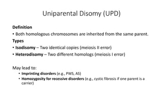
Uniparental Disomy (UPD)
Definition
•Both homologous chromosomes are inherited from the same parent.
Types
• Isodisomy – Two identical copies (meiosis II error)
• Heterodisomy – Two different homologs (meiosis I error)
May lead to:
• Imprinting disorders (e.g., PWS, AS)
• Homozygosity for recessive disorders (e.g., cystic fibrosis if one parent is a
carrier)
12.
Imprinting Disorders &Syndromes
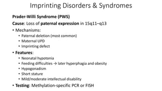
Prader-Willi Syndrome (PWS)
Cause: Loss of paternal expression in 15q11–q13
• Mechanisms:
• Paternal deletion (most common)
• Maternal UPD
• Imprinting defect
• Features:
• Neonatal hypotonia
• Feeding difficulties → later hyperphagia and obesity
• Hypogonadism
• Short stature
• Mild/moderate intellectual disability
• Testing: Methylation-specific PCR or FISH
13.
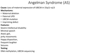
Angelman Syndrome (AS)
Cause:Loss of maternal expression of UBE3A in 15q11–q13
Mechanisms:
• Maternal deletion
• Paternal UPD
• UBE3A mutation
• Imprinting defect
Features:
Severe intellectual disability
Minimal speech
Ataxic gait,
jerky movements
Happy disposition,
frequent laughter
Seizures
Testing:
DNA methylation, UBE3A sequencing
14.
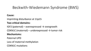
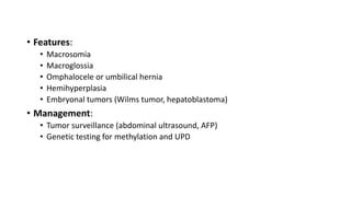
Beckwith-Wiedemann Syndrome (BWS)
Cause:
Imprintingdisturbance at 11p15
Two critical domains:
IGF2 (paternal) – overexpressed → overgrowth
CDKN1C (maternal) – underexpressed → tumor risk
Mechanisms:
Paternal UPD
Loss of maternal methylation
CDKN1C mutations


![SecurityBoat_Service_Pitch_Deck[24158].pdf](https://cdn.slidesharecdn.com/ss_thumbnails/securityboatservicepitchdeck24158-260121113056-452683e3-thumbnail.jpg?width=640&height=640&fit=bounds)


