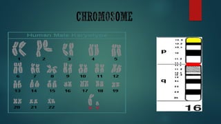
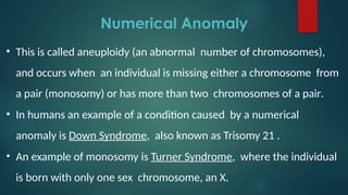
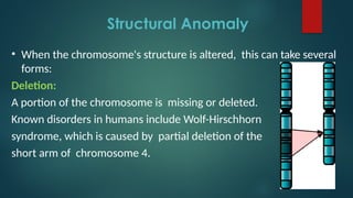
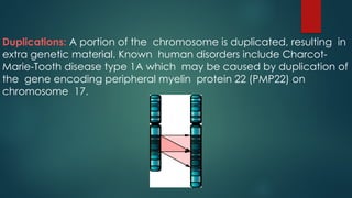
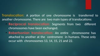
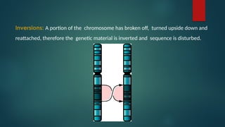
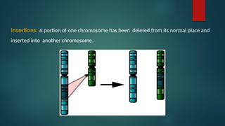
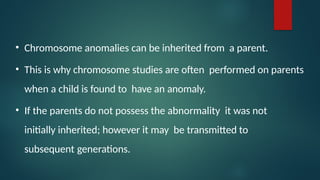
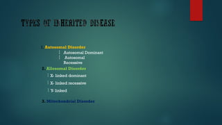
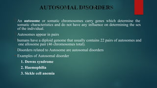
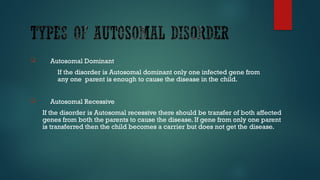
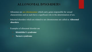
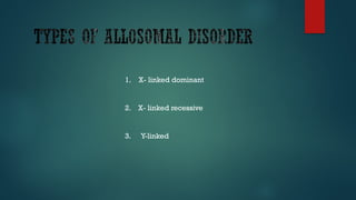
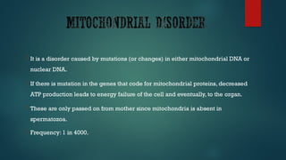
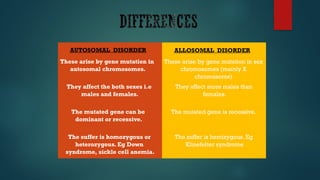
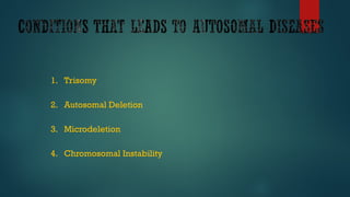
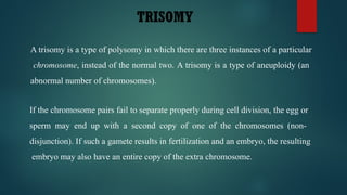
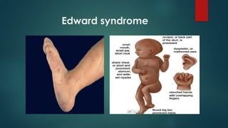
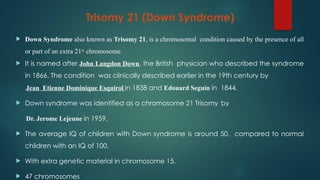
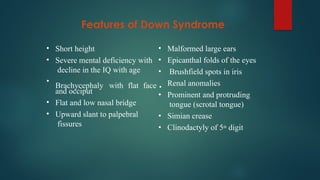
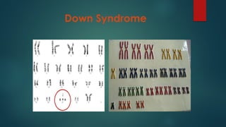
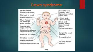
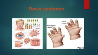
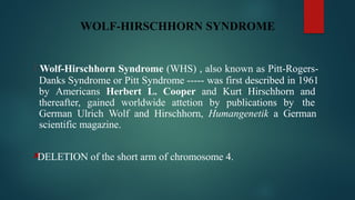
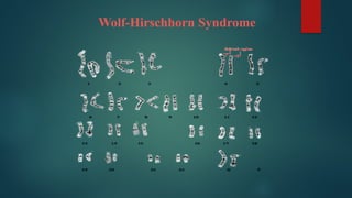
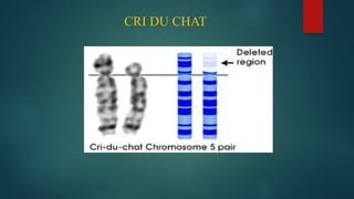
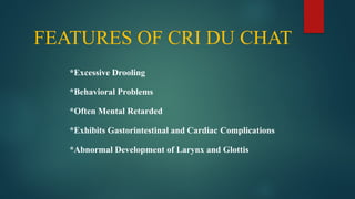
Genetic Disorders and
Chromosomal anomalies
Presented by:
MARRAH
SCS
 Genetic disorder is a disease that is caused by an abnormality
in an individual’s DNA. Abnormalities can range from a small
mutation in DNA or addition or subtraction of an entire
chromosome or set of chromosomes.
 Most Genetic disorders are quite rare and affect one person
in every several thousands or millions.
 Genetic disorders may results by
 Point mutation
 Deletion of a gene or genes
 Whole chromosome extra, missing, or both
• A chromosome anomaly, abnormality or aberration reflects on a
typical number of chromosome or a structural abnormality in one
or more chromosomes.
• Chromosome anomalies usually occur when there is an error in
cell division following meiosis or mitosis.
• There are many types of chromosome anomalies.
• They can be organized into two basic groups,
i. Numerical anomaly
ii. Structural anomaly
Numerical Anomaly
• This is called aneuploidy (an abnormal number of chromosomes),
and occurs when an individual is missing either a chromosome from
a pair (monosomy) or has more than two chromosomes of a pair.
• In humans an example of a condition caused by a numerical
anomaly is Down Syndrome, also known as Trisomy 21 .
• An example of monosomy is Turner Syndrome, where the individual
is born with only one sex chromosome, an X.
Structural Anomaly
• When the chromosome's structure is altered, this can take several
forms:
Deletion:
A portion of the chromosome is missing or deleted.
Known disorders in humans include Wolf-Hirschhorn
syndrome, which is caused by partial deletion of the
short arm of chromosome 4.
Duplications: A portion of the chromosome is duplicated, resulting in
extra genetic material. Known human disorders include Charcot-
Marie-Tooth disease type 1A which may be caused by duplication of
the gene encoding peripheral myelin protein 22 (PMP22) on
chromosome 17.
Translocation: A portion of one chromosome is transferred to
another chromosome. There are two main types of translocations:
• Reciprocal translocation: Segments from two different
chromosomes have been exchanged.
• Robertsonian translocation: An entire chromosome has
attached to another at the centromere in humans. These only
occur with chromosomes 13, 14, 15, 21 and 22.
Inversions: A portion of the chromosome has broken off, turned upside down and
reattached, therefore the genetic material is inverted and sequence is disturbed.
Insertions: A portion of one chromosome has been deleted from its normal place and
inserted into another chromosome.
• Chromosome anomalies can be inherited from a parent.
• This is why chromosome studies are often performed on parents
when a child is found to have an anomaly.
• If the parents do not possess the abnormality it was not
initially inherited; however it may be transmitted to
subsequent generations.
1. Autosomal Disorder


Autosomal Dominant
Autosomal
Recessive
2. Allosomal Disorder
 X- linked dominant
 X- linked recessive
 Y
- linked
3. Mitochondrial Disorder
 An autosome or somatic chromosomes carry genes which determine the
somatic characteristics and do not have any influence on determining the sex
of the individual.
 Autosomes appear in pairs
 humans have a diploid genome that usually contains 22 pairs of autosomes and
one allosome pair (46 chromosomes total).
 Disorders related to Autosome are autosomal disorders
• Examples of Autosomal disorder
1. Downs syndrome
2. Haemophilia
3. Sickle cell anemia
 Autosomal Dominant
If the disorder is Autosomal dominant only one infected gene from
any one parent is enough to cause the disease in the child.
 Autosomal Recessive
If the disorder is Autosomal recessive there should be transfer of both affected
genes from both the parents to cause the disease. If gene from only one parent
is transferred then the child becomes a carrier but does not get the disease.
• Allosomes are sex chromosomes which carry genes responsible for sexual
characteristics and as such have a significant role in the determination of sex.
• Inherited disorders which are related to sex chromosomes are called as Allosomal
disorders
• Examples of allosomal disorder are
1. Kleinfelder's syndrome
2. Turners syndrome
1. X- linked dominant
2. X- linked recessive
3. Y-linked
 It is a disorder caused by mutations (or changes) in either mitochondrial DNA or
nuclear DNA.
 If there is mutation in the genes that code for mitochondrial proteins, decreased
ATP production leads to energy failure of the cell and eventually, to the organ.
 These are only passed on from mother since mitochondria is absent in
spermatozoa.
 Frequency: 1 in 4000.
AUTOSOMAL DISORDER ALLOSOMAL DISORDER
These arise by gene mutation in
autosomal chromosomes.
These arise by gene mutation in sex
chromosomes (mainly X
chromosome)
They affect the both sexes i.e
males and females.
They affect more males than
females.
The mutated gene can be
dominant or recessive.
The mutated gene is recessive.
The suffer is homozygous or
heterozygous. Eg Down
syndrome, sickle cell anemia.
The suffer is hemizygous. Eg
Klinefelter syndrome
1. Trisomy
2. Autosomal Deletion
3. Microdeletion
4. Chromosomal Instability
TRISOMY
A trisomy is a type of polysomy in which there are three instances of a particular
chromosome, instead of the normal two. A trisomy is a type of aneuploidy (an
abnormal number of chromosomes).
If the chromosome pairs fail to separate properly during cell division, the egg or
sperm may end up with a second copy of one of the chromosomes (non-
disjunction). If such a gamete results in fertilization and an embryo, the resulting
embryo may also have an entire copy of the extra chromosome.
TRISOMY
The most common types of autosomal trisomy that survive to birth in humans are:
• Trisomy 21 (Down syndrome)
• Trisomy 18 (Edwards syndrome)
• Trisomy 13 (Patau syndrome)
TRISOMY 13 (PATAU SYNDROME)
 Patau Sundrome, also known as Trisomy 13 and Trisomy D.
 Is a chromosomal abnormality, a syndrome in which a patient had an additional
chromosome 13 due to non-disjunction of chromosomes during meiosis. Some are
caused by Robertsonian Translocations. The extra chromosome 13 disrupts the
normal course of development, causing heart and kidney defects, amongst other
features characteristic of Patau syndrome.
 Patau syndrome affects somewhere between 1 in 10,000 and 1 in 21,700 live births.
 TRISOMIC in the chromosome 13.
 47 chromosomes
FEATURES OF PATAU SYNDROME
•
•
•
•
•
•
•
Mental deficiency
Low birth weight
Abnormal development
of frontal lobe
Absence of corpus
callosum Hypoplasia of
cerebellum
Sloping
forehead Scalp
defects
•
•
•
•
•
•
•
Malformed ears
Congenital heart defects
Renal tract anomalies
Microphthalmia
Bilateral cleft lip/palate
Polydactyly with
rudimentary digits
Rocker-bottom heel
Patau syndrome
Patau syndrome
Trisomy 18 (Edward Syndrome)
• Edward’s Syndrome also kwnon as Trisomy 18 (T18) or Trisome E.
• It is a genetic disorder caused by the presence of all of an extra
18th
chromosome (Trisomy 18) due to meiotic nondisjunction.
• It is named after John H. Edwards, who first described the syndrome in
1960. It is the second most common autosomal trisomy, after Down
Syndrome, that carries to term.
• Edward’s Syndrome occurs in around 1 in 6,000 live births and around 80 %
of those affected are female.
Features of Edward Syndrome
•
•
•
•
•
•
Mental deficiency
Growth retardation
Prominent occiput with
elongated head
Webbing of the neck
Short sternum
Micrognathia
•
•
•
•
•
Low-set malformed ears
Ventricular septal defects
Renal anomalies
Clenched fists with overlapping
of fingers
Hypoplastic nails
Edward syndrome
Trisomy 21 (Down Syndrome)
 Down Syndrome also known as Trisomy 21, is a chromosomal condition caused by the presence of all
or part of an extra 21st chromosome.
 It is named after John Langdon Down, the British physician who described the syndrome
in 1866. The condition was clinically described earlier in the 19th century by
Jean Etienne Dominique Esquirol in 1838 and Edouard Seguin in 1844.
 Down syndrome was identified as a chromosome 21 Trisomy by
Dr. Jerome Lejeune in 1959.
 The average IQ of children with Down syndrome is around 50, compared to normal
children with an IQ of 100.
 With extra genetic material in chromosome 15.
 47 chromosomes
Features of Down Syndrome
•
•
Short height
Severe mental deficiency with
decline in the IQ with age
•
•
•
and occiput
Flat and low nasal bridge
Upward slant to palpebral
fissures
•
•
•
Brachycephaly with flat face •
•
•
•
Malformed large ears
Epicanthal folds of the eyes
Brushfield spots in iris
Renal anomalies
Prominent and protruding
tongue (scrotal tongue)
Simian crease
Clinodactyly of 5th digit
Down Syndrome
Down syndrome
Down syndrome
AUTOSOMAL DELETION
 Autosomal: involves one of the numbered (ie, non-sex chromosomes)
 Deletion: an abnormality of DNA that involves missing material. These can
range from very small (as little as 1 base pair) to very large (involving millions
of base pairs of DNA)
AUTOSOMAL DELETION
 The most common types of autosomal deletion syndromes are:
1.Wolf-Hirschhorn Syndrome
2.Cri Du Chat
3.Langer Giedion Syndrome
Wolf-Hirschhorn Syndrome
WOLF-HIRSCHHORN SYNDROME
 Wolf-Hirschhorn Syndrome (WHS) , also known as Pitt-Rogers-
Danks Syndrome or Pitt Syndrome ----- was first described in 1961
by Americans Herbert L. Cooper and Kurt Hirschhorn and
thereafter, gained worldwide attetion by publications by the
German Ulrich Wolf and Hirschhorn, Humangenetik a German
scientific magazine.
DELETION of the short arm of chromosome 4.
Wolf-Hirschhorn Syndrome
FEATURES OF WOLF-HIRSCHHORN SYNDROME
*Short Philtrum
*Immunodeficient
*Microcephaly (Small Head Size)
*Seizures
*Muscle Hypotonia
*Renal Anomalies
*Deafness
Cri Du Chat
CRI DU CHAT
 CRI DU CHAT also known as chromosome 5p deletion syndrome, 5p minus
syndrome or Lejeune’s syndrome.
 Is a rare genetic disorder due to a missing part of chromosome 5. Its name is a
French term (cat-cry or call of the cat) referring to the characteristic cat-like
cry of affected children.
 It was first described by Jerome Lejeune in 1963. The condition affects an 1
in 50,000 live births, strikes all ethnicities, and is more common in females by
a 4:3 ratio.
DELETION on the short arm of chromosome 5.
Cries like a CAT
CRI DU CHAT
FEATURES OF CRI DU CHAT
*Excessive Drooling
*Behavioral Problems
*Often Mental Retarded
*Exhibits Gastorintestinal and Cardiac Complications
*Abnormal Development of Larynx and Glottis
Thank you
for your
Patience.

geneticdisorderchromosomalabnormalities.pptx

  • 1.
    Genetic Disorders and Chromosomalanomalies Presented by: MARRAH SCS
  • 2.
     Genetic disorderis a disease that is caused by an abnormality in an individual’s DNA. Abnormalities can range from a small mutation in DNA or addition or subtraction of an entire chromosome or set of chromosomes.  Most Genetic disorders are quite rare and affect one person in every several thousands or millions.  Genetic disorders may results by  Point mutation  Deletion of a gene or genes  Whole chromosome extra, missing, or both
  • 4.
    • A chromosomeanomaly, abnormality or aberration reflects on a typical number of chromosome or a structural abnormality in one or more chromosomes. • Chromosome anomalies usually occur when there is an error in cell division following meiosis or mitosis. • There are many types of chromosome anomalies. • They can be organized into two basic groups, i. Numerical anomaly ii. Structural anomaly
  • 5.
    Numerical Anomaly • Thisis called aneuploidy (an abnormal number of chromosomes), and occurs when an individual is missing either a chromosome from a pair (monosomy) or has more than two chromosomes of a pair. • In humans an example of a condition caused by a numerical anomaly is Down Syndrome, also known as Trisomy 21 . • An example of monosomy is Turner Syndrome, where the individual is born with only one sex chromosome, an X.
  • 6.
    Structural Anomaly • Whenthe chromosome's structure is altered, this can take several forms: Deletion: A portion of the chromosome is missing or deleted. Known disorders in humans include Wolf-Hirschhorn syndrome, which is caused by partial deletion of the short arm of chromosome 4.
  • 7.
    Duplications: A portionof the chromosome is duplicated, resulting in extra genetic material. Known human disorders include Charcot- Marie-Tooth disease type 1A which may be caused by duplication of the gene encoding peripheral myelin protein 22 (PMP22) on chromosome 17.
  • 8.
    Translocation: A portionof one chromosome is transferred to another chromosome. There are two main types of translocations: • Reciprocal translocation: Segments from two different chromosomes have been exchanged. • Robertsonian translocation: An entire chromosome has attached to another at the centromere in humans. These only occur with chromosomes 13, 14, 15, 21 and 22.
  • 9.
    Inversions: A portionof the chromosome has broken off, turned upside down and reattached, therefore the genetic material is inverted and sequence is disturbed.
  • 10.
    Insertions: A portionof one chromosome has been deleted from its normal place and inserted into another chromosome.
  • 11.
    • Chromosome anomaliescan be inherited from a parent. • This is why chromosome studies are often performed on parents when a child is found to have an anomaly. • If the parents do not possess the abnormality it was not initially inherited; however it may be transmitted to subsequent generations.
  • 12.
    1. Autosomal Disorder   AutosomalDominant Autosomal Recessive 2. Allosomal Disorder  X- linked dominant  X- linked recessive  Y - linked 3. Mitochondrial Disorder
  • 13.
     An autosomeor somatic chromosomes carry genes which determine the somatic characteristics and do not have any influence on determining the sex of the individual.  Autosomes appear in pairs  humans have a diploid genome that usually contains 22 pairs of autosomes and one allosome pair (46 chromosomes total).  Disorders related to Autosome are autosomal disorders • Examples of Autosomal disorder 1. Downs syndrome 2. Haemophilia 3. Sickle cell anemia
  • 14.
     Autosomal Dominant Ifthe disorder is Autosomal dominant only one infected gene from any one parent is enough to cause the disease in the child.  Autosomal Recessive If the disorder is Autosomal recessive there should be transfer of both affected genes from both the parents to cause the disease. If gene from only one parent is transferred then the child becomes a carrier but does not get the disease.
  • 15.
    • Allosomes aresex chromosomes which carry genes responsible for sexual characteristics and as such have a significant role in the determination of sex. • Inherited disorders which are related to sex chromosomes are called as Allosomal disorders • Examples of allosomal disorder are 1. Kleinfelder's syndrome 2. Turners syndrome
  • 16.
    1. X- linkeddominant 2. X- linked recessive 3. Y-linked
  • 17.
     It isa disorder caused by mutations (or changes) in either mitochondrial DNA or nuclear DNA.  If there is mutation in the genes that code for mitochondrial proteins, decreased ATP production leads to energy failure of the cell and eventually, to the organ.  These are only passed on from mother since mitochondria is absent in spermatozoa.  Frequency: 1 in 4000.
  • 18.
    AUTOSOMAL DISORDER ALLOSOMALDISORDER These arise by gene mutation in autosomal chromosomes. These arise by gene mutation in sex chromosomes (mainly X chromosome) They affect the both sexes i.e males and females. They affect more males than females. The mutated gene can be dominant or recessive. The mutated gene is recessive. The suffer is homozygous or heterozygous. Eg Down syndrome, sickle cell anemia. The suffer is hemizygous. Eg Klinefelter syndrome
  • 19.
    1. Trisomy 2. AutosomalDeletion 3. Microdeletion 4. Chromosomal Instability
  • 20.
    TRISOMY A trisomy isa type of polysomy in which there are three instances of a particular chromosome, instead of the normal two. A trisomy is a type of aneuploidy (an abnormal number of chromosomes). If the chromosome pairs fail to separate properly during cell division, the egg or sperm may end up with a second copy of one of the chromosomes (non- disjunction). If such a gamete results in fertilization and an embryo, the resulting embryo may also have an entire copy of the extra chromosome.
  • 21.
    TRISOMY The most commontypes of autosomal trisomy that survive to birth in humans are: • Trisomy 21 (Down syndrome) • Trisomy 18 (Edwards syndrome) • Trisomy 13 (Patau syndrome)
  • 22.
    TRISOMY 13 (PATAUSYNDROME)  Patau Sundrome, also known as Trisomy 13 and Trisomy D.  Is a chromosomal abnormality, a syndrome in which a patient had an additional chromosome 13 due to non-disjunction of chromosomes during meiosis. Some are caused by Robertsonian Translocations. The extra chromosome 13 disrupts the normal course of development, causing heart and kidney defects, amongst other features characteristic of Patau syndrome.  Patau syndrome affects somewhere between 1 in 10,000 and 1 in 21,700 live births.  TRISOMIC in the chromosome 13.  47 chromosomes
  • 23.
    FEATURES OF PATAUSYNDROME • • • • • • • Mental deficiency Low birth weight Abnormal development of frontal lobe Absence of corpus callosum Hypoplasia of cerebellum Sloping forehead Scalp defects • • • • • • • Malformed ears Congenital heart defects Renal tract anomalies Microphthalmia Bilateral cleft lip/palate Polydactyly with rudimentary digits Rocker-bottom heel
  • 24.
  • 25.
  • 26.
    Trisomy 18 (EdwardSyndrome) • Edward’s Syndrome also kwnon as Trisomy 18 (T18) or Trisome E. • It is a genetic disorder caused by the presence of all of an extra 18th chromosome (Trisomy 18) due to meiotic nondisjunction. • It is named after John H. Edwards, who first described the syndrome in 1960. It is the second most common autosomal trisomy, after Down Syndrome, that carries to term. • Edward’s Syndrome occurs in around 1 in 6,000 live births and around 80 % of those affected are female.
  • 28.
    Features of EdwardSyndrome • • • • • • Mental deficiency Growth retardation Prominent occiput with elongated head Webbing of the neck Short sternum Micrognathia • • • • • Low-set malformed ears Ventricular septal defects Renal anomalies Clenched fists with overlapping of fingers Hypoplastic nails
  • 29.
  • 30.
    Trisomy 21 (DownSyndrome)  Down Syndrome also known as Trisomy 21, is a chromosomal condition caused by the presence of all or part of an extra 21st chromosome.  It is named after John Langdon Down, the British physician who described the syndrome in 1866. The condition was clinically described earlier in the 19th century by Jean Etienne Dominique Esquirol in 1838 and Edouard Seguin in 1844.  Down syndrome was identified as a chromosome 21 Trisomy by Dr. Jerome Lejeune in 1959.  The average IQ of children with Down syndrome is around 50, compared to normal children with an IQ of 100.  With extra genetic material in chromosome 15.  47 chromosomes
  • 31.
    Features of DownSyndrome • • Short height Severe mental deficiency with decline in the IQ with age • • • and occiput Flat and low nasal bridge Upward slant to palpebral fissures • • • Brachycephaly with flat face • • • • Malformed large ears Epicanthal folds of the eyes Brushfield spots in iris Renal anomalies Prominent and protruding tongue (scrotal tongue) Simian crease Clinodactyly of 5th digit
  • 32.
  • 33.
  • 34.
  • 35.
    AUTOSOMAL DELETION  Autosomal:involves one of the numbered (ie, non-sex chromosomes)  Deletion: an abnormality of DNA that involves missing material. These can range from very small (as little as 1 base pair) to very large (involving millions of base pairs of DNA)
  • 36.
    AUTOSOMAL DELETION  Themost common types of autosomal deletion syndromes are: 1.Wolf-Hirschhorn Syndrome 2.Cri Du Chat 3.Langer Giedion Syndrome
  • 37.
  • 38.
    WOLF-HIRSCHHORN SYNDROME  Wolf-HirschhornSyndrome (WHS) , also known as Pitt-Rogers- Danks Syndrome or Pitt Syndrome ----- was first described in 1961 by Americans Herbert L. Cooper and Kurt Hirschhorn and thereafter, gained worldwide attetion by publications by the German Ulrich Wolf and Hirschhorn, Humangenetik a German scientific magazine. DELETION of the short arm of chromosome 4.
  • 39.
  • 40.
    FEATURES OF WOLF-HIRSCHHORNSYNDROME *Short Philtrum *Immunodeficient *Microcephaly (Small Head Size) *Seizures *Muscle Hypotonia *Renal Anomalies *Deafness
  • 41.
  • 42.
    CRI DU CHAT CRI DU CHAT also known as chromosome 5p deletion syndrome, 5p minus syndrome or Lejeune’s syndrome.  Is a rare genetic disorder due to a missing part of chromosome 5. Its name is a French term (cat-cry or call of the cat) referring to the characteristic cat-like cry of affected children.  It was first described by Jerome Lejeune in 1963. The condition affects an 1 in 50,000 live births, strikes all ethnicities, and is more common in females by a 4:3 ratio. DELETION on the short arm of chromosome 5. Cries like a CAT
  • 43.
  • 44.
    FEATURES OF CRIDU CHAT *Excessive Drooling *Behavioral Problems *Often Mental Retarded *Exhibits Gastorintestinal and Cardiac Complications *Abnormal Development of Larynx and Glottis
  • 45.