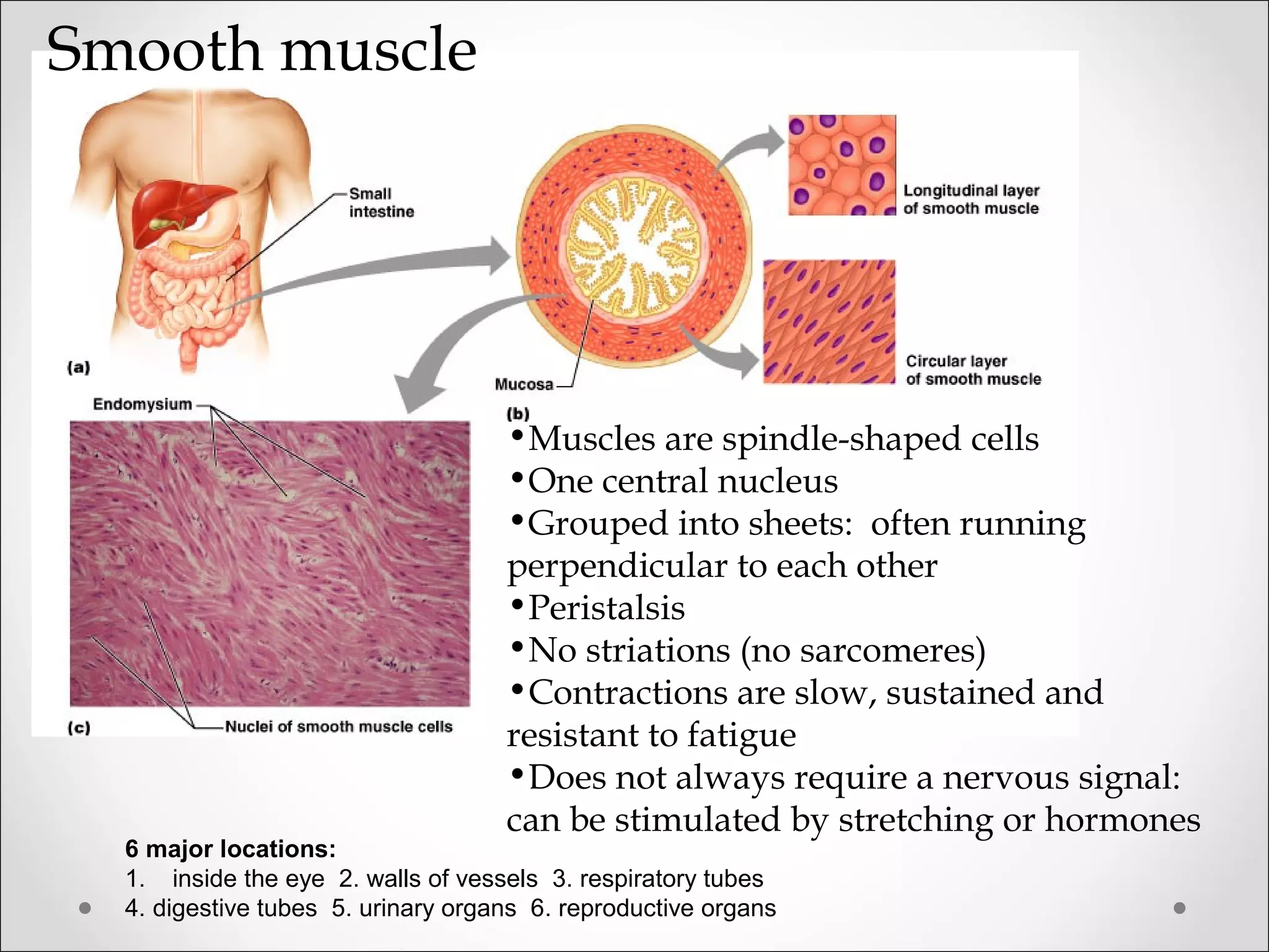
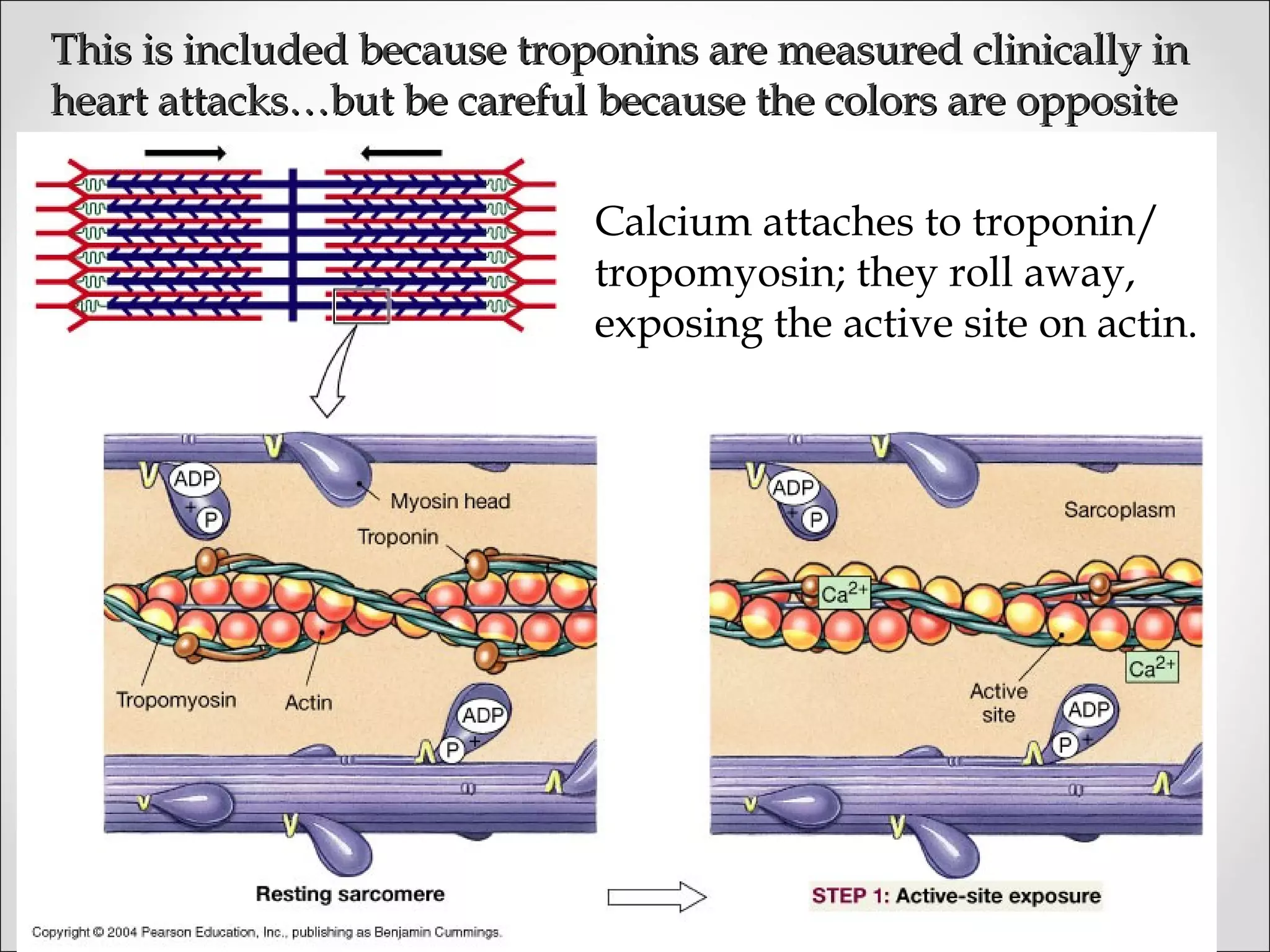
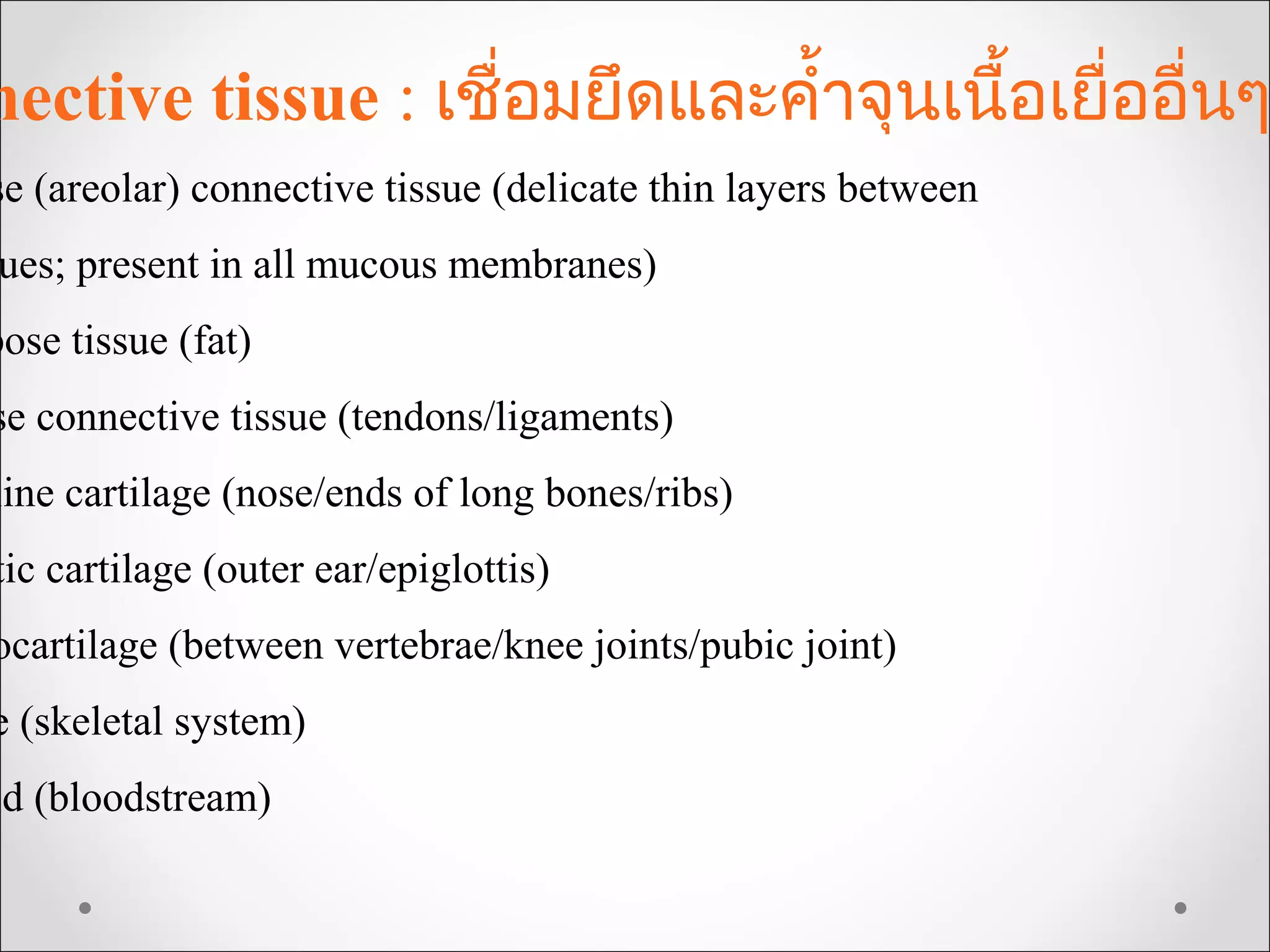
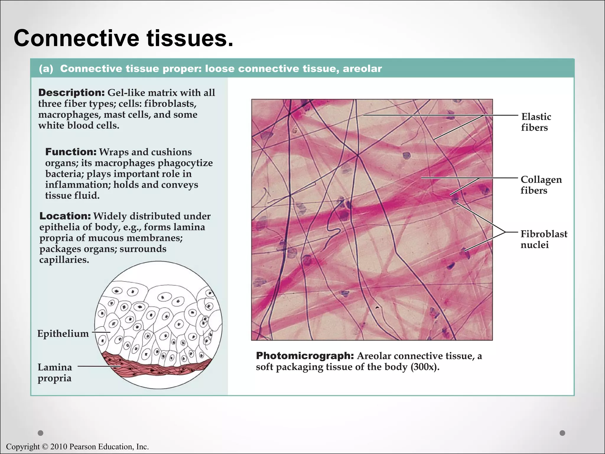
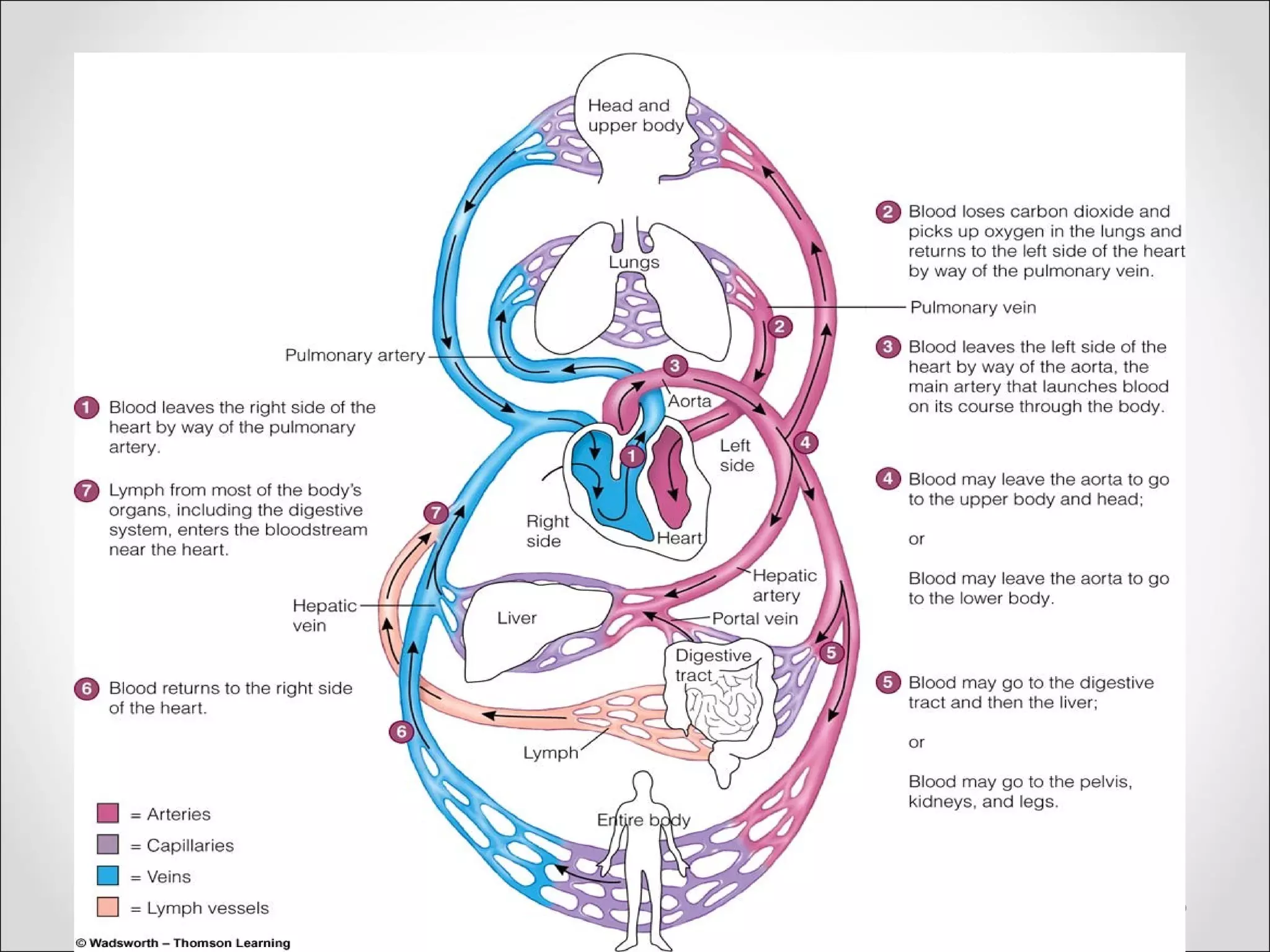
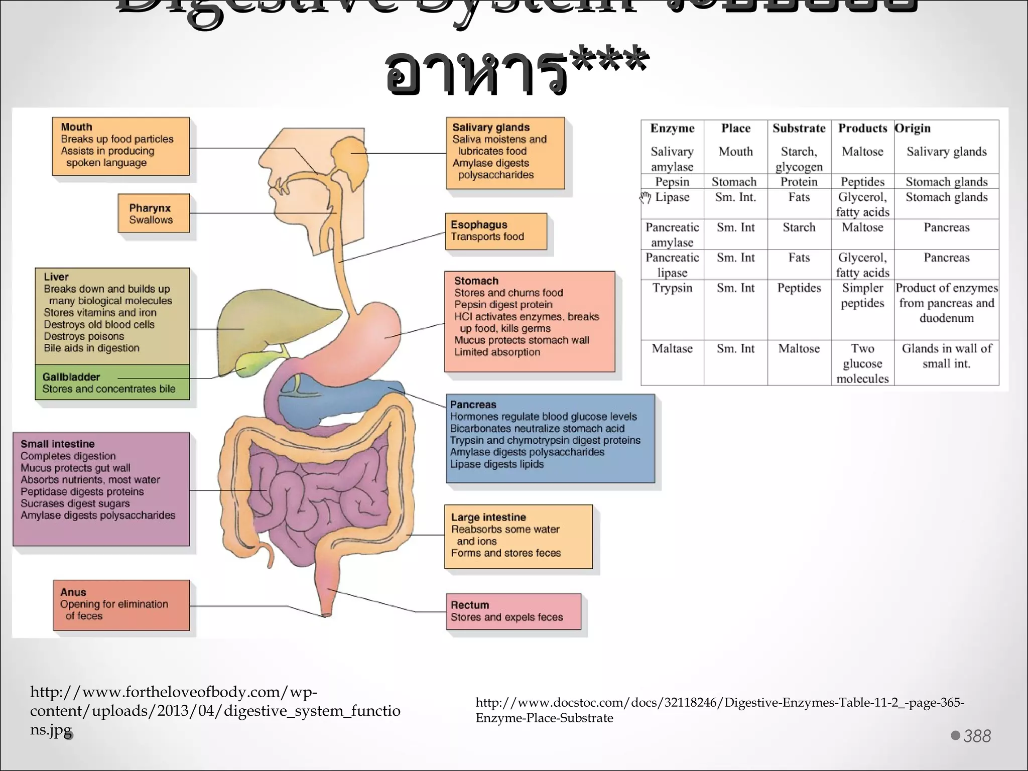
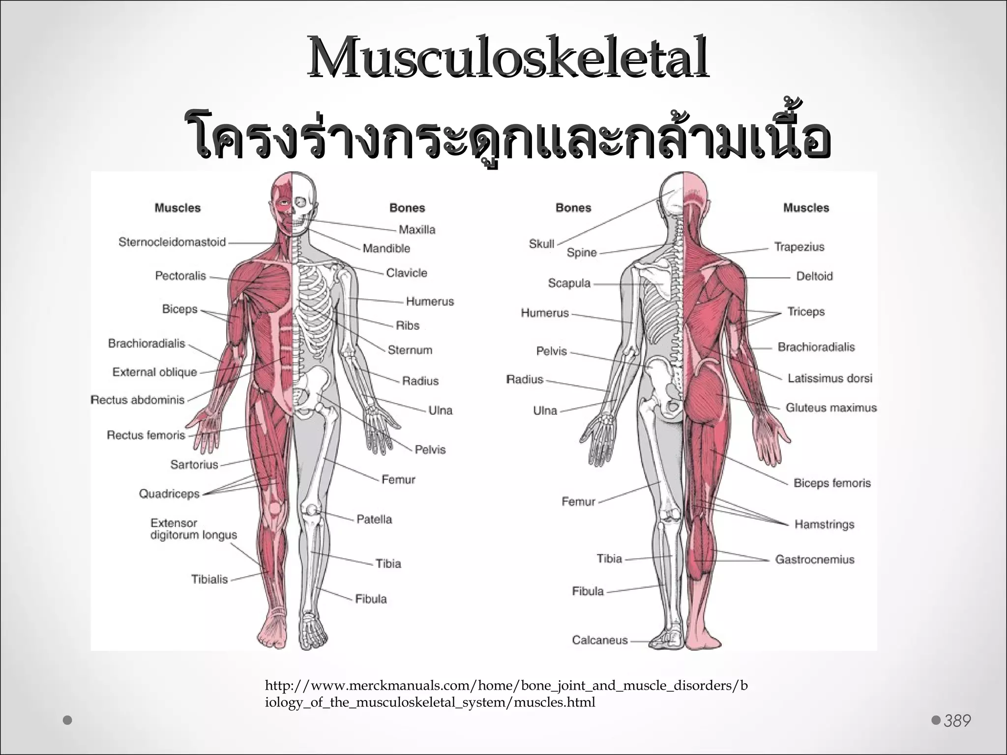
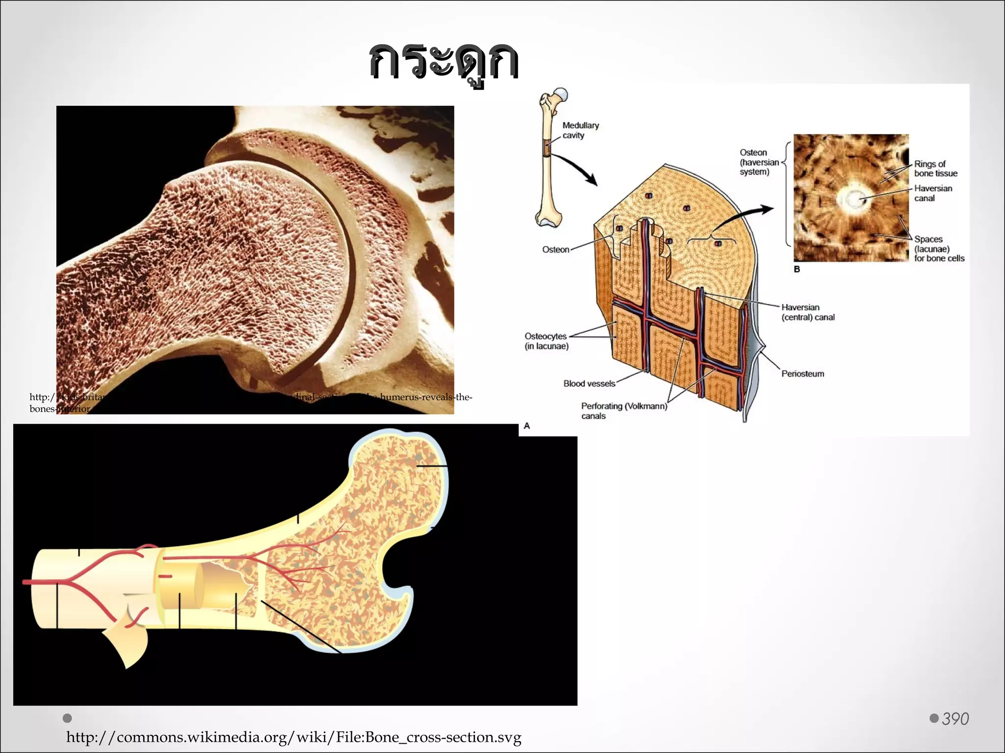
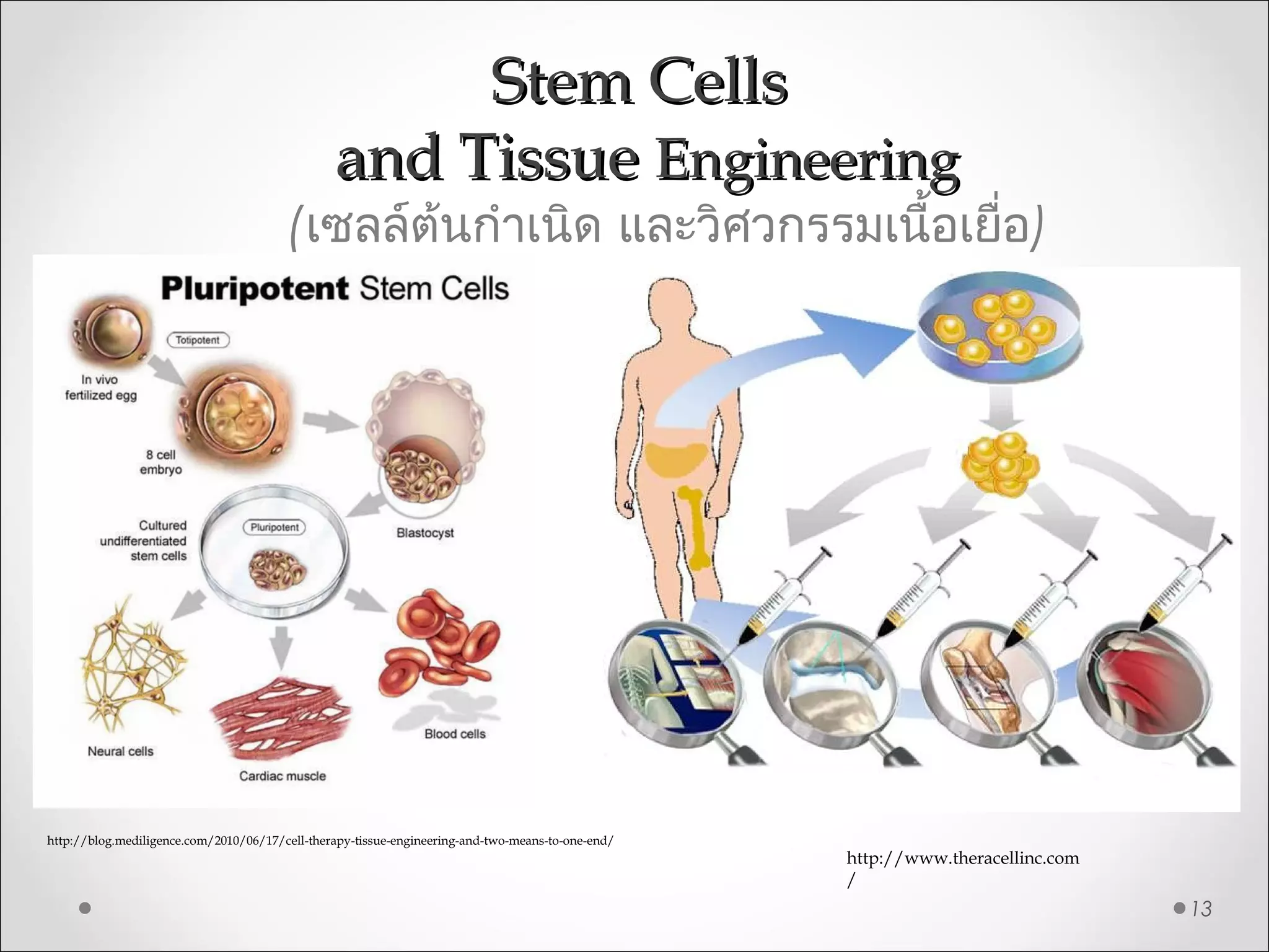
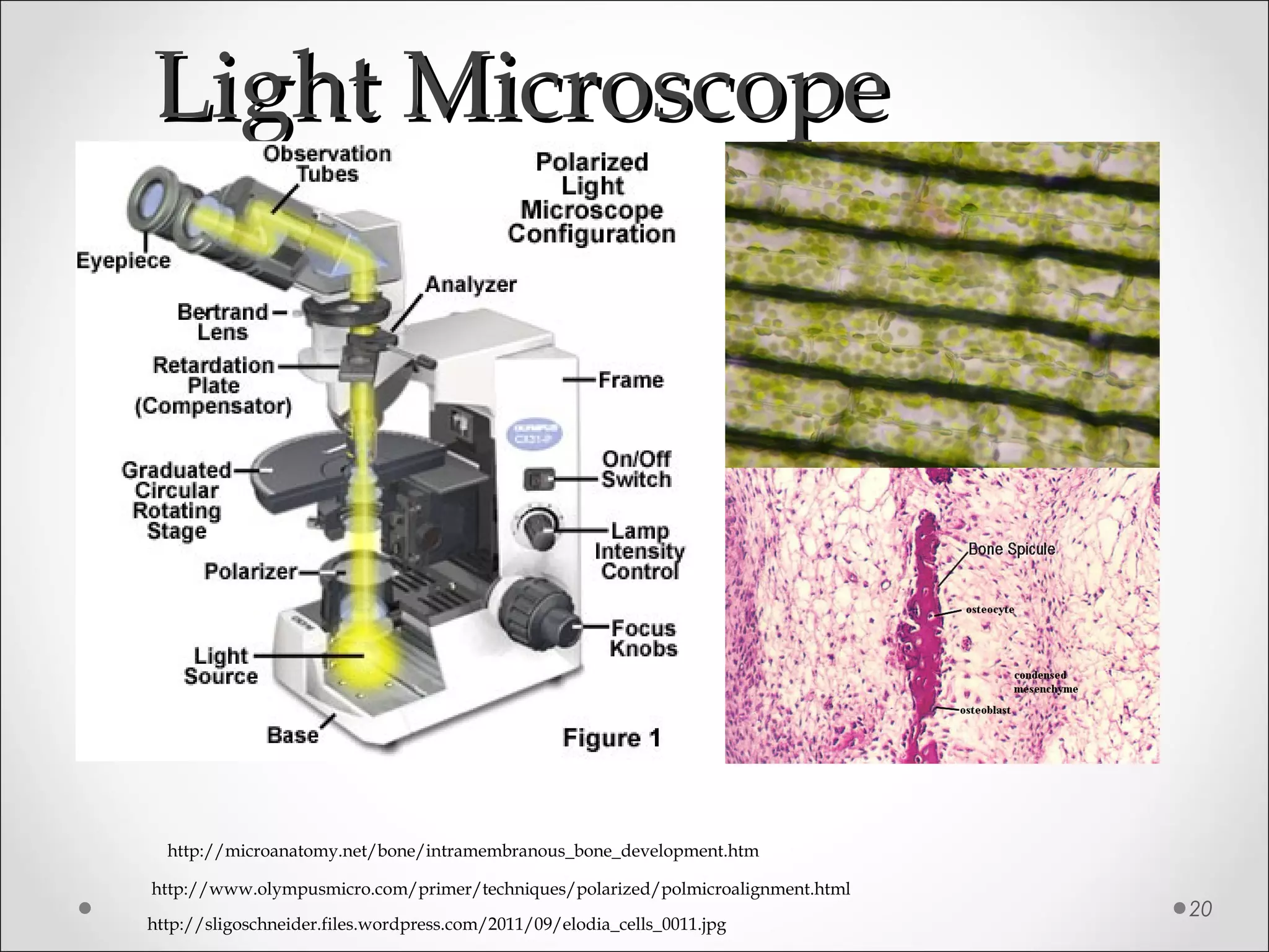
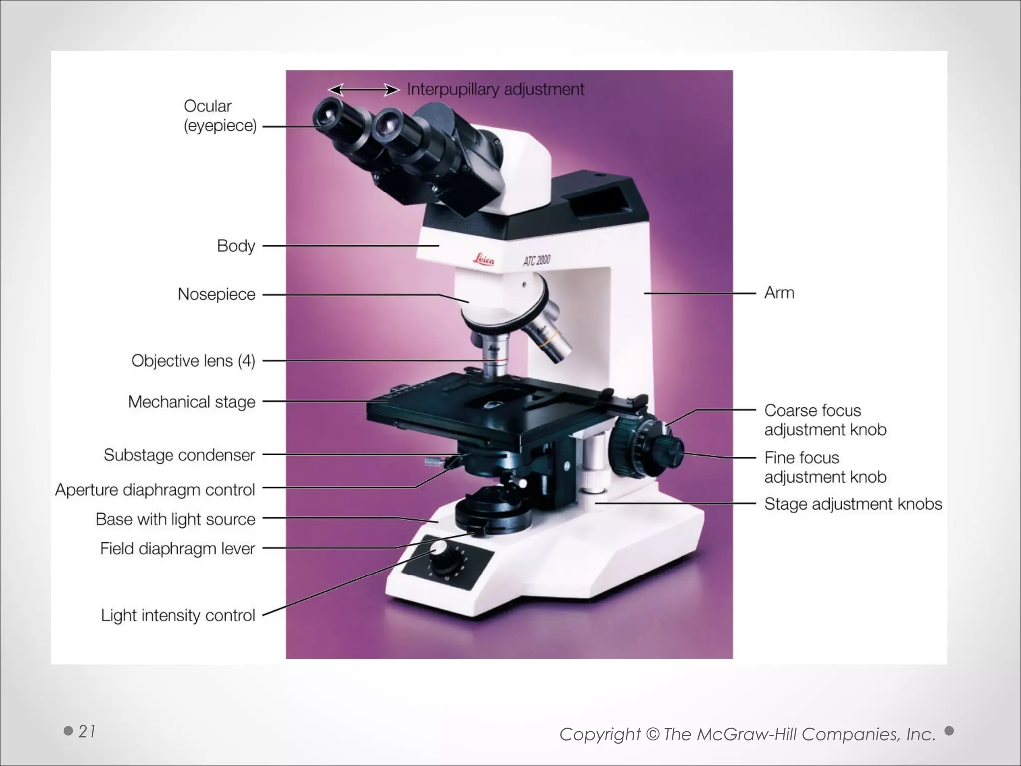
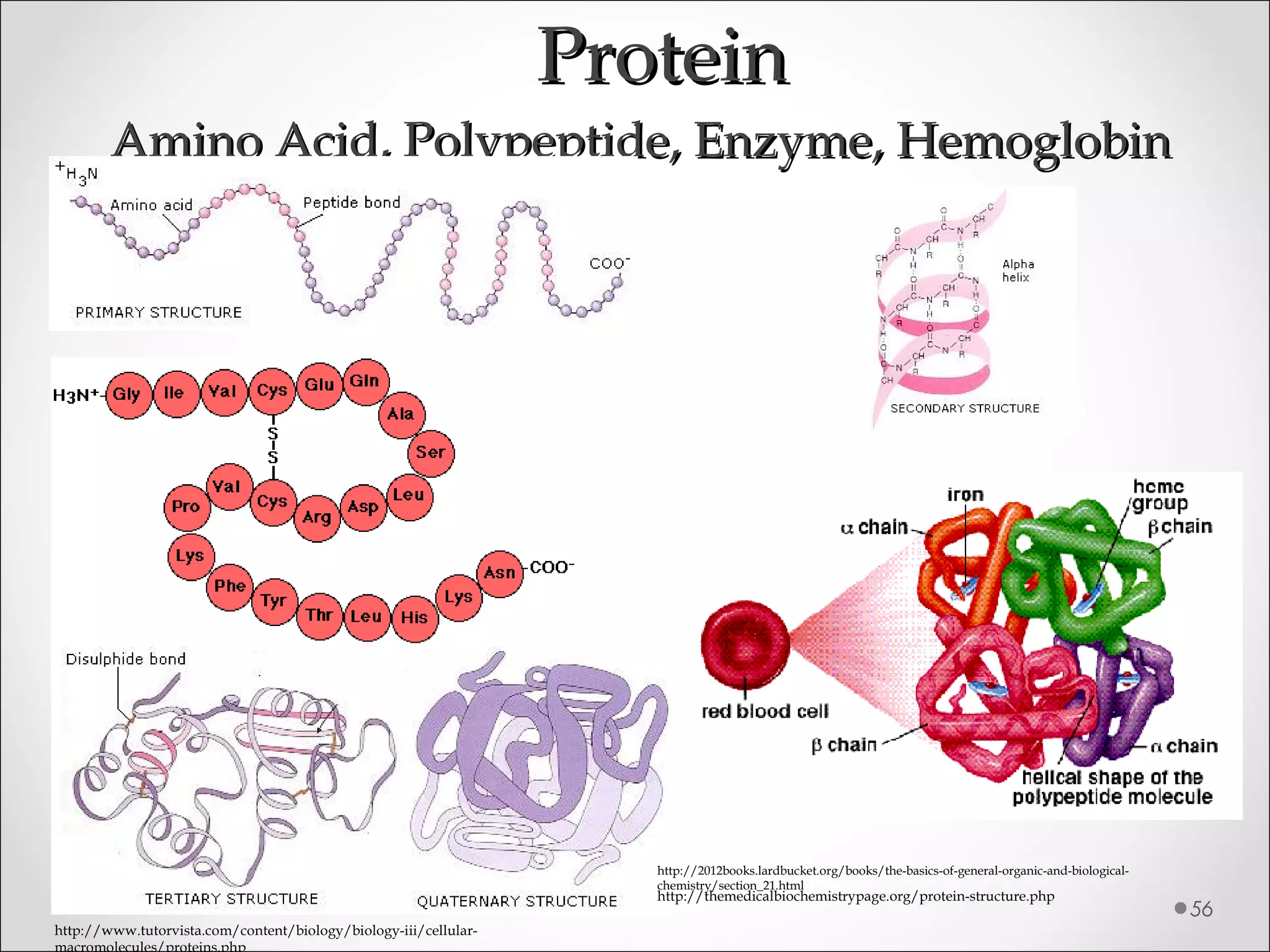
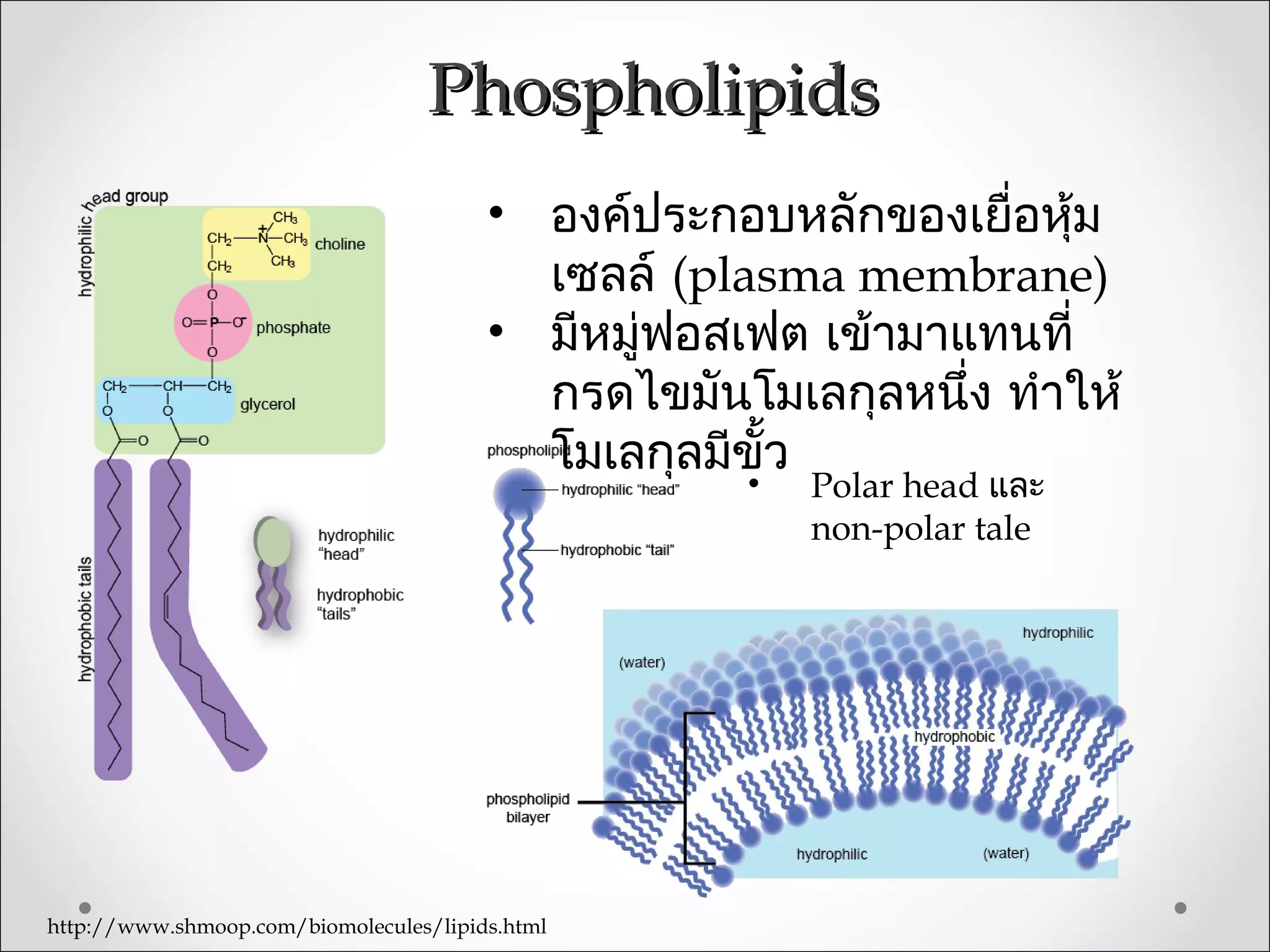
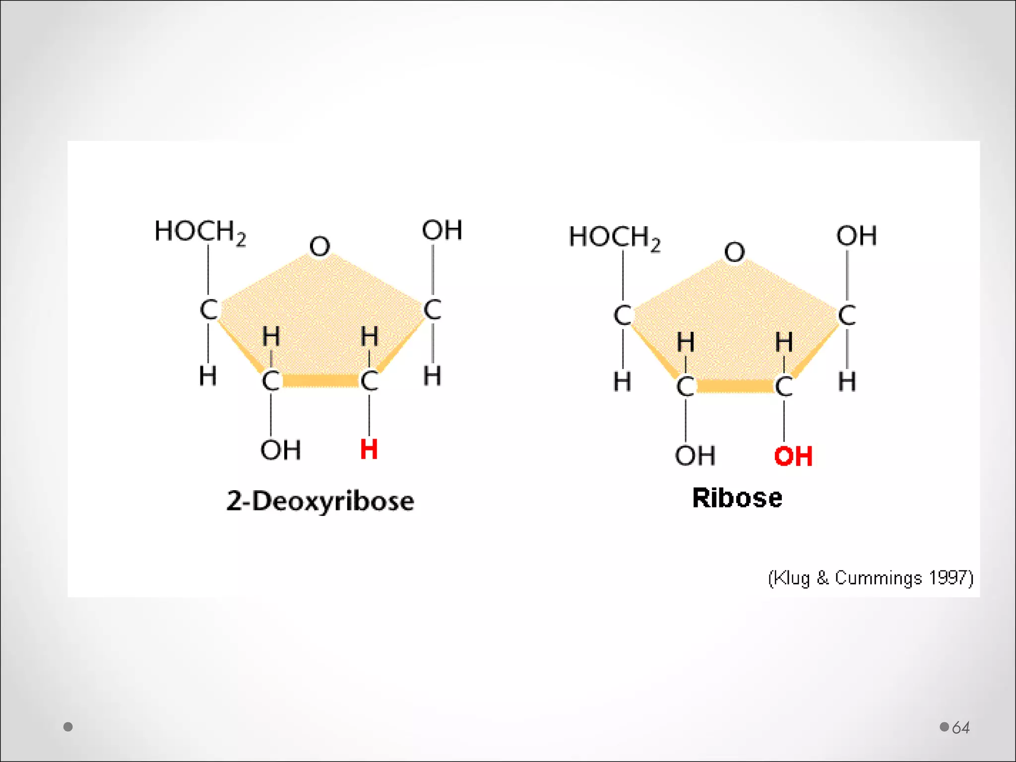
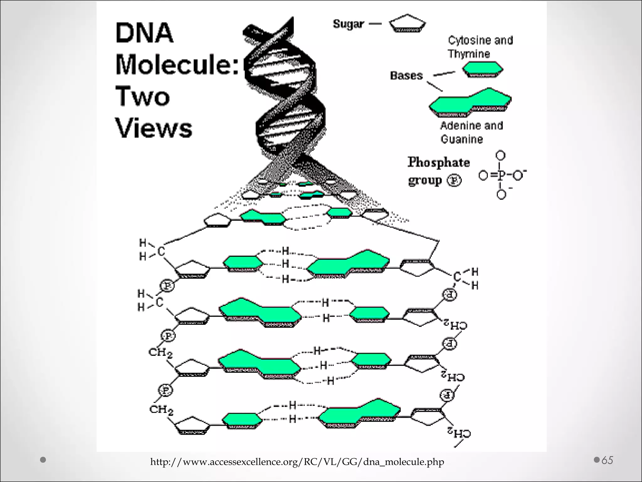
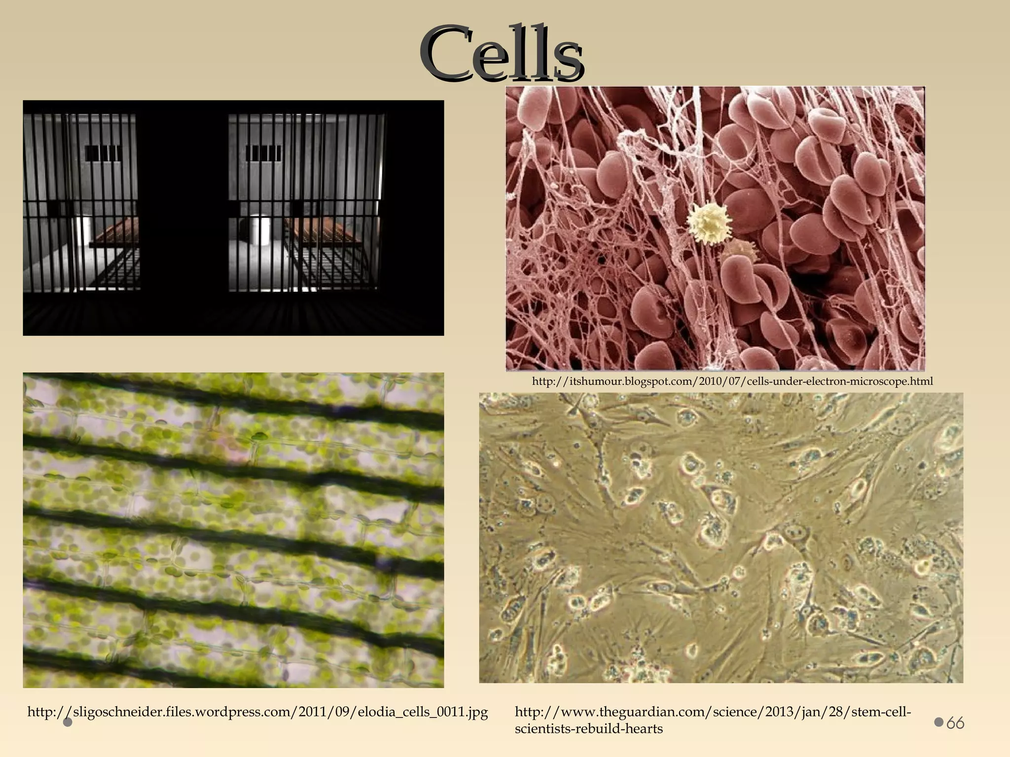
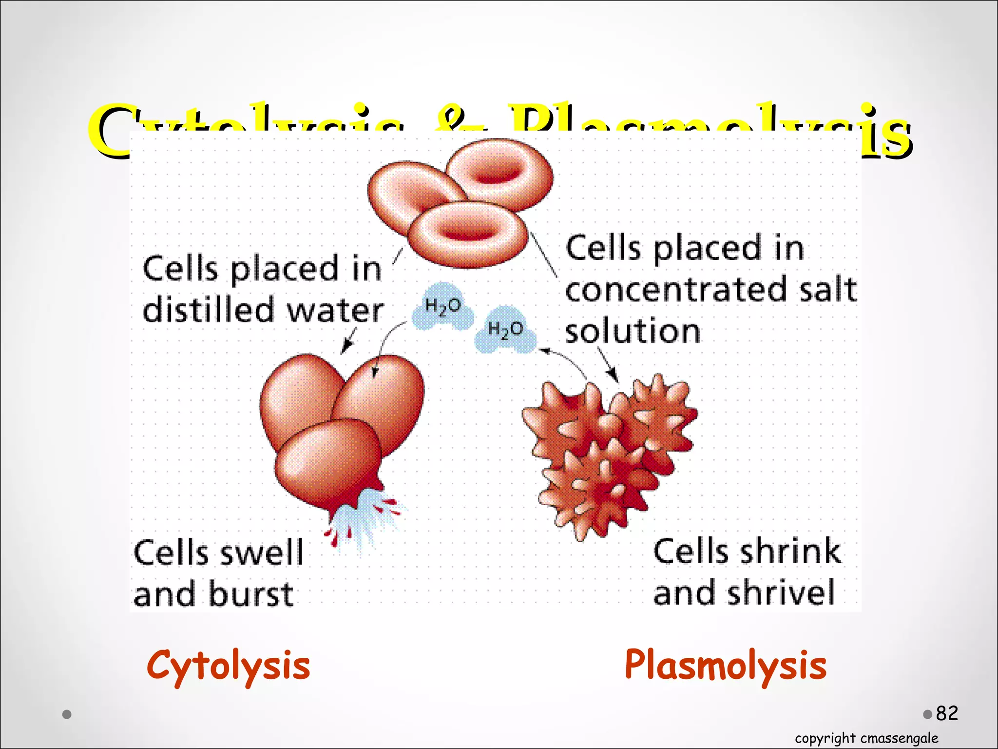
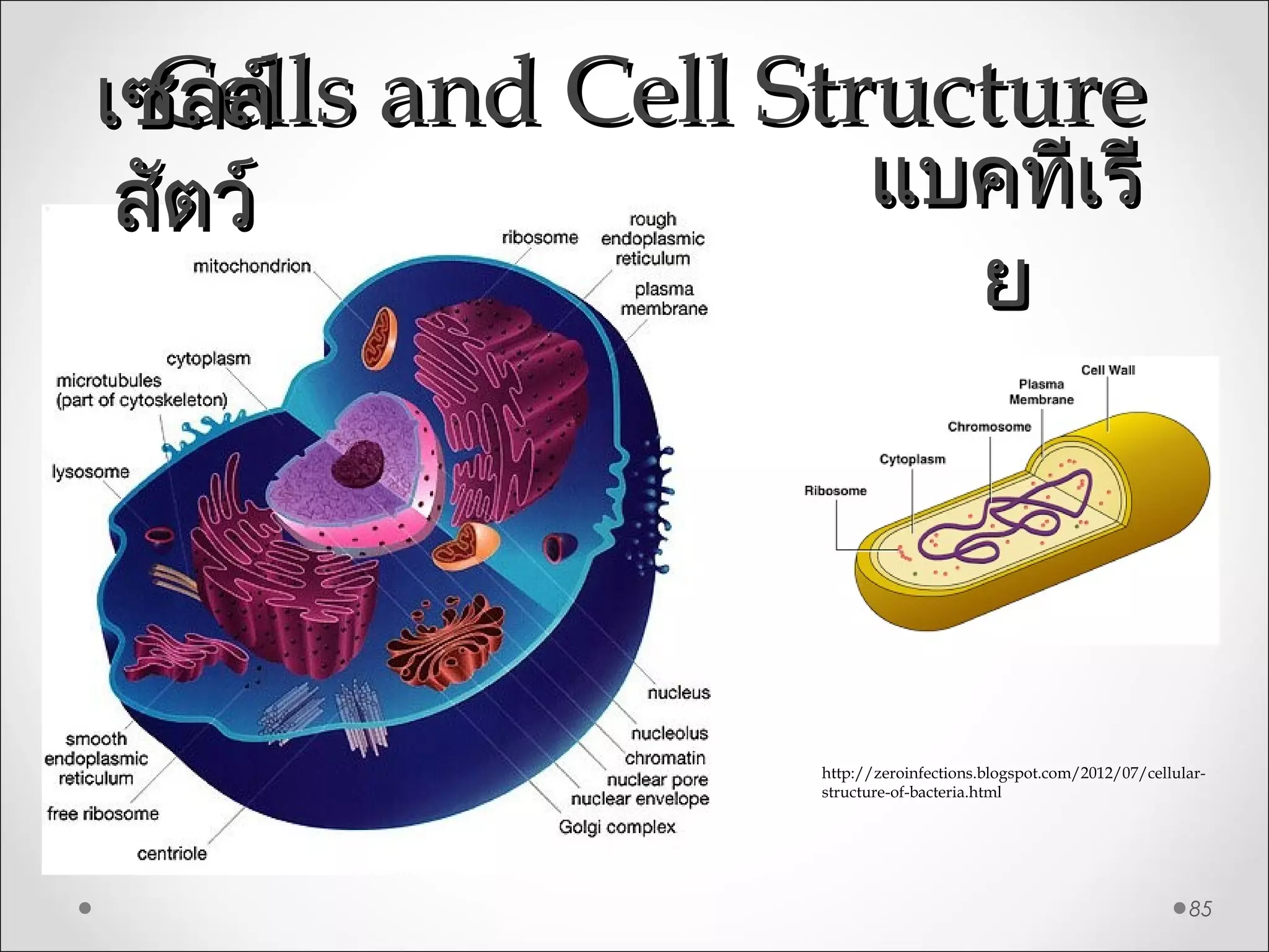
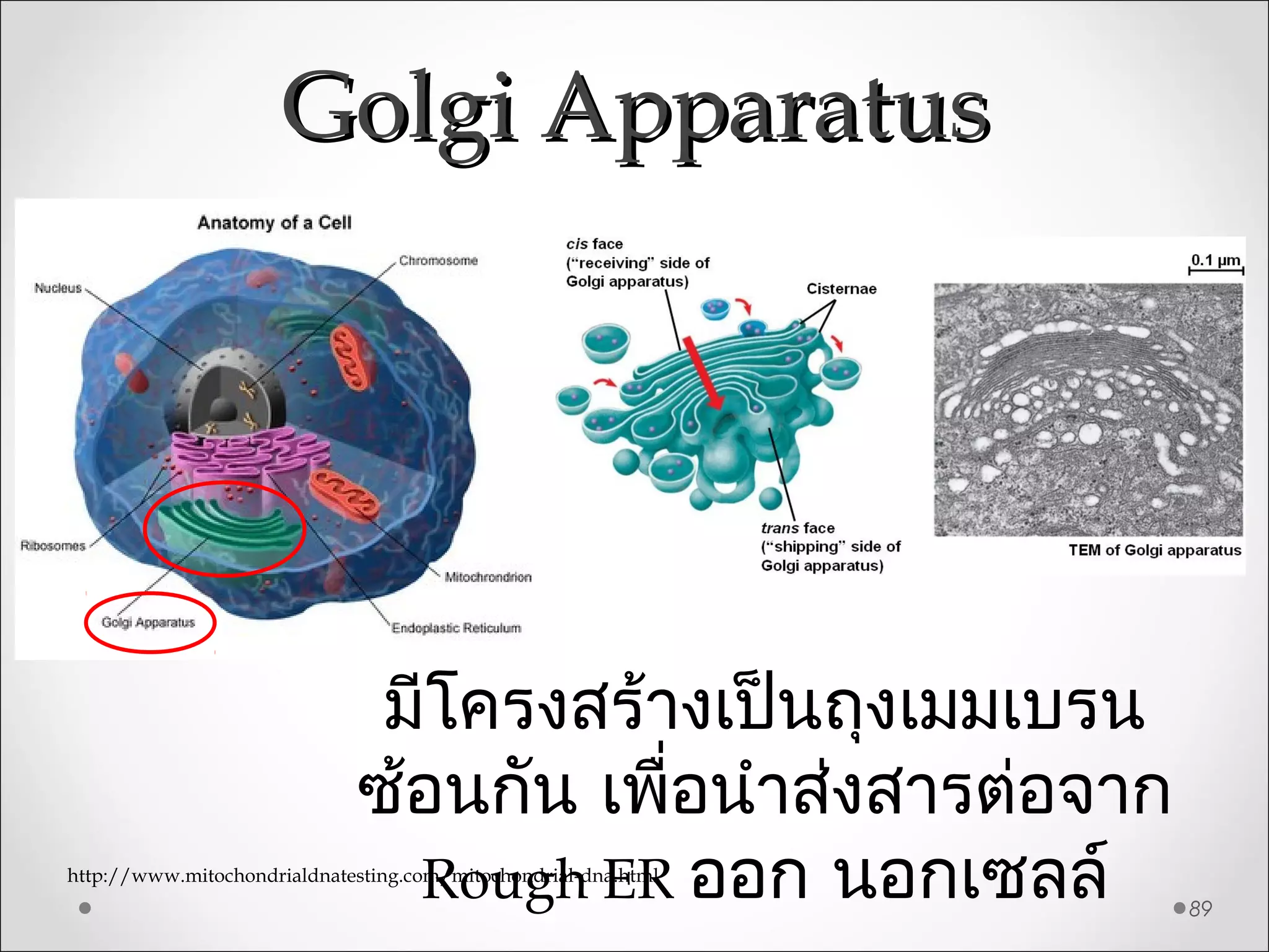
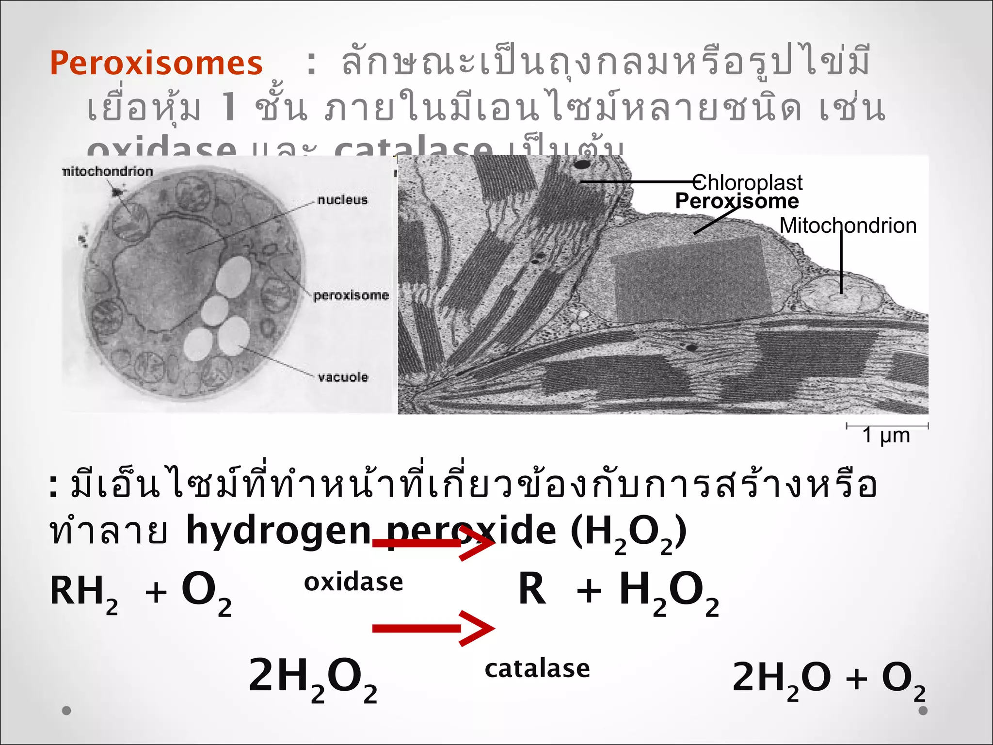
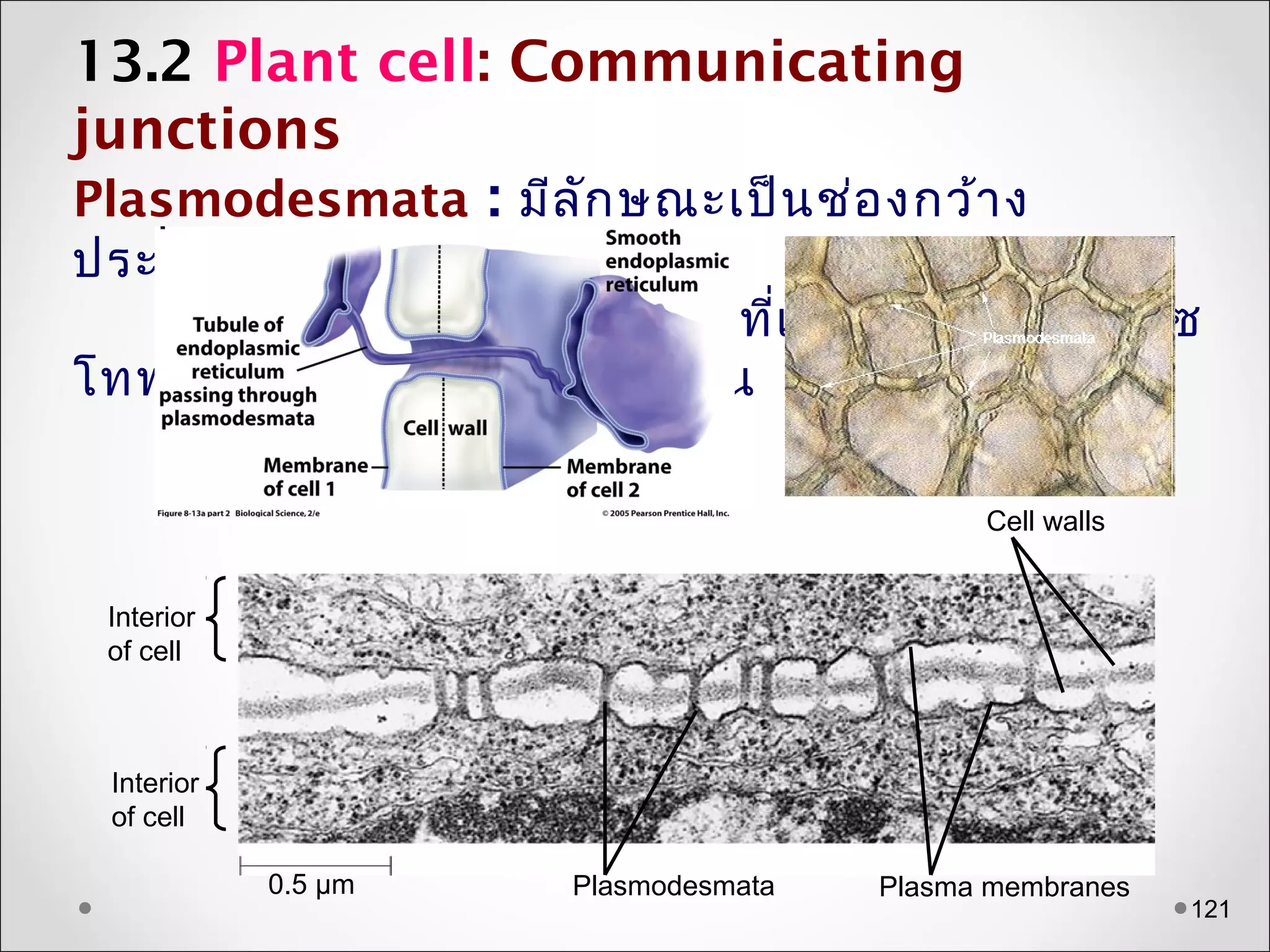
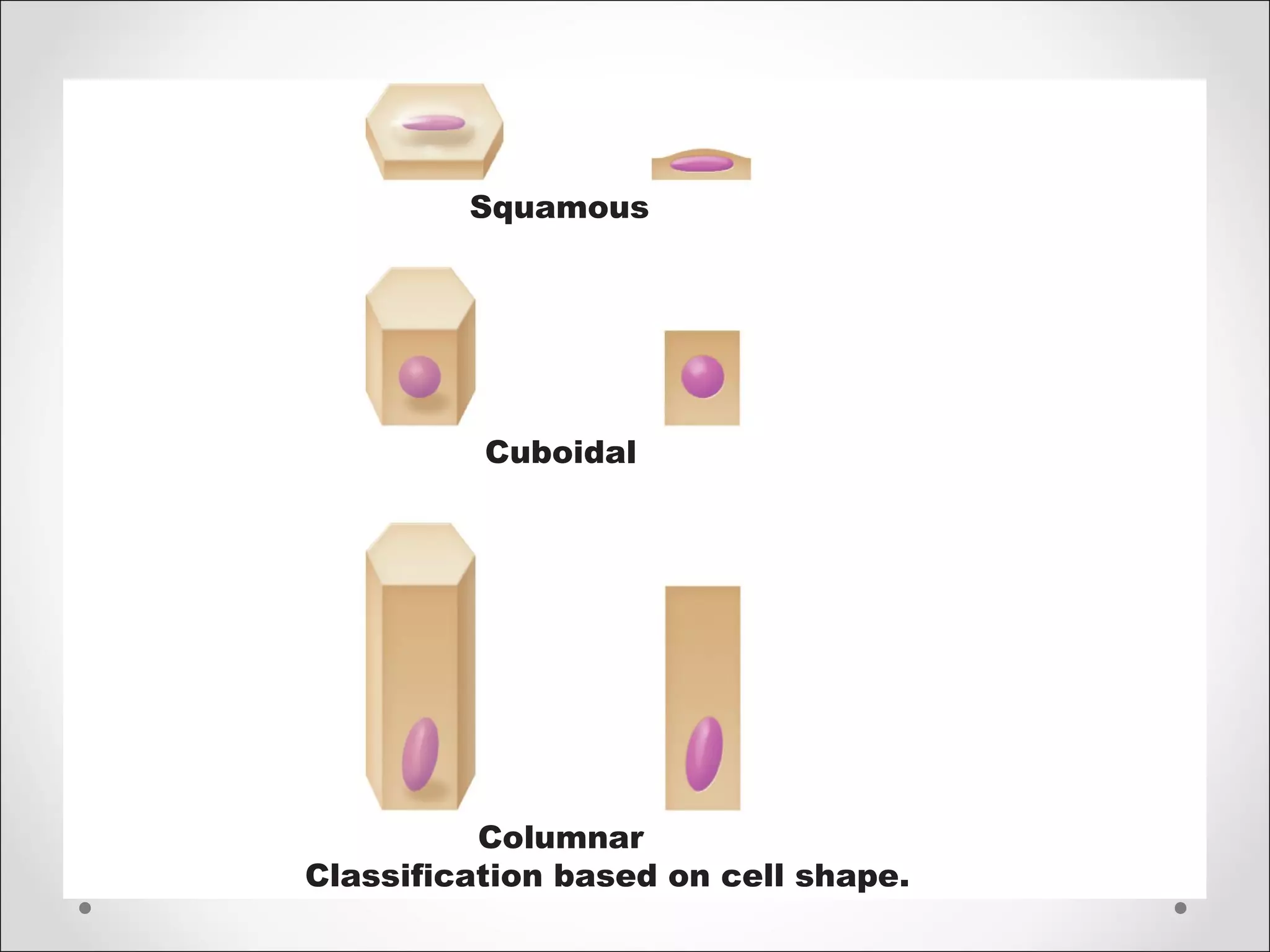
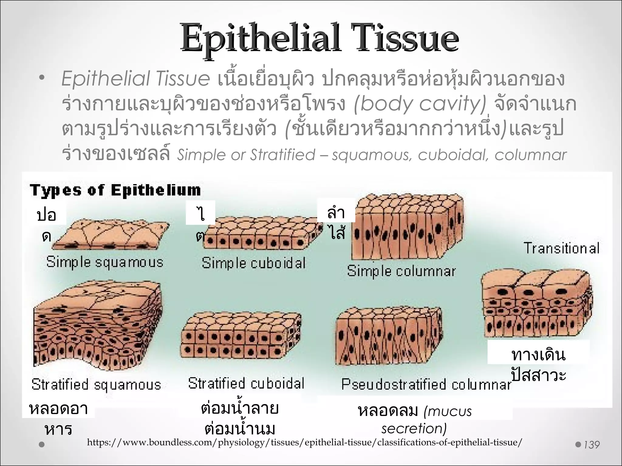
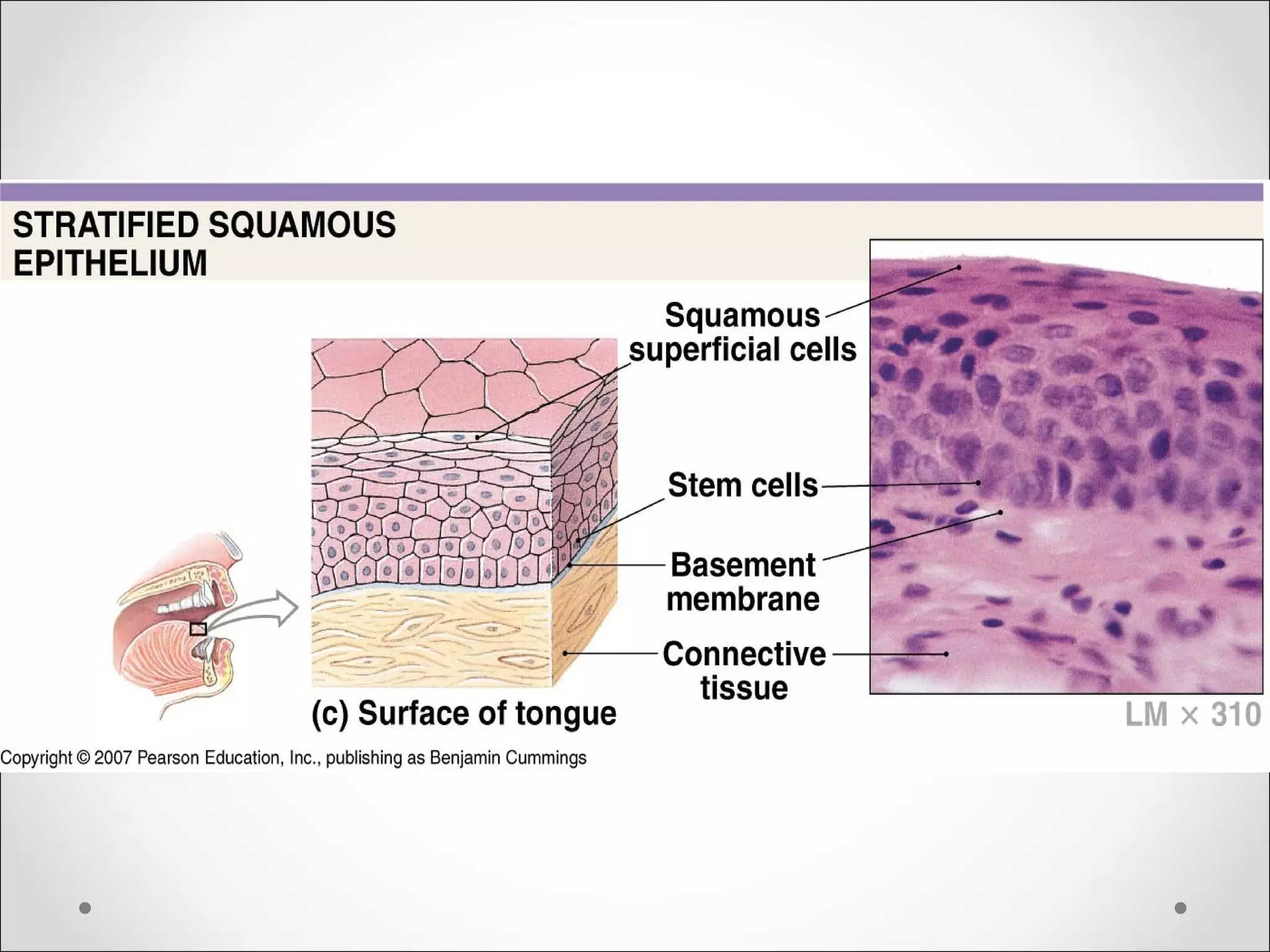
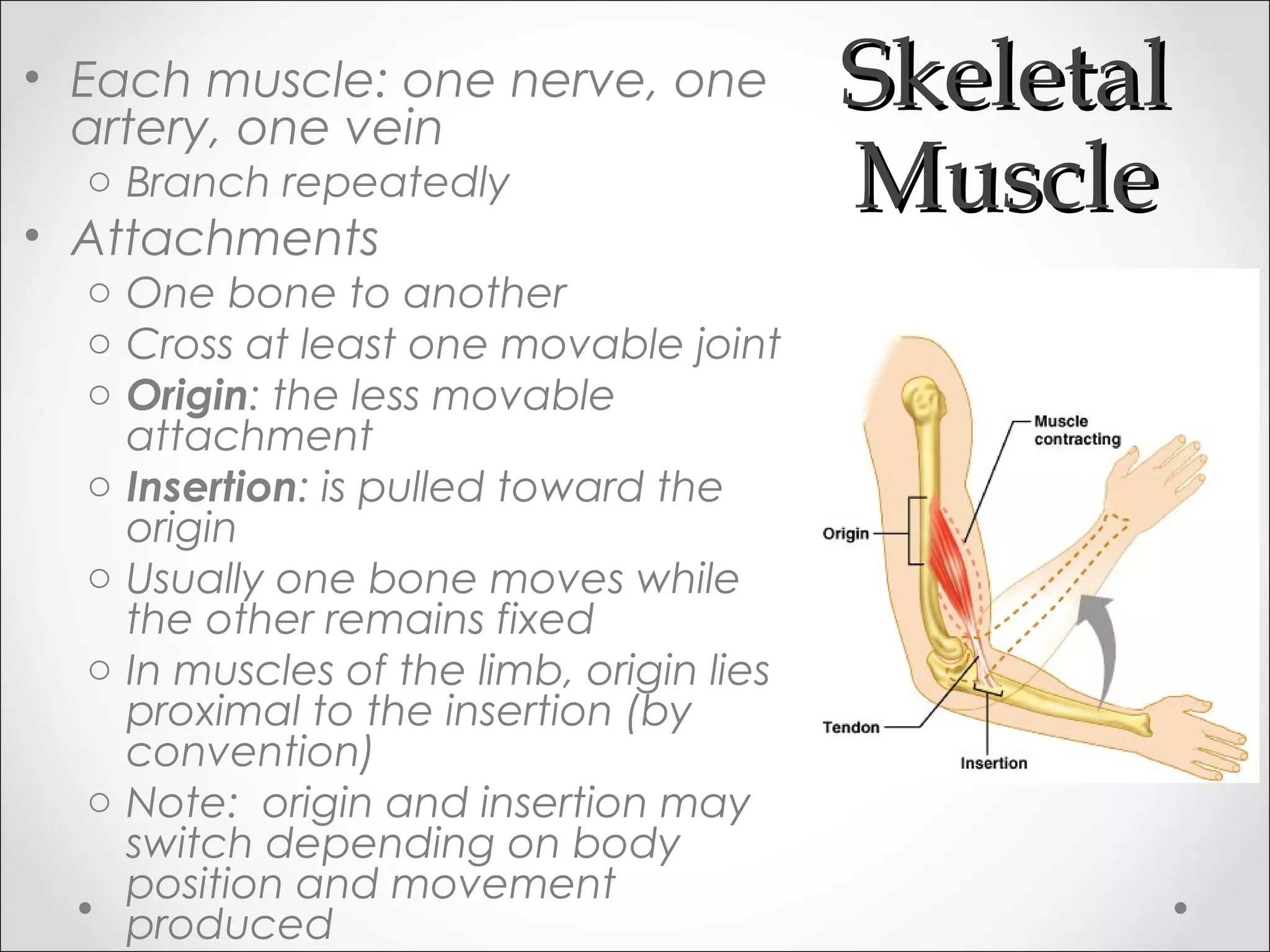
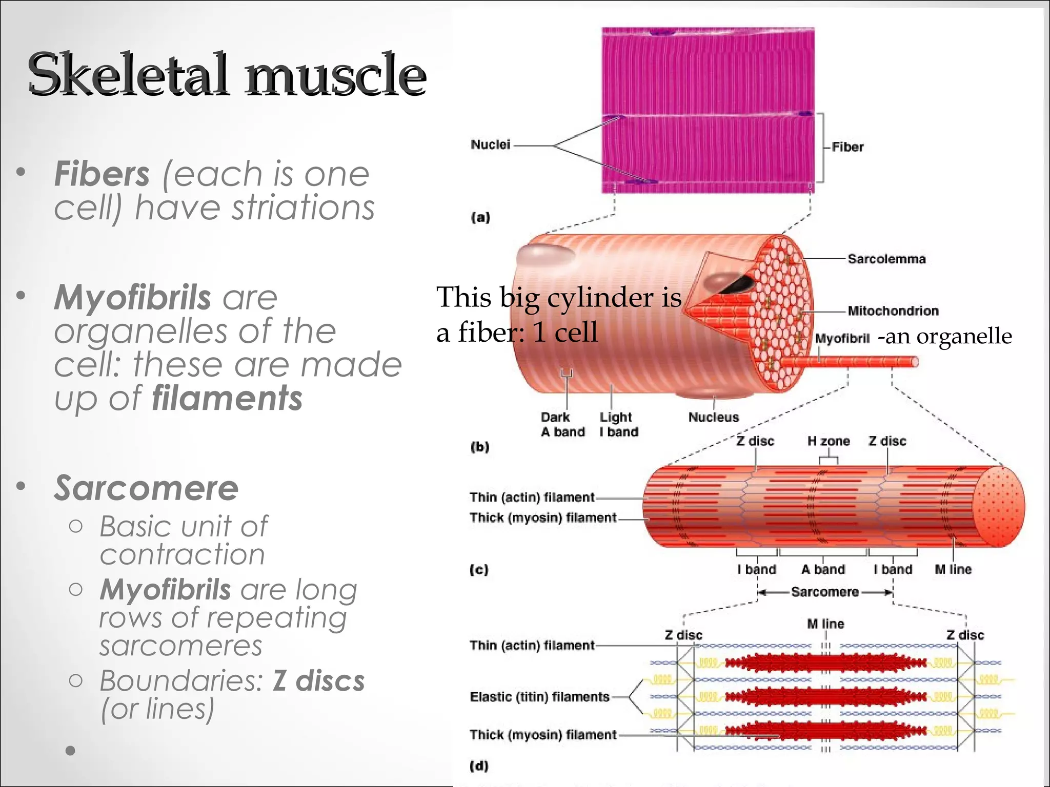
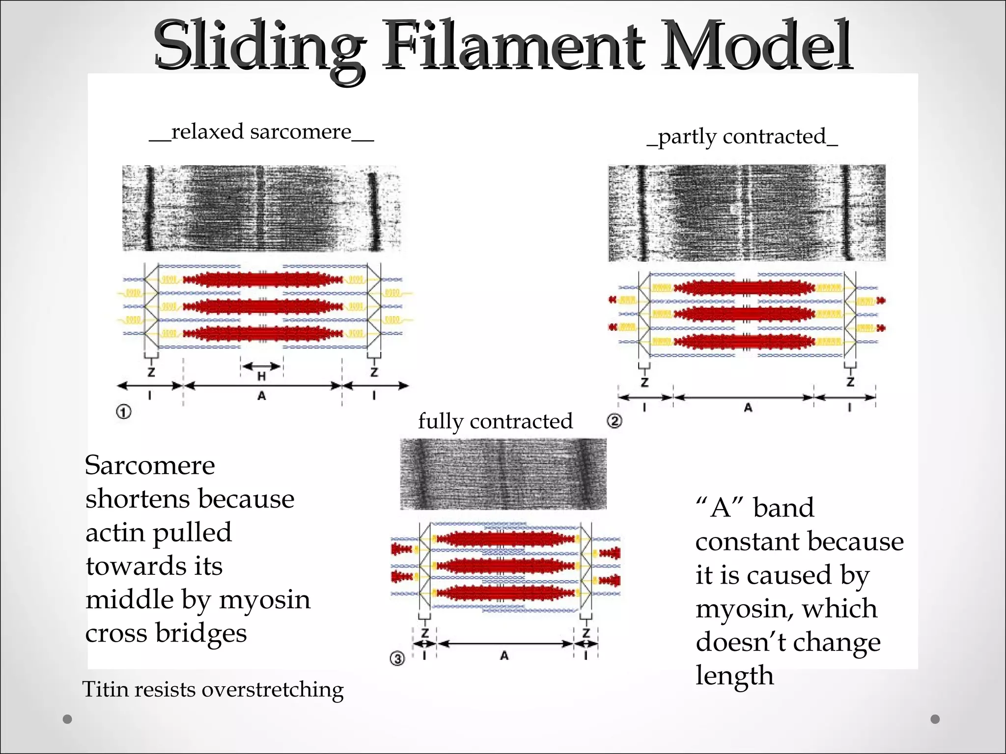
This document provides an overview of general biology topics including introductions to modern technologies, biological study tools and methods, biomolecules, cell structure and functions, reproduction, genetics and technology, animal tissue development, and human physiology. Key points covered include the structure and functions of carbohydrates, proteins, lipids, and nucleic acids as important biomolecules, as well as methods used in biological studies such as microscopes, centrifuges, and incubators.


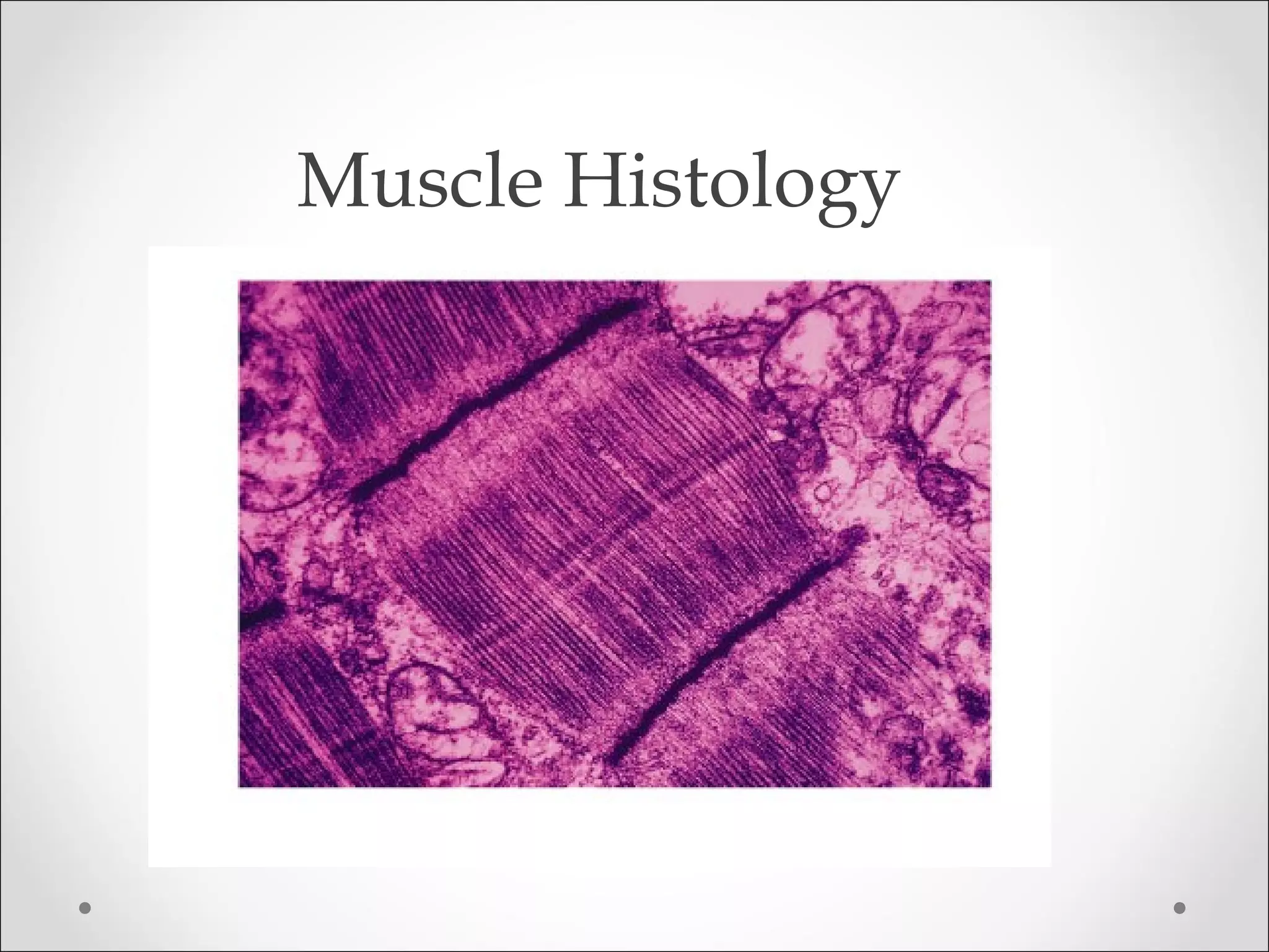


![• Muscle hypertrophy
o Weight training (repeated intense workouts): increases
diameter and strength of “fast” muscle fibers by increasing
production of
• Mitochondria
• Actin and myosin protein
• Myofilaments containing these contractile proteins
• The myofibril organelles these myofilaments form
o Fibers enlarge (hypertrophy) as number and size of myofibrils
increase
[Muscle fibers (=muscle cells) don’t increase in number but
increase in diameter producing large muscles]
• Endurance training (aerobic): doesn’t produce
hypertrophy
• Muscle atrophy: loss of tone and mass from lack of
stimulation
o Muscle becomes smaller and weaker
Note on terminology: in general, increased size is hypertrophy; increased number](https://image.slidesharecdn.com/generalbiology8-2-14-2-140803185226-phpapp02/75/General-biology-8-2-14-2-171-2048.jpg)