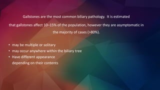
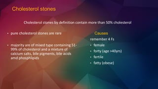
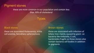
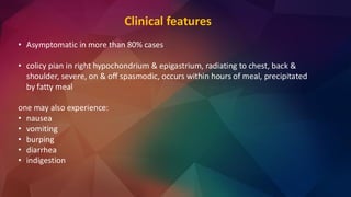
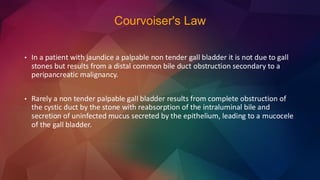
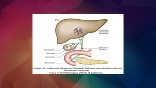
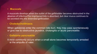
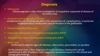
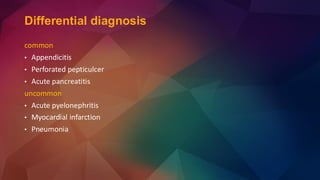
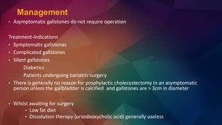
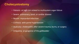
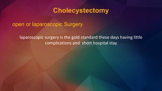
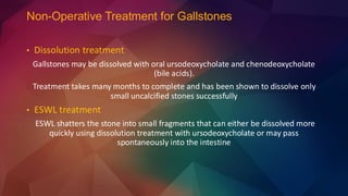
Gall stones
(cholelithiasis)
Dr. Umer Raiz & Dr. Hina Khan
Gallstones are the most common biliary pathology. It is estimated
that gallstones affect 10–15% of the population, however they are asymptomatic in
the majority of cases (>80%).
• may be multiple or solitary
• may occur anywhere within the biliary tree
• Have different appearance
depending on their contents
Pathogenesis
• Cholesterol supersaturation in bile
• Crystal nucleation
• Stone growth
Bile is composed of bile salts, phospholipids, cholesterol. If there is imbalance
between those components cholesterol will precipitate out of solution (cholesterol
super-saturation)
Types
• cholesterol stones
• pigment (brown/black) stones
• mixed stones
Cholesterol stones
Causes
remember 4 Fs
• female
• forty (age >40yrs)
• fertile
• fatty (obese)
• pure cholesterol stones are rare
• majority are of mixed type containing 51-
99% of cholesterol and a mixture of
calcium salts, bile pigments, bile acids
amd phosphlipids
Cholesterol stones by definition contain more than 50% cholesterol
Black stones
these are associated thalassemia, sickle
cell anemia, hereditary spherocytosis
Brown stones
these are associated with infection of
biliary tree mainly caused by gram -ve
bacteria like klebsiella, E coli,
bacteroides fragilis so these stones also
contain bacteria cell bodies in addition
to pigments
Pigment stones
these are more common in our population and contain less
than 30% of cholesterol
Types
Clinical features
• Asymptomatic in more than 80% cases
• colicy pian in right hypochondrium & epigastrium, radiating to chest, back &
shoulder, severe, on & off spasmodic, occurs within hours of meal, precipitated
by fatty meal
one may also experience:
• nausea
• vomiting
• burping
• diarrhea
• indigestion
Courvoiser's Law
• In a patient with jaundice a palpable non tender gall bladder it is not due to gall
stones but results from a distal common bile duct obstruction secondary to a
peripancreatic malignancy.
• Rarely a non tender palpable gall bladder results from complete obstruction of
the cystic duct by the stone with reabsorption of the intraluminal bile and
secretion of uninfected mucus secreted by the epithelium, leading to a mucocele
of the gall bladder.
Complications
in gall bladder
• Asymptomatic
• acute cholecystitis
• chronic cholecystitis
• empyema of gall bladder
• mucocele
• perforation of gall bladder
• gangrene of gall bladder
• CA gall bladder
in bile duct
• biliary obstruction
causing obstructive
jaundice
• acute cholangitis
• acute pancreatitis
• cholangiocarcinoma
in intestine
• gall stone ileus
• occurs due to obstruction of the neck of gallbladder or cystic duct by a stone
resulting in a chemical inflammatory reaction. Bacteria are cultured from the bile in
approximately 1/2 of patients with gallstones and unrelieved obstruction
• in the presence of this infected bile may produce an empyema
Boas' sign
hyperaesthesia below the right scapula in cholecystitis.
• Acute cholecystitis
MURPHY'S SIGN
Patient winces in pain with catch of breath when inflamed gall bladder strikes
palpating fingers on inspiration
Tokyo Classification
• Repeated bouts of biliary colic or acute cholecystitis culminate in fibrosis, contraction
of the gallbladder. However chronic inflammatory change my be present in the
absence of gallstones
• The incidence of carcinoma of the gallbladder is increased in patients with long
standing gallstones
• Chronic cholecystitis
• Mucocele
A mucocele develops when the outlet of the gallbladder becomes obstructed in the
absence of infectionThe imprisoned bile is absorbed, but clear mucus continues to
be secreted into the distended gallbladder.
• Choledocholithiasis
When gallstones enter the common bile duct, they may pass spontaneously
or give rise to obstructive jaundice, cholangitis or acute pancreatitis
• Gallstone pancreatitis
It most commonly occurs when a small stone becomes temporarily arrested
at the ampulla of vater.
• Cholangitis
Nonsuppurative acute cholangitis is most common and respond rapidly to antibiotics
Suppurative acute cholangitis Pus in completely obstructed ductal system symptoms of
severe toxicity such as mental confusion and septic shock Poor response to antibiotics and
mortality is 100% unless prompt endoscopic or surgical relief of the obstruction and
drainage of infected bile are carried out.
Mirizzi's syndrome
Rarely, a gallstone gets impacted in the GB wall,
mostly in the Hartman's pouch or fundus of GB and
compresses it causing pressure necrosis and its
adherence to the common bile duct, and occasionally
it can leads to cholecysto-choledochal fistula.
Diagnosis
• Ultrasound
Ultrasonography is the initial investigation of anypatient suspected of disease of
the biliary tree.
• Computerized tomography (CT) scan
complementary to US being valuable in the assessment of complications, in particular
emphysematous cholecystitis and perforation of the gallbladder
• Cholescintigraphy (HIDA scan)
• Endoscopic Retrograde Cholangiopancreatography (ERCP)
• Megnetic Resonance Cholangiopancreatography (ERCP
• Blood tests
Performed to look for signs of infection, obstruction, pancreatitis, or jaundice
At the present time, a firm diagnosis of acute calculous cholecystitis can be
established in 90% of patients with suggestive symptoms based on the clinical and
sonographic findings.
Differential diagnosis
common
• Appendicitis
• Perforated pepticulcer
• Acute pancreatitis
uncommon
• Acute pyelonephritis
• Myocardial infarction
• Pneumonia
• Asymptomatic gallstones do not require operation
Treatment-Indications
• Symptomatic gallstones
• Complicated gallstones
• Silent gallstones
Diabetics
Patients undergoing bariatric surgery
• There is generally no reason for prophylactic cholecystectomy in an asymptomatic
person unless the gallbladder is calcified and gallstones are > 3cm in diameter
• Whilst awaiting for surgery
• Low fat diet
• Dissolution therapy (ursodeoxycholic acid) generally useless
Management
• Cholecystostomy
• Subtotal cholecystectomy
• Open cholecystectomy
• Laparoscopic cholecystectomy
Surgical options
Cholecystostomy
• Patients at high risk related to multisystem organ failure
• Severe pulmonary, renal, or cardiac disease
• Recent myocardial infarction
• Cirrhosis with portal hypertension
• Acalculus cholecystitis after severe trauma, burns, or surgery
• Empyema or gangrene of the gallbladder
Subtotal Cholecystectomy
Severe inflammation renders identification of the anatomy impossible, eg.
Gangrenous cholecystitis
Cholecystectomy
open or laparoscopic Surgery
laparoscopic surgery is the gold standard these days having little
complications and short hospital stay
Non-Operative Treatment for Gallstones
• Dissolution treatment
Gallstones may be dissolved with oral ursodeoxycholate and chenodeoxycholate
(bile acids).
Treatment takes many months to complete and has been shown to dissolve only
small uncalcified stones successfully
• ESWL treatment
ESWL shatters the stone into small fragments that can either be dissolved more
quickly using dissolution treatment with ursodeoxycholate or may pass
spontaneously into the intestine
T H A N K S
Gall stones surgery presentation final.pdf

Gall stones surgery presentation final.pdf

  • 1.
  • 2.
    Gallstones are themost common biliary pathology. It is estimated that gallstones affect 10–15% of the population, however they are asymptomatic in the majority of cases (>80%). • may be multiple or solitary • may occur anywhere within the biliary tree • Have different appearance depending on their contents
  • 3.
    Pathogenesis • Cholesterol supersaturationin bile • Crystal nucleation • Stone growth Bile is composed of bile salts, phospholipids, cholesterol. If there is imbalance between those components cholesterol will precipitate out of solution (cholesterol super-saturation)
  • 4.
    Types • cholesterol stones •pigment (brown/black) stones • mixed stones
  • 5.
    Cholesterol stones Causes remember 4Fs • female • forty (age >40yrs) • fertile • fatty (obese) • pure cholesterol stones are rare • majority are of mixed type containing 51- 99% of cholesterol and a mixture of calcium salts, bile pigments, bile acids amd phosphlipids Cholesterol stones by definition contain more than 50% cholesterol
  • 6.
    Black stones these areassociated thalassemia, sickle cell anemia, hereditary spherocytosis Brown stones these are associated with infection of biliary tree mainly caused by gram -ve bacteria like klebsiella, E coli, bacteroides fragilis so these stones also contain bacteria cell bodies in addition to pigments Pigment stones these are more common in our population and contain less than 30% of cholesterol Types
  • 7.
    Clinical features • Asymptomaticin more than 80% cases • colicy pian in right hypochondrium & epigastrium, radiating to chest, back & shoulder, severe, on & off spasmodic, occurs within hours of meal, precipitated by fatty meal one may also experience: • nausea • vomiting • burping • diarrhea • indigestion
  • 8.
    Courvoiser's Law • Ina patient with jaundice a palpable non tender gall bladder it is not due to gall stones but results from a distal common bile duct obstruction secondary to a peripancreatic malignancy. • Rarely a non tender palpable gall bladder results from complete obstruction of the cystic duct by the stone with reabsorption of the intraluminal bile and secretion of uninfected mucus secreted by the epithelium, leading to a mucocele of the gall bladder.
  • 9.
    Complications in gall bladder •Asymptomatic • acute cholecystitis • chronic cholecystitis • empyema of gall bladder • mucocele • perforation of gall bladder • gangrene of gall bladder • CA gall bladder in bile duct • biliary obstruction causing obstructive jaundice • acute cholangitis • acute pancreatitis • cholangiocarcinoma in intestine • gall stone ileus
  • 11.
    • occurs dueto obstruction of the neck of gallbladder or cystic duct by a stone resulting in a chemical inflammatory reaction. Bacteria are cultured from the bile in approximately 1/2 of patients with gallstones and unrelieved obstruction • in the presence of this infected bile may produce an empyema Boas' sign hyperaesthesia below the right scapula in cholecystitis. • Acute cholecystitis
  • 12.
    MURPHY'S SIGN Patient wincesin pain with catch of breath when inflamed gall bladder strikes palpating fingers on inspiration
  • 13.
  • 14.
    • Repeated boutsof biliary colic or acute cholecystitis culminate in fibrosis, contraction of the gallbladder. However chronic inflammatory change my be present in the absence of gallstones • The incidence of carcinoma of the gallbladder is increased in patients with long standing gallstones • Chronic cholecystitis
  • 15.
    • Mucocele A mucoceledevelops when the outlet of the gallbladder becomes obstructed in the absence of infectionThe imprisoned bile is absorbed, but clear mucus continues to be secreted into the distended gallbladder. • Choledocholithiasis When gallstones enter the common bile duct, they may pass spontaneously or give rise to obstructive jaundice, cholangitis or acute pancreatitis • Gallstone pancreatitis It most commonly occurs when a small stone becomes temporarily arrested at the ampulla of vater.
  • 16.
    • Cholangitis Nonsuppurative acutecholangitis is most common and respond rapidly to antibiotics Suppurative acute cholangitis Pus in completely obstructed ductal system symptoms of severe toxicity such as mental confusion and septic shock Poor response to antibiotics and mortality is 100% unless prompt endoscopic or surgical relief of the obstruction and drainage of infected bile are carried out. Mirizzi's syndrome Rarely, a gallstone gets impacted in the GB wall, mostly in the Hartman's pouch or fundus of GB and compresses it causing pressure necrosis and its adherence to the common bile duct, and occasionally it can leads to cholecysto-choledochal fistula.
  • 17.
    Diagnosis • Ultrasound Ultrasonography isthe initial investigation of anypatient suspected of disease of the biliary tree. • Computerized tomography (CT) scan complementary to US being valuable in the assessment of complications, in particular emphysematous cholecystitis and perforation of the gallbladder • Cholescintigraphy (HIDA scan) • Endoscopic Retrograde Cholangiopancreatography (ERCP) • Megnetic Resonance Cholangiopancreatography (ERCP • Blood tests Performed to look for signs of infection, obstruction, pancreatitis, or jaundice At the present time, a firm diagnosis of acute calculous cholecystitis can be established in 90% of patients with suggestive symptoms based on the clinical and sonographic findings.
  • 18.
    Differential diagnosis common • Appendicitis •Perforated pepticulcer • Acute pancreatitis uncommon • Acute pyelonephritis • Myocardial infarction • Pneumonia
  • 19.
    • Asymptomatic gallstonesdo not require operation Treatment-Indications • Symptomatic gallstones • Complicated gallstones • Silent gallstones Diabetics Patients undergoing bariatric surgery • There is generally no reason for prophylactic cholecystectomy in an asymptomatic person unless the gallbladder is calcified and gallstones are > 3cm in diameter • Whilst awaiting for surgery • Low fat diet • Dissolution therapy (ursodeoxycholic acid) generally useless Management
  • 20.
    • Cholecystostomy • Subtotalcholecystectomy • Open cholecystectomy • Laparoscopic cholecystectomy Surgical options
  • 21.
    Cholecystostomy • Patients athigh risk related to multisystem organ failure • Severe pulmonary, renal, or cardiac disease • Recent myocardial infarction • Cirrhosis with portal hypertension • Acalculus cholecystitis after severe trauma, burns, or surgery • Empyema or gangrene of the gallbladder
  • 22.
    Subtotal Cholecystectomy Severe inflammationrenders identification of the anatomy impossible, eg. Gangrenous cholecystitis
  • 23.
    Cholecystectomy open or laparoscopicSurgery laparoscopic surgery is the gold standard these days having little complications and short hospital stay
  • 24.
    Non-Operative Treatment forGallstones • Dissolution treatment Gallstones may be dissolved with oral ursodeoxycholate and chenodeoxycholate (bile acids). Treatment takes many months to complete and has been shown to dissolve only small uncalcified stones successfully • ESWL treatment ESWL shatters the stone into small fragments that can either be dissolved more quickly using dissolution treatment with ursodeoxycholate or may pass spontaneously into the intestine
  • 25.
    T H AN K S