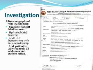
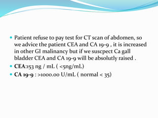
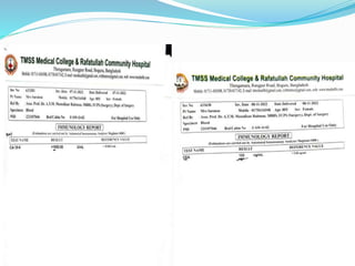
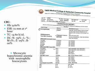
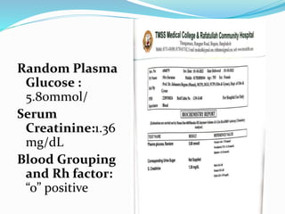
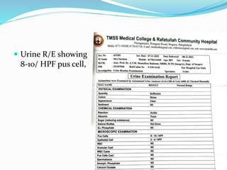
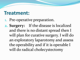
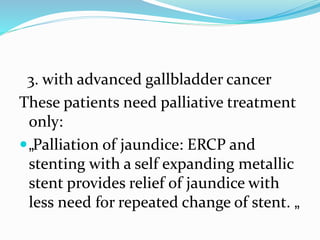
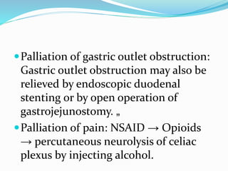
Presented By :
Intern doctor Amit Kumar Shah
Intern doctor TythiKundo
Department of surgery
Particulars of the patient:
 Name: Mrs. Suraton
 Age : 80 years
 Sex: Female
 Adress: bangla Heli, Hakimpur,DinajpurSadar
 Marital status: Widow
 Religion: Islam
 Date of admission: 07-11-2022@7:14 PM
 Date of examination: 07-11-2022@7:30 PM
Chief Complaints:
1. Pain in the upper abdomen for 4
months.
2. Loss of appetite for 3 months.
3. Weight loss for 3 months.
History of present illness:
She was apparently alright 4 months
ago when she start experiencing
episodes of sudden onset of pain in
right upper abdomen which is colicy in
nature radiating to back in the
shoulder occurring around 20 to 30
minutes after meals and subsides on its
own after an hour or two.
She experiences this episodes daily
associated with feeling of fullness in
upper abdomen after taking meals but
no complaints of heartburn. From the
last1 month she start having dull
aching pain in the right abdomen
which was continuous in nature not
radiating to back, more after taking
meals and do not subsides completely.
She also gives history of loss of appetite
which let her to lose weight of 8-10 k kg
in last 3 months but this was not
associated with abdominal distension,
nausea or vomiting .There is no history
of yellow colouration of eyes or skin,
high colour rine or itching over the
body
There is no history of fever , no history
of altered bowel habit, no history of
blood in the stool or black colored
stool. There is no history of lump in the
abdomen or swelling in the neck or
other part of body . No history
breathlessness, cough, or chest
tightness.
History of past illness:
Hysterectomy was done for uterine prolapse 2
months back.
She is hypertensive and on medication for
last 5 years and has no history of DM,
asthma, tuberculosis.
Immunization History:
She is immunized as per EPI schedule. She is
immunized against HBV. (For surgeon’s
safety)
Personal history:
No addiction
Bowel and bladder habit normal
Sleep and appetite decreases
Mixed bengali diet
Family History
No history of gallbladder stone disease
in any family member
No history of malignancies in her any
family member
No history of liver disease
Menstrual history
She attains menopause at the age of 47
years and previous menopausal history
is unremarkable .
Obstetric history
she have 5 childrens and age of last
child born is 45 years.
Drug history:
She took anti- Hypertensive drug and
also took some tablets from local quack
but could not mention the names.
General Examination
Patient was examined in well lit room
after obtaining consent in presence of
female attendant
 Conscious, co operative and well oriented to
time, place and person
 Lean, thin,well hydrated, lying confortably
in bed with iv canulla in left hand and
cathetrized
 Temperature: 98.6 (Right axilla)
Pulse : 76min regular rhythm (right
radial artery)
BP: 130/80mmhg ( right arm supine
position )
RR: 14/ min
Anemia:mild anemic
 Non icterus, cyanosis, clubbing, koilonychias,
leuconychia were absent
 Lymphoneds are not enlarged , no bipedal edema
Abdominal Examination:
Examined on the supine position with
arm kept on sides and exposed from
lower chest to pubic tubercle( after
taking consent again and in presence of
female attendant)
INSPECTION:
Abdomen was normal in shape with a
central and inverted umbilicus and
vertically slitted.
Overlying skin was normal with an scar
of previous surgery ( lower midline
incision),there was no dilated veins,
visible pulsation , peristalsis absent
All quadrant moves equally with
respiration and is thoraco- Abdominal
No visible lump in any of the quadrant
nor was any swelling at hernial site
Palpation:
Temperature was not raised
There is no superficial tenderness
The abdomen is soft
There is a single globular lump
palpable in the right hypochondriac
region around 4cm X 3 cm extending
1cm lateral and 3 cm medial to right
midclavicular line and extending 3cm
below the right costal margin. The
surface is irregular, margins are well
defined , is hard in consistency and
moves well with respiration. It become
palpable when patient is asked to
raised a head.
There is no evidence of hepatomegaly and
spleenomegaly or any other lump. Hernia
sites are normal.
Percussion:
Abdomen is tympanic on percussion. But
the lump is dull on percussion and is in
continuation with the dullness of liver
which ends at the 6th ICS in the mid
clavicular line
Auscultation:
 Normal bowel sound are audible and no
hepatic bruit
On D/R/E: Yellow staining of examining
figure is noticed and no mass detected
 On cardiovascular examination and respiratory
examination revels no abnormalities , nervous
system ……
Case summery :
Mrs. Suraton, 80 years old hypertensive,
non diabetic lady resident of Bangla
Heli, Hakimpur, Dinajpur sadar got
admitted to TMC & RCH with the
complaints of episodes of colicy right
upper quadrant pain for last 4 months
which change to dull aching
continuous pain for last 1 month
without any jaundice or bowel habit
alter.
She also gives history of loss of appetite
which let her to lose weight of 8-10 k kg
in last 3 months but this was not
associated with abdominal distension,
nausea or vomiting
On examination she is lean but pale
lady without icterus , single hard lump
in the right hypochondrium 4X3 cm
with well defined margins and irregular
surface moving well with respiration ,
lump was dull on percussion. There is
no clinical evidences of ascitis and this
mass is likely to arise from gall bladder.
Provisional Diagnosis:
Gall bladder malignancy
Differential Diagnosis:
„Mucocele of gallbladder
 „Empyema of gallbladder „„
Periampullary carcinoma „
Cholangiocarcinoma involving the
distal bile duct „
Investigation:
„Ultrasonography of
whole abdomen :
 Suggestive of gall
bladder mass.
 Hydronephosis(
bilateral)
 And H/O
hysterectomy with
inflammed stump.
And patient is
adiviced to do CT
abdomen but
patient refuse.
 Patient refuse to pay test for CT scan of abdomen, so
we advice the patient CEA and CA 19-9 , it is increased
in other GI malinancy but if we suscpect Ca gall
bladder CEA and CA 19-9 will be absolutly raised .
 CEA:153 ng / mL ( <5ng/mL)
 CA 19-9 : >1000.00 U/mL ( normal < 35)
CBC:
 Hb: 9.60%
 ESR: 72 mm at 1st
hour
 TC: 14.60 k/uL
 DC: N- 79% ; L- % ;
M-1% ; E- 05% ; B-
00%
 Microcytic
hypochromic anemia
with neutrophilic
leucocytosis.
Random Plasma
Glucose :
5.80mmol/
Serum
Creatinine:1.36
mg/dL
Blood Grouping
and Rh factor:
“o” positive
 Urine R/E showing
8-10/ HPF pus cell,
Confirm Diagnosis:
Gall bladder carcinoma
Treatment:
1. Pre-operative preparation.
2. Surgery: If the disease is localized
and there is no distant spread then I
will plan for curative surgery. I will do
an exploratory laparotomy and assess
the operability and if it is operable I
will do radical cholecystectomy
3. with advanced gallbladder cancer
These patients need palliative treatment
only:
„Palliation of jaundice: ERCP and
stenting with a self expanding metallic
stent provides relief of jaundice with
less need for repeated change of stent. „
Palliation of gastric outlet obstruction:
Gastric outlet obstruction may also be
relieved by endoscopic duodenal
stenting or by open operation of
gastrojejunostomy. „
Palliation of pain: NSAID → Opioids
→ percutaneous neurolysis of celiac
plexus by injecting alcohol.
Chemotherapy is not very effective in
carcinoma gallbladder. Most frequently
5-fluorouracil and mitomycin have
been used. Cisplatin has shown better
results
Radiation therapy has been used after
resectional surgery as an adjuvant.
No survival advantage has been
reported. Radiation sensitizer 5-
fluorouracil along with radiation
provides some benefit. Advanced
unresectable disease radiotherapy is
not useful.
Prognosis: 5-year survival rate < 5%
Thank you

gall bladder carcinoma Long case surgery

  • 1.
    Presented By : Interndoctor Amit Kumar Shah Intern doctor TythiKundo Department of surgery
  • 2.
    Particulars of thepatient:  Name: Mrs. Suraton  Age : 80 years  Sex: Female  Adress: bangla Heli, Hakimpur,DinajpurSadar  Marital status: Widow  Religion: Islam  Date of admission: 07-11-2022@7:14 PM  Date of examination: 07-11-2022@7:30 PM
  • 3.
    Chief Complaints: 1. Painin the upper abdomen for 4 months. 2. Loss of appetite for 3 months. 3. Weight loss for 3 months.
  • 4.
    History of presentillness: She was apparently alright 4 months ago when she start experiencing episodes of sudden onset of pain in right upper abdomen which is colicy in nature radiating to back in the shoulder occurring around 20 to 30 minutes after meals and subsides on its own after an hour or two.
  • 5.
    She experiences thisepisodes daily associated with feeling of fullness in upper abdomen after taking meals but no complaints of heartburn. From the last1 month she start having dull aching pain in the right abdomen which was continuous in nature not radiating to back, more after taking meals and do not subsides completely.
  • 6.
    She also giveshistory of loss of appetite which let her to lose weight of 8-10 k kg in last 3 months but this was not associated with abdominal distension, nausea or vomiting .There is no history of yellow colouration of eyes or skin, high colour rine or itching over the body
  • 7.
    There is nohistory of fever , no history of altered bowel habit, no history of blood in the stool or black colored stool. There is no history of lump in the abdomen or swelling in the neck or other part of body . No history breathlessness, cough, or chest tightness.
  • 8.
    History of pastillness: Hysterectomy was done for uterine prolapse 2 months back. She is hypertensive and on medication for last 5 years and has no history of DM, asthma, tuberculosis. Immunization History: She is immunized as per EPI schedule. She is immunized against HBV. (For surgeon’s safety)
  • 9.
    Personal history: No addiction Boweland bladder habit normal Sleep and appetite decreases Mixed bengali diet
  • 10.
    Family History No historyof gallbladder stone disease in any family member No history of malignancies in her any family member No history of liver disease
  • 11.
    Menstrual history She attainsmenopause at the age of 47 years and previous menopausal history is unremarkable . Obstetric history she have 5 childrens and age of last child born is 45 years.
  • 12.
    Drug history: She tookanti- Hypertensive drug and also took some tablets from local quack but could not mention the names.
  • 13.
    General Examination Patient wasexamined in well lit room after obtaining consent in presence of female attendant  Conscious, co operative and well oriented to time, place and person  Lean, thin,well hydrated, lying confortably in bed with iv canulla in left hand and cathetrized  Temperature: 98.6 (Right axilla)
  • 14.
    Pulse : 76minregular rhythm (right radial artery) BP: 130/80mmhg ( right arm supine position ) RR: 14/ min Anemia:mild anemic
  • 15.
     Non icterus,cyanosis, clubbing, koilonychias, leuconychia were absent  Lymphoneds are not enlarged , no bipedal edema
  • 16.
    Abdominal Examination: Examined onthe supine position with arm kept on sides and exposed from lower chest to pubic tubercle( after taking consent again and in presence of female attendant) INSPECTION: Abdomen was normal in shape with a central and inverted umbilicus and vertically slitted.
  • 17.
    Overlying skin wasnormal with an scar of previous surgery ( lower midline incision),there was no dilated veins, visible pulsation , peristalsis absent All quadrant moves equally with respiration and is thoraco- Abdominal No visible lump in any of the quadrant nor was any swelling at hernial site
  • 18.
    Palpation: Temperature was notraised There is no superficial tenderness The abdomen is soft There is a single globular lump palpable in the right hypochondriac region around 4cm X 3 cm extending 1cm lateral and 3 cm medial to right
  • 19.
    midclavicular line andextending 3cm below the right costal margin. The surface is irregular, margins are well defined , is hard in consistency and moves well with respiration. It become palpable when patient is asked to raised a head.
  • 20.
    There is noevidence of hepatomegaly and spleenomegaly or any other lump. Hernia sites are normal. Percussion: Abdomen is tympanic on percussion. But the lump is dull on percussion and is in continuation with the dullness of liver which ends at the 6th ICS in the mid clavicular line
  • 21.
    Auscultation:  Normal bowelsound are audible and no hepatic bruit On D/R/E: Yellow staining of examining figure is noticed and no mass detected
  • 22.
     On cardiovascularexamination and respiratory examination revels no abnormalities , nervous system ……
  • 23.
    Case summery : Mrs.Suraton, 80 years old hypertensive, non diabetic lady resident of Bangla Heli, Hakimpur, Dinajpur sadar got admitted to TMC & RCH with the complaints of episodes of colicy right upper quadrant pain for last 4 months which change to dull aching continuous pain for last 1 month without any jaundice or bowel habit alter.
  • 24.
    She also giveshistory of loss of appetite which let her to lose weight of 8-10 k kg in last 3 months but this was not associated with abdominal distension, nausea or vomiting
  • 25.
    On examination sheis lean but pale lady without icterus , single hard lump in the right hypochondrium 4X3 cm with well defined margins and irregular surface moving well with respiration , lump was dull on percussion. There is no clinical evidences of ascitis and this mass is likely to arise from gall bladder.
  • 26.
  • 27.
    Differential Diagnosis: „Mucocele ofgallbladder  „Empyema of gallbladder „„ Periampullary carcinoma „ Cholangiocarcinoma involving the distal bile duct „
  • 28.
    Investigation: „Ultrasonography of whole abdomen:  Suggestive of gall bladder mass.  Hydronephosis( bilateral)  And H/O hysterectomy with inflammed stump. And patient is adiviced to do CT abdomen but patient refuse.
  • 29.
     Patient refuseto pay test for CT scan of abdomen, so we advice the patient CEA and CA 19-9 , it is increased in other GI malinancy but if we suscpect Ca gall bladder CEA and CA 19-9 will be absolutly raised .  CEA:153 ng / mL ( <5ng/mL)  CA 19-9 : >1000.00 U/mL ( normal < 35)
  • 31.
    CBC:  Hb: 9.60% ESR: 72 mm at 1st hour  TC: 14.60 k/uL  DC: N- 79% ; L- % ; M-1% ; E- 05% ; B- 00%  Microcytic hypochromic anemia with neutrophilic leucocytosis.
  • 32.
  • 33.
     Urine R/Eshowing 8-10/ HPF pus cell,
  • 34.
  • 35.
    Treatment: 1. Pre-operative preparation. 2.Surgery: If the disease is localized and there is no distant spread then I will plan for curative surgery. I will do an exploratory laparotomy and assess the operability and if it is operable I will do radical cholecystectomy
  • 36.
    3. with advancedgallbladder cancer These patients need palliative treatment only: „Palliation of jaundice: ERCP and stenting with a self expanding metallic stent provides relief of jaundice with less need for repeated change of stent. „
  • 37.
    Palliation of gastricoutlet obstruction: Gastric outlet obstruction may also be relieved by endoscopic duodenal stenting or by open operation of gastrojejunostomy. „ Palliation of pain: NSAID → Opioids → percutaneous neurolysis of celiac plexus by injecting alcohol.
  • 38.
    Chemotherapy is notvery effective in carcinoma gallbladder. Most frequently 5-fluorouracil and mitomycin have been used. Cisplatin has shown better results Radiation therapy has been used after resectional surgery as an adjuvant.
  • 39.
    No survival advantagehas been reported. Radiation sensitizer 5- fluorouracil along with radiation provides some benefit. Advanced unresectable disease radiotherapy is not useful. Prognosis: 5-year survival rate < 5%
  • 40.