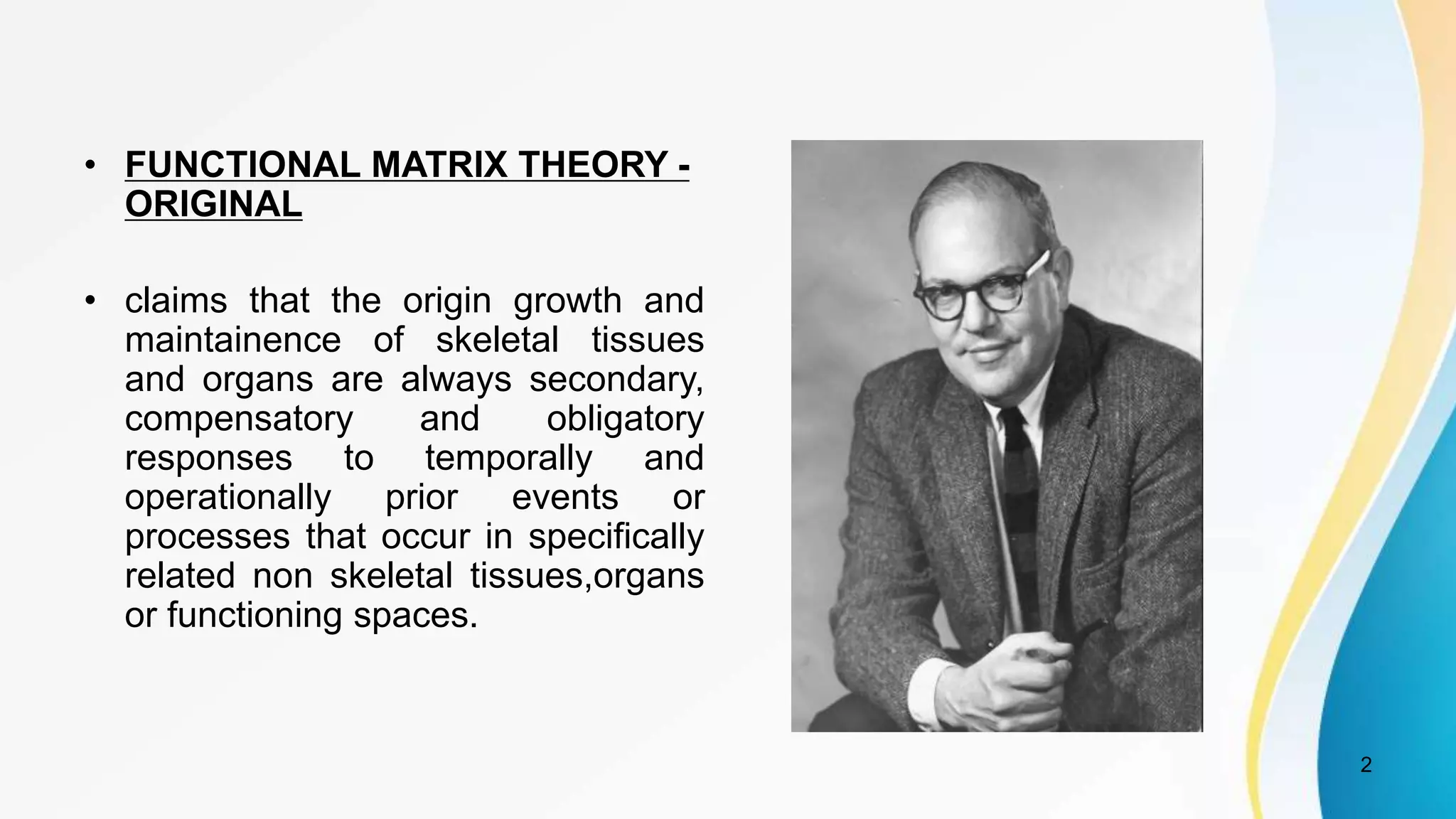
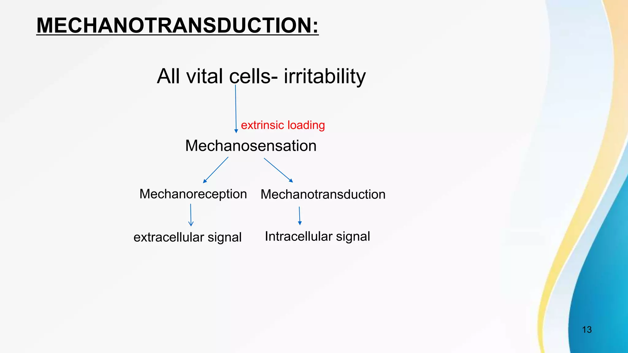
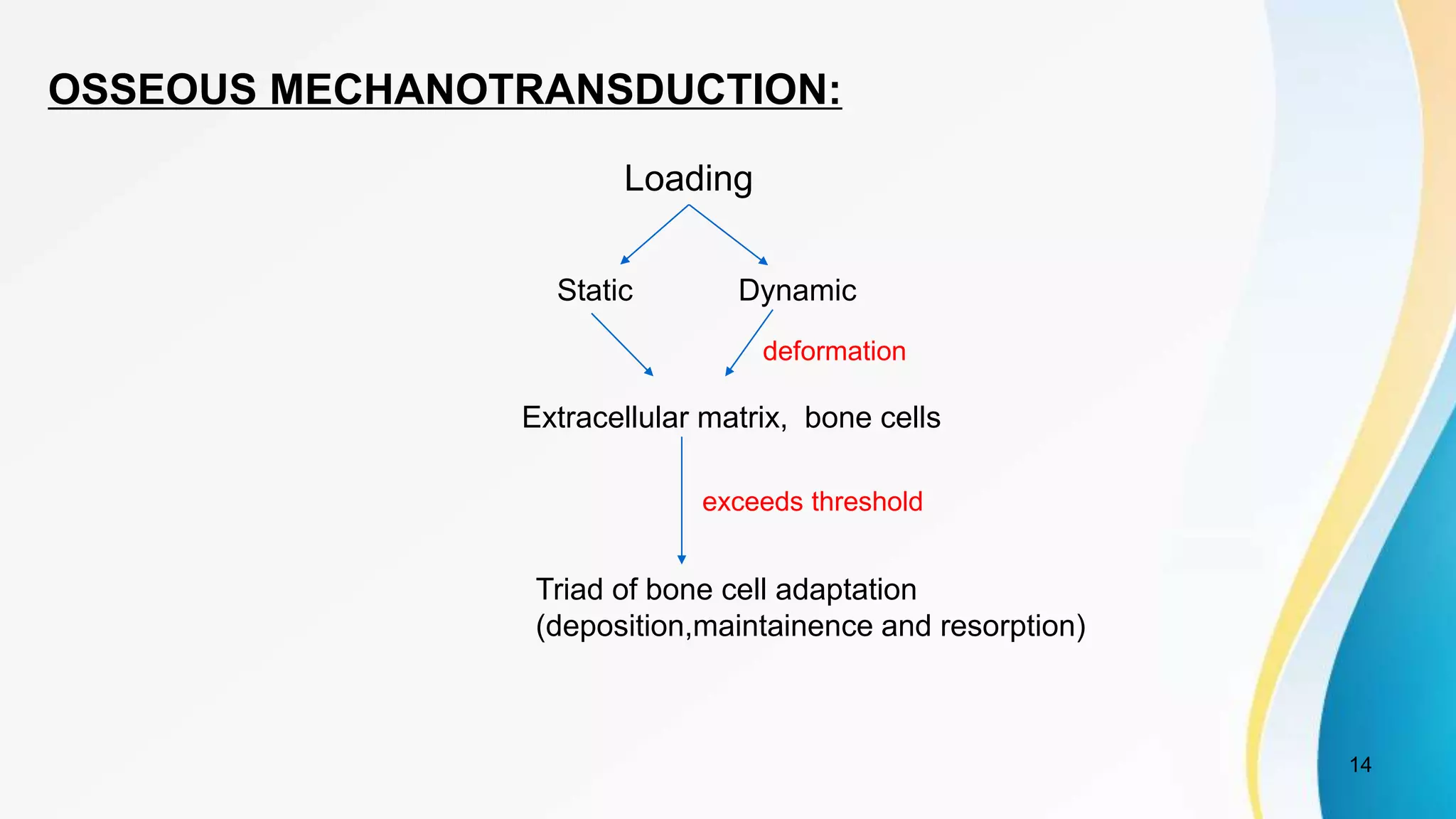
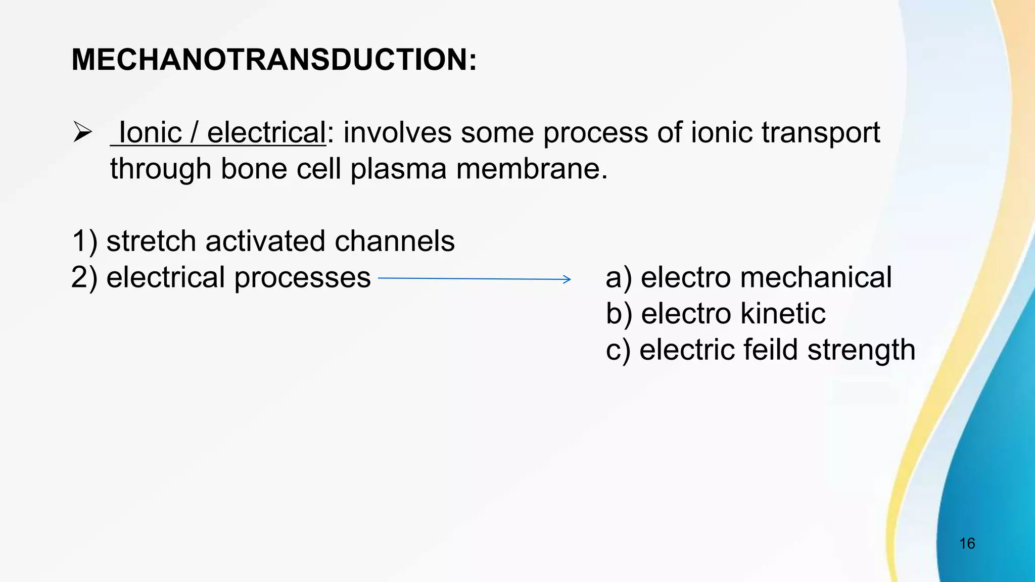
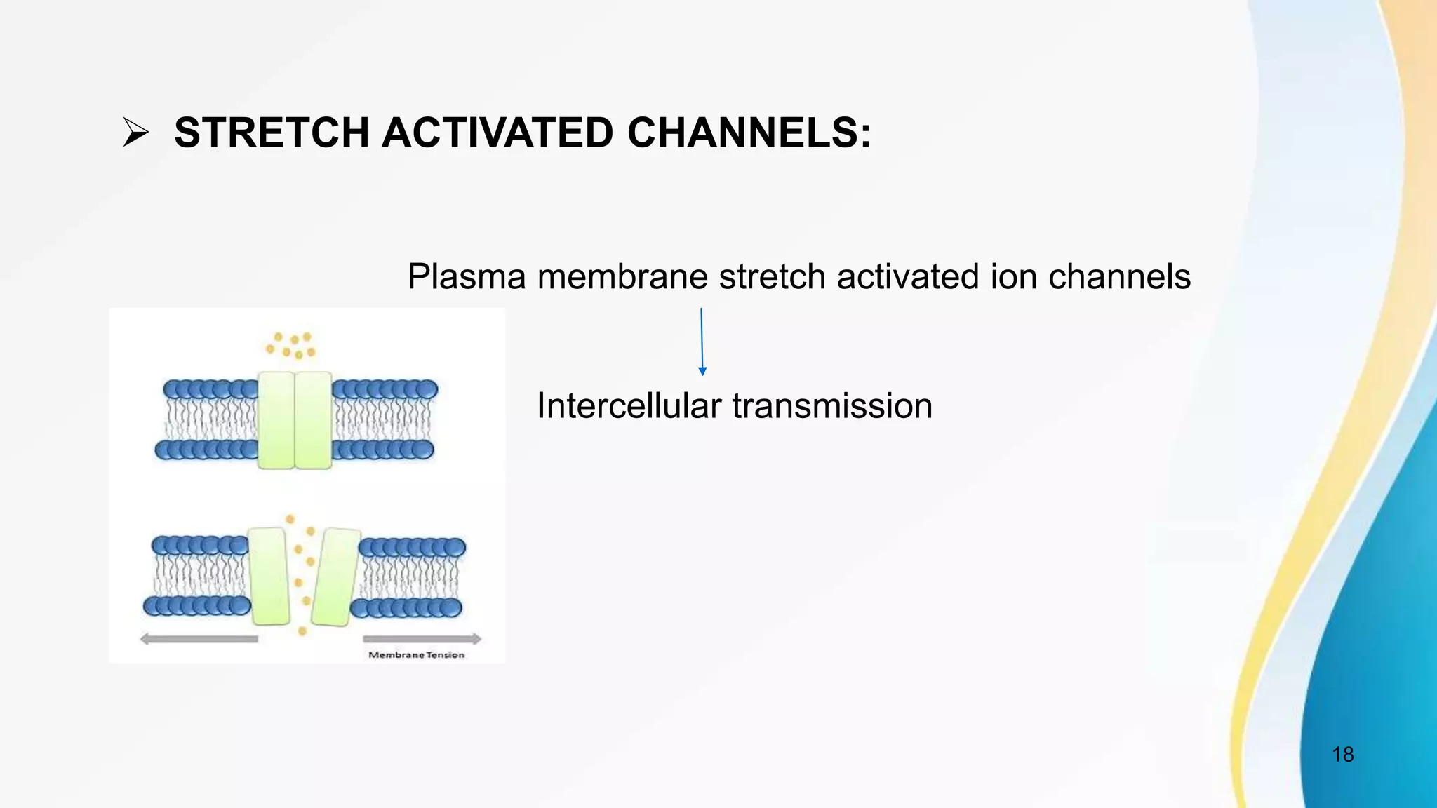
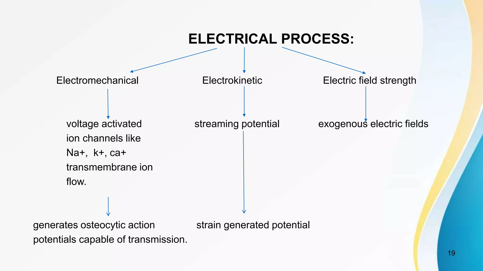
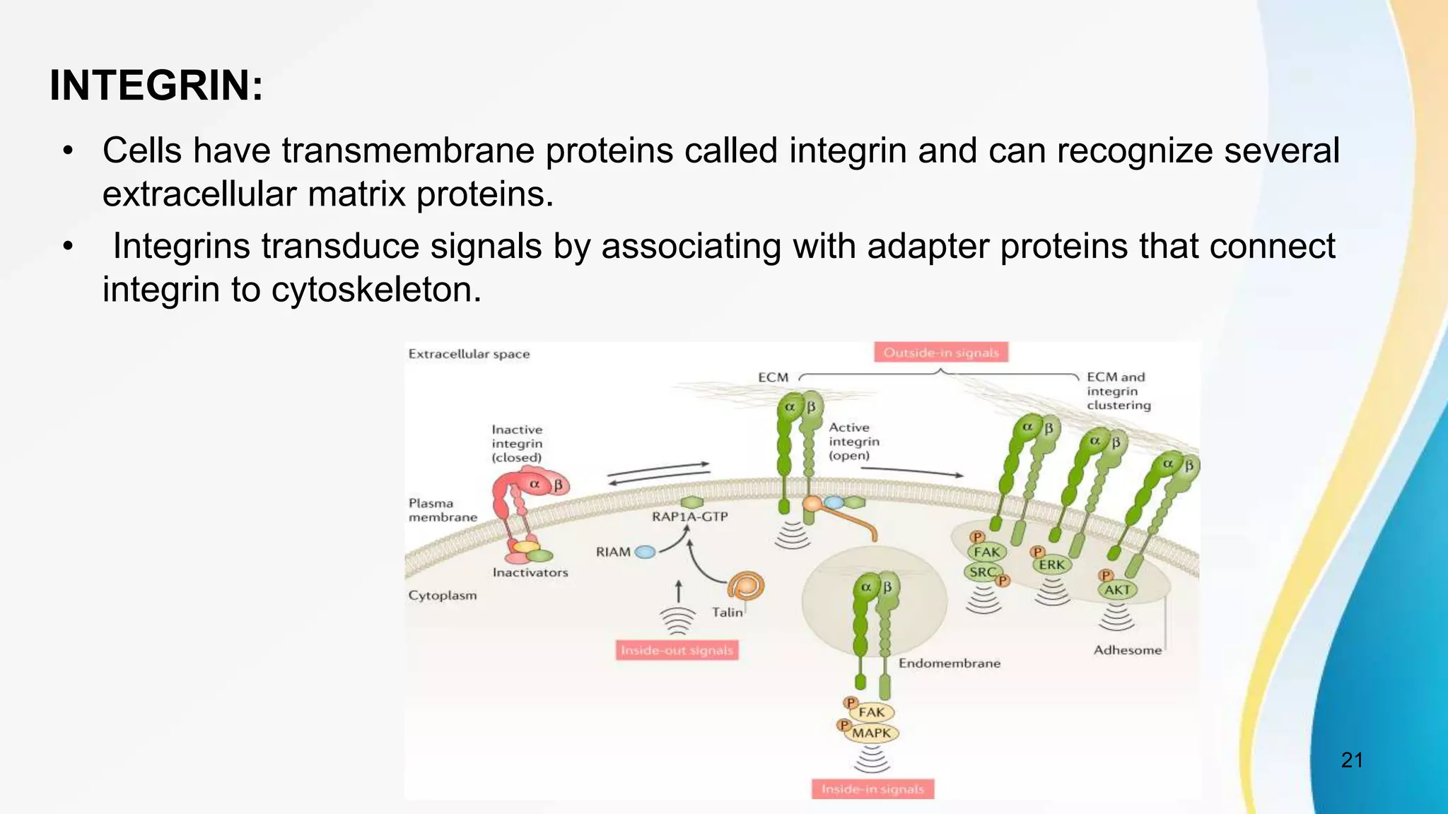
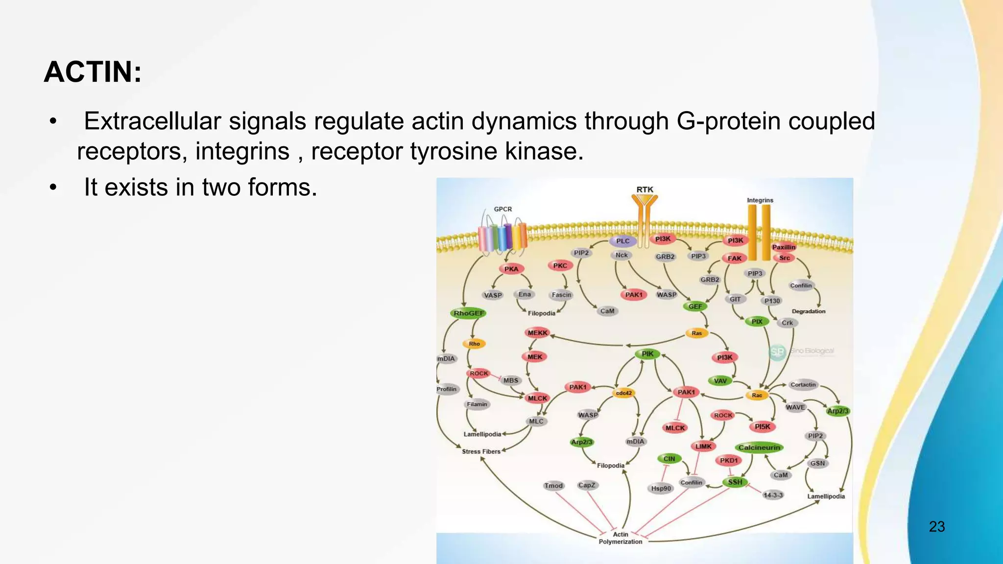
THE FUNCTIONAL MATRIX
THEORY REVISITED
1
Melvin L. Moss., AJODO 1997; vol 112:8-11
1. THE ROLE OF MECHANOTRANSDUCTION
Dr V Chandra Shekhar
PG Trainee
Department of Orthodontics and Dentofacial Orthopaedics
SBDC
Guide: Dr Shubhaker Rao sir
Co-guide: Dr Sravani ma’am
• FUNCTIONAL MATRIX THEORY -
ORIGINAL
• claims that the origin growth and
maintainence of skeletal tissues
and organs are always secondary,
compensatory and obligatory
responses to temporally and
operationally prior events or
processes that occur in specifically
related non skeletal tissues,organs
or functioning spaces.
2
 DEVELOPMENT OF FUNCTIONAL MATRIX HYPOTHESIS:
• A decades study of roles of intrinsic and extrinsic factors in cephalic growth
evolved into functional matrix hypothesis.
• A recent advances in biomedical, bioengineering and computer sciences
created more explanatory FMH versions.
3
 CONCEPTUAL AND ANATOMIC BASES OF FMH:
FUNCTIONAL CRANIAL COMPONENT
4
SKELETAL UNIT FUNCTIONAL MATRIX
MICRO
SKELETAL
Ex-
coronoid,angle,
condyle.
MACRO
SKELETAL
Ex- Maxilla,
Mandible.
PERIOSTEAL
Ex- teeth,
muscles.
CAPSULAR
Ex- neurocranial
capsule,
Orofacial capsule.
FUNCTIONAL CRANIAL COMPONENT
• A series of functions like respiration, vision, olfaction,
chewing,speech, etc. are carried out by a specific
Functional Cranial Component.
• FCC denominate all of those tissues and spaces
necessary to carry out a given function.
• skeletal units originate and remain completely embedded
within their respective functional matrices.
5
1) FUNCTIONAL MATRIX-
PERIOSTEAL MATRIX-
This corresponds to immediate local environment, which
acts directly and actively upon related skeletal units.
Ex- muscles, blood vessels, nerves, glands, teeth.
 The periosteal matrices stimulation causes direct growth of the microskeletal
units.
Ex- interaction between temporalis muscle and coronoid process of mandible.
6
CAPSULAR MATRIX-
• It is organ or space that occupy broader anatomical complex.
• Each capsule is an envelope.
• Capsules expand due to volumetric increase of capsular matrix.
Ex- Neurocranial capsule, orofacial capsule.
7
SKELETAL UNIT
• Micro skeletal unit:-
• single bone composed of many number of units termed as
micro skeletal units
• ex- condyle of mandible.
8
• Macro skeletal unit:-
• when adjoining portions of
bone united to function as
a single cranial unit.
• ex- internal surface of
calvarium.
9
 CONSTRAINTS OF FMH:
1. METHODOLOGIC CONSTRAINT:
Macroscopic measurements, which
use techniques of point mechanics and arbitrary reference frames.
Ex- CEPHALOMETRIC RADIOGRAPHY.
10
2.HIERARCHICAL CONSTRAINT:
• Earlier version’s description did not extend “downward” or extend
“upward” to multicellular process by which bone responds to lower level
signals.
• The FMH could describe:
1) how extrinsic FM stimuli transduced into regulatory signals by
individual bone cells.
2) how individual cells communicate to produce multicellular responses.
11
• FUNCTIONAL MATRIX HYPOTHESIS – REVISITED:
 ROLE OF MECHANOTRANSDUCTION- PART-1
 ROLE OF OSSEOUS CONNECTED CELLULAR NETWORK-PART-2
 GENOMIC THESIS- PART-3
 EPIGENETIC ANTITHESIS & RESOLVING SYNTHESIS- PART-4
12
MECHANOTRANSDUCTION:
All vital cells- irritability
13
Mechanosensation
Mechanoreception Mechanotransduction
Intracellular signal
extrinsic loading
extracellular signal
14
OSSEOUS MECHANOTRANSDUCTION:
Loading
Static Dynamic
Extracellular matrix, bone cells
Triad of bone cell adaptation
(deposition,maintainence and resorption)
deformation
exceeds threshold
15
 Osseous mechanotransduction unique in 4 ways:
1) Most mechanosensory cells are cytologically specialized but bone cells are not.
2) 1 bone loading stimulus evoke-3 adaptational responses.
3) osseous signal transmission is aneural.
4) adaptational processes are independent.
16
MECHANOTRANSDUCTION:
 Ionic / electrical: involves some process of ionic transport
through bone cell plasma membrane.
1) stretch activated channels
2) electrical processes a) electro mechanical
b) electro kinetic
c) electric feild strength
17
 IONIC PROCESS:
It involves ionic transport through ionic channels like voltage gated,
ligand gated in bone cell plasma membrane
Intercellular transmission
18
 STRETCH ACTIVATED CHANNELS:
Plasma membrane stretch activated ion channels
Intercellular transmission
ELECTRICAL PROCESS:
Electromechanical Electrokinetic Electric field strength
voltage activated streaming potential exogenous electric fields
ion channels like
Na+, k+, ca+
transmembrane ion
flow.
generates osteocytic action strain generated potential
potentials capable of transmission.
19
 MECHANICAL PROCESS:
• It is an alternative means by which periosteal functional matrix activity
may regulate hierarchically lower level bone cell genomic functions.
• The basis of this mechanism is physical continuity of transmembrane
molecule integrin.
• connected extracellularly to collagen and intra-
cellularly with cytoskeletal actin.
• actin is connected to nuclear membrane.
20
INTEGRIN:
• Cells have transmembrane proteins called integrin and can recognize several
extracellular matrix proteins.
• Integrins transduce signals by associating with adapter proteins that connect
integrin to cytoskeleton.
21
• RIAM- rap 1 GTP interacting molecule
• Rap 1a- GTP - ras related protein 1 GTP
• Ras proteins are small monomeric GTPases that cycle
between the GTP-bound active form and the GDP-bound
inactive form.
• RIAMisa Rap1 effector that is a member of
MRL(Mig10/RIAM/Lamellipodin) family of adaptor proteins.The
Ras subfamily members Rap1A and Rap1B stimulate integrin
activation.
• MRL proteins function as scaffolds that connect the membrane
targeting sequences in Ras GTPases to talin, thereby
recruiting talin to integrins at the plasma membrane. 22
ACTIN:
• Extracellular signals regulate actin dynamics through G-protein coupled
receptors, integrins , receptor tyrosine kinase.
• It exists in two forms.
23
• Signaling to the cytoskeleton through G protein-coupled
receptors (GPCRs), integrins, receptor tyrosine kinases
(RTKs) lead to diverse effects on cell activity, including
changes in cell shape, migration, proliferation, and
survival.
• Intracellular regulation of the cell’s response to external
cues occurs through a large number of signaling
cascades that include the Rho family of small GTPases
(Rho, Rac, and Cdc42) and their activators, guanine
nucleotide exchange factors (GEFs), their downstream
protein kinase effectors.
24
• These cascades converge on proteins that directly
regulate the behavior and organization of the actin
cytoskeleton, including actin interacting regulatory
proteins such as Arp2/3 complex, profilin, and gelsolin.
Signaling through different pathways can lead to the
formation of distinct actin-dependent structures whose
coordinated assembly/disassembly is important for
directed cell migration and other cellular behavior.
25
REFERENCES:
• Melvin L. Moss and letty Salentijn The primary role of
fuctional matrices in facial growth (june 1969)
• Moss ML. The functional matrix hypothesis revisited.1.
The role of mechanotransduction. American journal of
orthodontics and dentofacial orthopedics. 1997 Jul
1;112(1):8-11.
• The functional matrix hypothesis revisited. 2. The role of
an osseous connected cellular network
• Text book of contemporary orthodontics - William R Proffit
*
• Bone research 2020 8;23 - molecular mechanosensors in
osteocytes.
• Orthodontic mechanotransduction and the role of the
P2X7 receptor by Rodrigo F. Viecilli, Thomas R. Katona,
Jie Chen- AJODO 2009;135:694-5
• Cellular, molecular, and tissue-level reactions to
orthodontic force-Vinod Krishnan and Ze’ev Davidovitch -
AJODO -april 2006
27
Orthodontic mechanotransduction and the role ofthe P2X7
receptor by Rodrigo F. Viecilli, Thomas R. Katona, Jie Chen
28
• The P2X7 receptor promotes necrotic tissue metabolism
by ensuring a normal acute-phase inflammatory
response.
• There were direct relationships between certain stress
magnitudes and root resorption and bone formation.
• Orthodontic responses are related to the principal stress
patterns in the periodontal ligament,and the P2X7
receptor plays a significant role in their
mechanotransduction.
29
Biomechanical and Molecular Regulation of Bone
Remodeling by
Alexander G. Robling, Alesha B. Castillo & charles
turner.
and Charles H. Turner
30
• when the bone is loaded, bending causes fluid movement
in the canalicular network, which leads to shear stress on
integrin and actin molecules mainly on the cell surface of
osteocytes.
• Fluid shear enhances ATP release and causes an influx of
Ca2+ via voltage-gated channels. ATP then binds to P2Y
(g-protein coupled) or P2X (ligand-gated ion channel)
receptors. The P2Y G protein pathway leads to Ca2+
release. ATP binds to P2X7 receptor increasing pore
formation and causing the release of PGE2, which in turn
binds to PGE receptor stimulating bone formation through
a pathway that is still unknown.
31
32
bone
bending and fluid movement
integrin and actin molecule
ATP release and influx of calcium ions
P2Y or P2X
calcium release
increase pore formation
release of PGE2
bone formation
load
stress on
binds to
Cellular, molecular, and tissue-level reactions
to orthodontic force by Vinod Krishnan and Ze’ev Davidovitch(Am J
Orthod Dentofacial Orthop 2006;129:469e.1-460e.32)
*
• In response to applied mechanical forces, there is
generation of electric potentials in the stressed tissues.
• Borgens - “stress-generated potentials or streaming
potentials”.
• In hydrated tissues, streaming potentials predominate as
the interstitial fluid moves.
• Pollack - electric double layer surrounds bone.
34
CONCLUSION:
35
• In addition to the FMH, the concepts of
mechanotransduction and computational bone biology
offers explanatory chain extending from epigenetic event of
skeletal muscle contraction, hierarchically downward, then
upward again.
• Analyzing size, shape changes by reference frame
invariant, FEM produces comprehensive and integrated
description of epigenetic regulation of bone form.

Functional matrix hypothesis revisited 1

  • 1.
    THE FUNCTIONAL MATRIX THEORYREVISITED 1 Melvin L. Moss., AJODO 1997; vol 112:8-11 1. THE ROLE OF MECHANOTRANSDUCTION Dr V Chandra Shekhar PG Trainee Department of Orthodontics and Dentofacial Orthopaedics SBDC Guide: Dr Shubhaker Rao sir Co-guide: Dr Sravani ma’am
  • 2.
    • FUNCTIONAL MATRIXTHEORY - ORIGINAL • claims that the origin growth and maintainence of skeletal tissues and organs are always secondary, compensatory and obligatory responses to temporally and operationally prior events or processes that occur in specifically related non skeletal tissues,organs or functioning spaces. 2
  • 3.
     DEVELOPMENT OFFUNCTIONAL MATRIX HYPOTHESIS: • A decades study of roles of intrinsic and extrinsic factors in cephalic growth evolved into functional matrix hypothesis. • A recent advances in biomedical, bioengineering and computer sciences created more explanatory FMH versions. 3
  • 4.
     CONCEPTUAL ANDANATOMIC BASES OF FMH: FUNCTIONAL CRANIAL COMPONENT 4 SKELETAL UNIT FUNCTIONAL MATRIX MICRO SKELETAL Ex- coronoid,angle, condyle. MACRO SKELETAL Ex- Maxilla, Mandible. PERIOSTEAL Ex- teeth, muscles. CAPSULAR Ex- neurocranial capsule, Orofacial capsule.
  • 5.
    FUNCTIONAL CRANIAL COMPONENT •A series of functions like respiration, vision, olfaction, chewing,speech, etc. are carried out by a specific Functional Cranial Component. • FCC denominate all of those tissues and spaces necessary to carry out a given function. • skeletal units originate and remain completely embedded within their respective functional matrices. 5
  • 6.
    1) FUNCTIONAL MATRIX- PERIOSTEALMATRIX- This corresponds to immediate local environment, which acts directly and actively upon related skeletal units. Ex- muscles, blood vessels, nerves, glands, teeth.  The periosteal matrices stimulation causes direct growth of the microskeletal units. Ex- interaction between temporalis muscle and coronoid process of mandible. 6
  • 7.
    CAPSULAR MATRIX- • Itis organ or space that occupy broader anatomical complex. • Each capsule is an envelope. • Capsules expand due to volumetric increase of capsular matrix. Ex- Neurocranial capsule, orofacial capsule. 7
  • 8.
    SKELETAL UNIT • Microskeletal unit:- • single bone composed of many number of units termed as micro skeletal units • ex- condyle of mandible. 8
  • 9.
    • Macro skeletalunit:- • when adjoining portions of bone united to function as a single cranial unit. • ex- internal surface of calvarium. 9
  • 10.
     CONSTRAINTS OFFMH: 1. METHODOLOGIC CONSTRAINT: Macroscopic measurements, which use techniques of point mechanics and arbitrary reference frames. Ex- CEPHALOMETRIC RADIOGRAPHY. 10
  • 11.
    2.HIERARCHICAL CONSTRAINT: • Earlierversion’s description did not extend “downward” or extend “upward” to multicellular process by which bone responds to lower level signals. • The FMH could describe: 1) how extrinsic FM stimuli transduced into regulatory signals by individual bone cells. 2) how individual cells communicate to produce multicellular responses. 11
  • 12.
    • FUNCTIONAL MATRIXHYPOTHESIS – REVISITED:  ROLE OF MECHANOTRANSDUCTION- PART-1  ROLE OF OSSEOUS CONNECTED CELLULAR NETWORK-PART-2  GENOMIC THESIS- PART-3  EPIGENETIC ANTITHESIS & RESOLVING SYNTHESIS- PART-4 12
  • 13.
    MECHANOTRANSDUCTION: All vital cells-irritability 13 Mechanosensation Mechanoreception Mechanotransduction Intracellular signal extrinsic loading extracellular signal
  • 14.
    14 OSSEOUS MECHANOTRANSDUCTION: Loading Static Dynamic Extracellularmatrix, bone cells Triad of bone cell adaptation (deposition,maintainence and resorption) deformation exceeds threshold
  • 15.
    15  Osseous mechanotransductionunique in 4 ways: 1) Most mechanosensory cells are cytologically specialized but bone cells are not. 2) 1 bone loading stimulus evoke-3 adaptational responses. 3) osseous signal transmission is aneural. 4) adaptational processes are independent.
  • 16.
    16 MECHANOTRANSDUCTION:  Ionic /electrical: involves some process of ionic transport through bone cell plasma membrane. 1) stretch activated channels 2) electrical processes a) electro mechanical b) electro kinetic c) electric feild strength
  • 17.
    17  IONIC PROCESS: Itinvolves ionic transport through ionic channels like voltage gated, ligand gated in bone cell plasma membrane Intercellular transmission
  • 18.
    18  STRETCH ACTIVATEDCHANNELS: Plasma membrane stretch activated ion channels Intercellular transmission
  • 19.
    ELECTRICAL PROCESS: Electromechanical ElectrokineticElectric field strength voltage activated streaming potential exogenous electric fields ion channels like Na+, k+, ca+ transmembrane ion flow. generates osteocytic action strain generated potential potentials capable of transmission. 19
  • 20.
     MECHANICAL PROCESS: •It is an alternative means by which periosteal functional matrix activity may regulate hierarchically lower level bone cell genomic functions. • The basis of this mechanism is physical continuity of transmembrane molecule integrin. • connected extracellularly to collagen and intra- cellularly with cytoskeletal actin. • actin is connected to nuclear membrane. 20
  • 21.
    INTEGRIN: • Cells havetransmembrane proteins called integrin and can recognize several extracellular matrix proteins. • Integrins transduce signals by associating with adapter proteins that connect integrin to cytoskeleton. 21
  • 22.
    • RIAM- rap1 GTP interacting molecule • Rap 1a- GTP - ras related protein 1 GTP • Ras proteins are small monomeric GTPases that cycle between the GTP-bound active form and the GDP-bound inactive form. • RIAMisa Rap1 effector that is a member of MRL(Mig10/RIAM/Lamellipodin) family of adaptor proteins.The Ras subfamily members Rap1A and Rap1B stimulate integrin activation. • MRL proteins function as scaffolds that connect the membrane targeting sequences in Ras GTPases to talin, thereby recruiting talin to integrins at the plasma membrane. 22
  • 23.
    ACTIN: • Extracellular signalsregulate actin dynamics through G-protein coupled receptors, integrins , receptor tyrosine kinase. • It exists in two forms. 23
  • 24.
    • Signaling tothe cytoskeleton through G protein-coupled receptors (GPCRs), integrins, receptor tyrosine kinases (RTKs) lead to diverse effects on cell activity, including changes in cell shape, migration, proliferation, and survival. • Intracellular regulation of the cell’s response to external cues occurs through a large number of signaling cascades that include the Rho family of small GTPases (Rho, Rac, and Cdc42) and their activators, guanine nucleotide exchange factors (GEFs), their downstream protein kinase effectors. 24
  • 25.
    • These cascadesconverge on proteins that directly regulate the behavior and organization of the actin cytoskeleton, including actin interacting regulatory proteins such as Arp2/3 complex, profilin, and gelsolin. Signaling through different pathways can lead to the formation of distinct actin-dependent structures whose coordinated assembly/disassembly is important for directed cell migration and other cellular behavior. 25
  • 26.
    REFERENCES: • Melvin L.Moss and letty Salentijn The primary role of fuctional matrices in facial growth (june 1969) • Moss ML. The functional matrix hypothesis revisited.1. The role of mechanotransduction. American journal of orthodontics and dentofacial orthopedics. 1997 Jul 1;112(1):8-11. • The functional matrix hypothesis revisited. 2. The role of an osseous connected cellular network • Text book of contemporary orthodontics - William R Proffit *
  • 27.
    • Bone research2020 8;23 - molecular mechanosensors in osteocytes. • Orthodontic mechanotransduction and the role of the P2X7 receptor by Rodrigo F. Viecilli, Thomas R. Katona, Jie Chen- AJODO 2009;135:694-5 • Cellular, molecular, and tissue-level reactions to orthodontic force-Vinod Krishnan and Ze’ev Davidovitch - AJODO -april 2006 27
  • 28.
    Orthodontic mechanotransduction andthe role ofthe P2X7 receptor by Rodrigo F. Viecilli, Thomas R. Katona, Jie Chen 28
  • 29.
    • The P2X7receptor promotes necrotic tissue metabolism by ensuring a normal acute-phase inflammatory response. • There were direct relationships between certain stress magnitudes and root resorption and bone formation. • Orthodontic responses are related to the principal stress patterns in the periodontal ligament,and the P2X7 receptor plays a significant role in their mechanotransduction. 29
  • 30.
    Biomechanical and MolecularRegulation of Bone Remodeling by Alexander G. Robling, Alesha B. Castillo & charles turner. and Charles H. Turner 30
  • 31.
    • when thebone is loaded, bending causes fluid movement in the canalicular network, which leads to shear stress on integrin and actin molecules mainly on the cell surface of osteocytes. • Fluid shear enhances ATP release and causes an influx of Ca2+ via voltage-gated channels. ATP then binds to P2Y (g-protein coupled) or P2X (ligand-gated ion channel) receptors. The P2Y G protein pathway leads to Ca2+ release. ATP binds to P2X7 receptor increasing pore formation and causing the release of PGE2, which in turn binds to PGE receptor stimulating bone formation through a pathway that is still unknown. 31
  • 32.
    32 bone bending and fluidmovement integrin and actin molecule ATP release and influx of calcium ions P2Y or P2X calcium release increase pore formation release of PGE2 bone formation load stress on binds to
  • 33.
    Cellular, molecular, andtissue-level reactions to orthodontic force by Vinod Krishnan and Ze’ev Davidovitch(Am J Orthod Dentofacial Orthop 2006;129:469e.1-460e.32) *
  • 34.
    • In responseto applied mechanical forces, there is generation of electric potentials in the stressed tissues. • Borgens - “stress-generated potentials or streaming potentials”. • In hydrated tissues, streaming potentials predominate as the interstitial fluid moves. • Pollack - electric double layer surrounds bone. 34
  • 35.
    CONCLUSION: 35 • In additionto the FMH, the concepts of mechanotransduction and computational bone biology offers explanatory chain extending from epigenetic event of skeletal muscle contraction, hierarchically downward, then upward again. • Analyzing size, shape changes by reference frame invariant, FEM produces comprehensive and integrated description of epigenetic regulation of bone form.