FROM LIGHT TOSUPER RESOLUTION:
Evolution of Optical Microscopy in Cell Imaging
Presented By: Ms. Farhana Parween
Roll No.: CGU251103
Registration No.: 2503060020
M.Sc. Biotechnology, Semester I
Course: Bioinstrumentation and Biotechniques
Under the Guidance of: Dr. Jyoti Prakash Sahoo
2.
OPTICAL MICROSCOPY
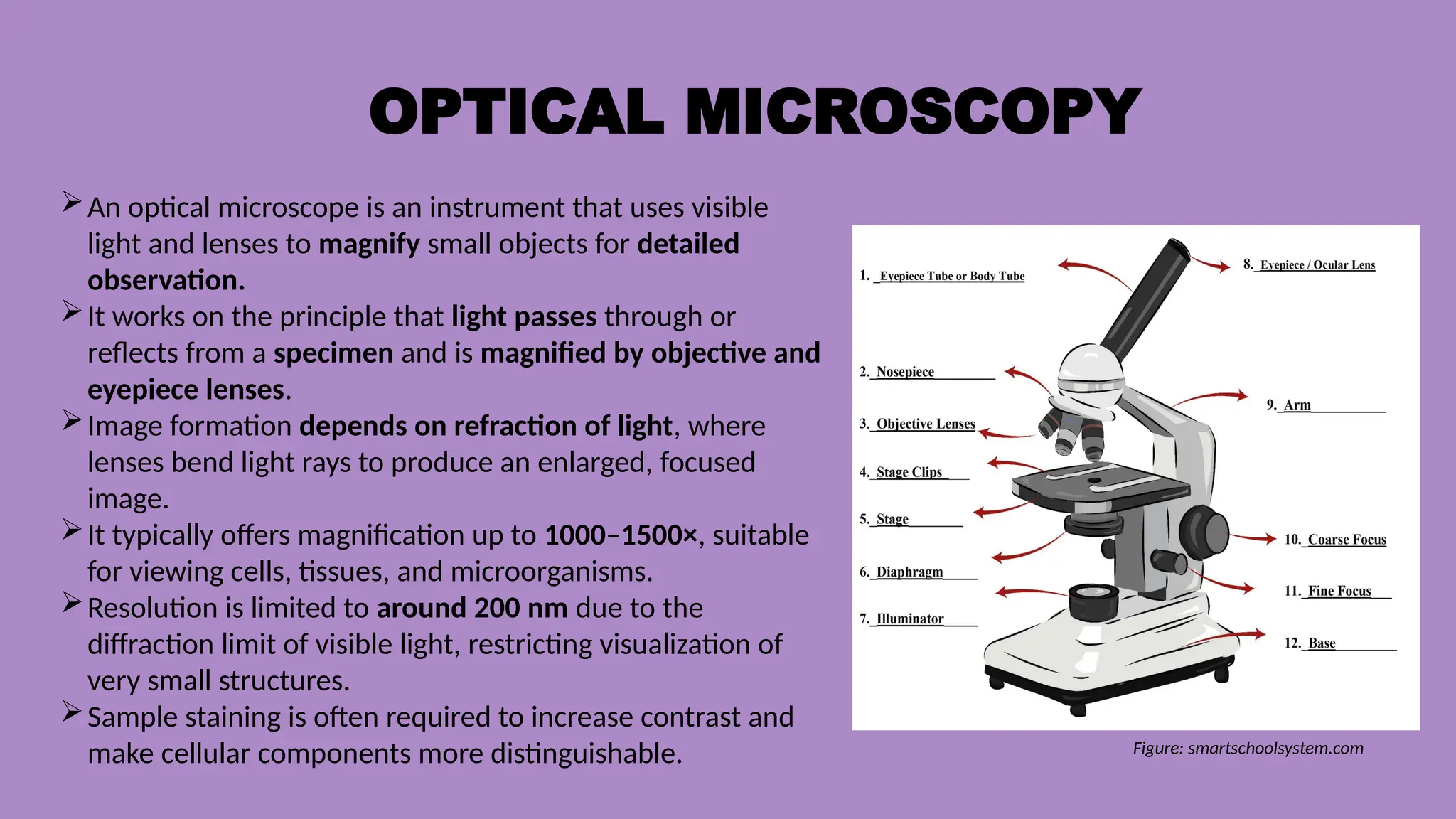
An opticalmicroscope is an instrument that uses visible
light and lenses to magnify small objects for detailed
observation.
It works on the principle that light passes through or
reflects from a specimen and is magnified by objective and
eyepiece lenses.
Image formation depends on refraction of light, where
lenses bend light rays to produce an enlarged, focused
image.
It typically offers magnification up to 1000–1500×, suitable
for viewing cells, tissues, and microorganisms.
Resolution is limited to around 200 nm due to the
diffraction limit of visible light, restricting visualization of
very small structures.
Sample staining is often required to increase contrast and
make cellular components more distinguishable. Figure: smartschoolsystem.com
3.
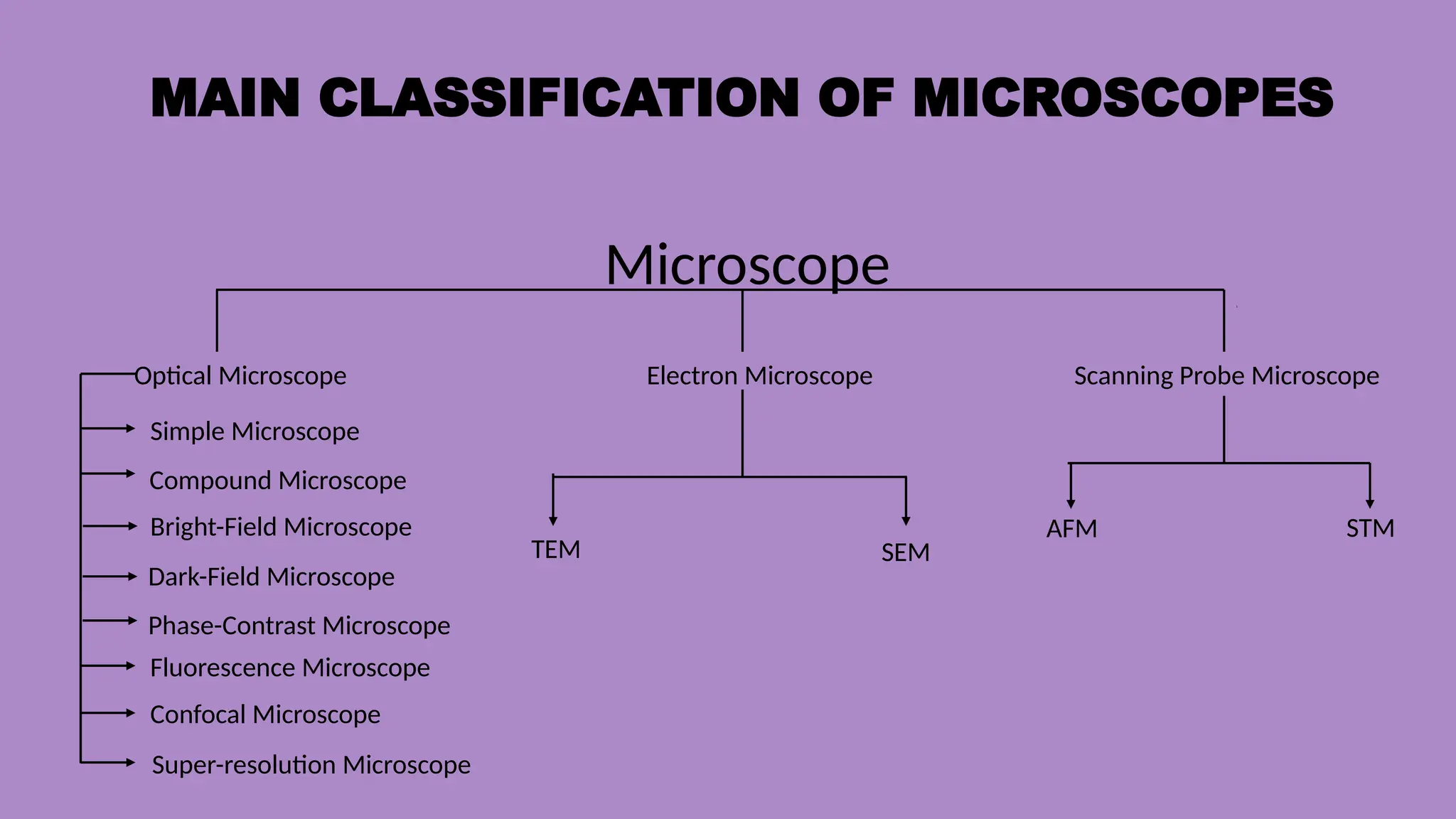
MAIN CLASSIFICATION OFMICROSCOPES
Microscope
Optical Microscope Electron Microscope Scanning Probe Microscope
Simple Microscope
Compound Microscope
Bright-Field Microscope
Dark-Field Microscope
Phase-Contrast Microscope
Super-resolution Microscope
Fluorescence Microscope
Confocal Microscope
STM
AFM
TEM SEM
4.
BRIGHT-FIELD MICROSCOPE
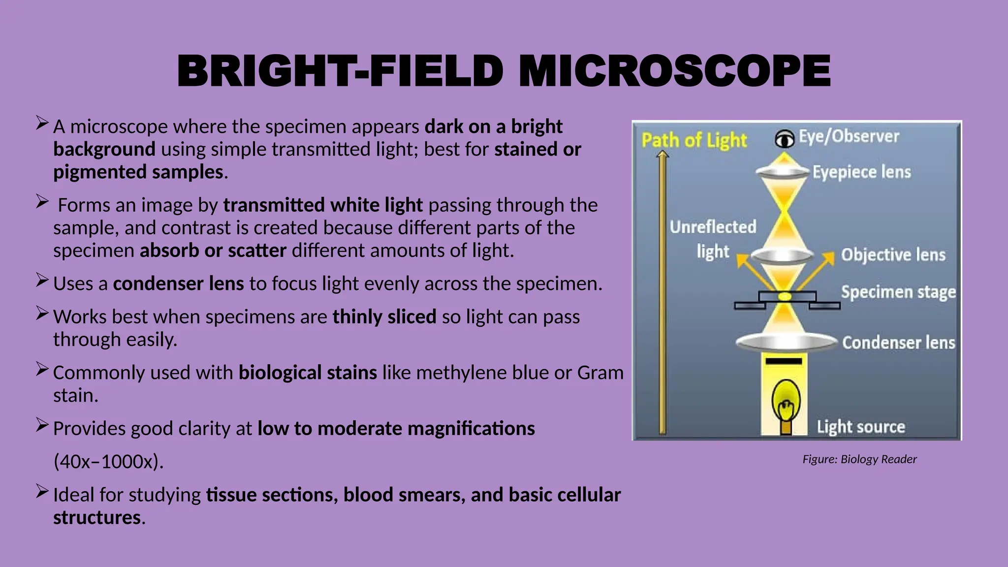
A microscopewhere the specimen appears dark on a bright
background using simple transmitted light; best for stained or
pigmented samples.
Forms an image by transmitted white light passing through the
sample, and contrast is created because different parts of the
specimen absorb or scatter different amounts of light.
Uses a condenser lens to focus light evenly across the specimen.
Works best when specimens are thinly sliced so light can pass
through easily.
Commonly used with biological stains like methylene blue or Gram
stain.
Provides good clarity at low to moderate magnifications
(40x–1000x).
Ideal for studying tissue sections, blood smears, and basic cellular
structures.
Figure: Biology Reader
5.
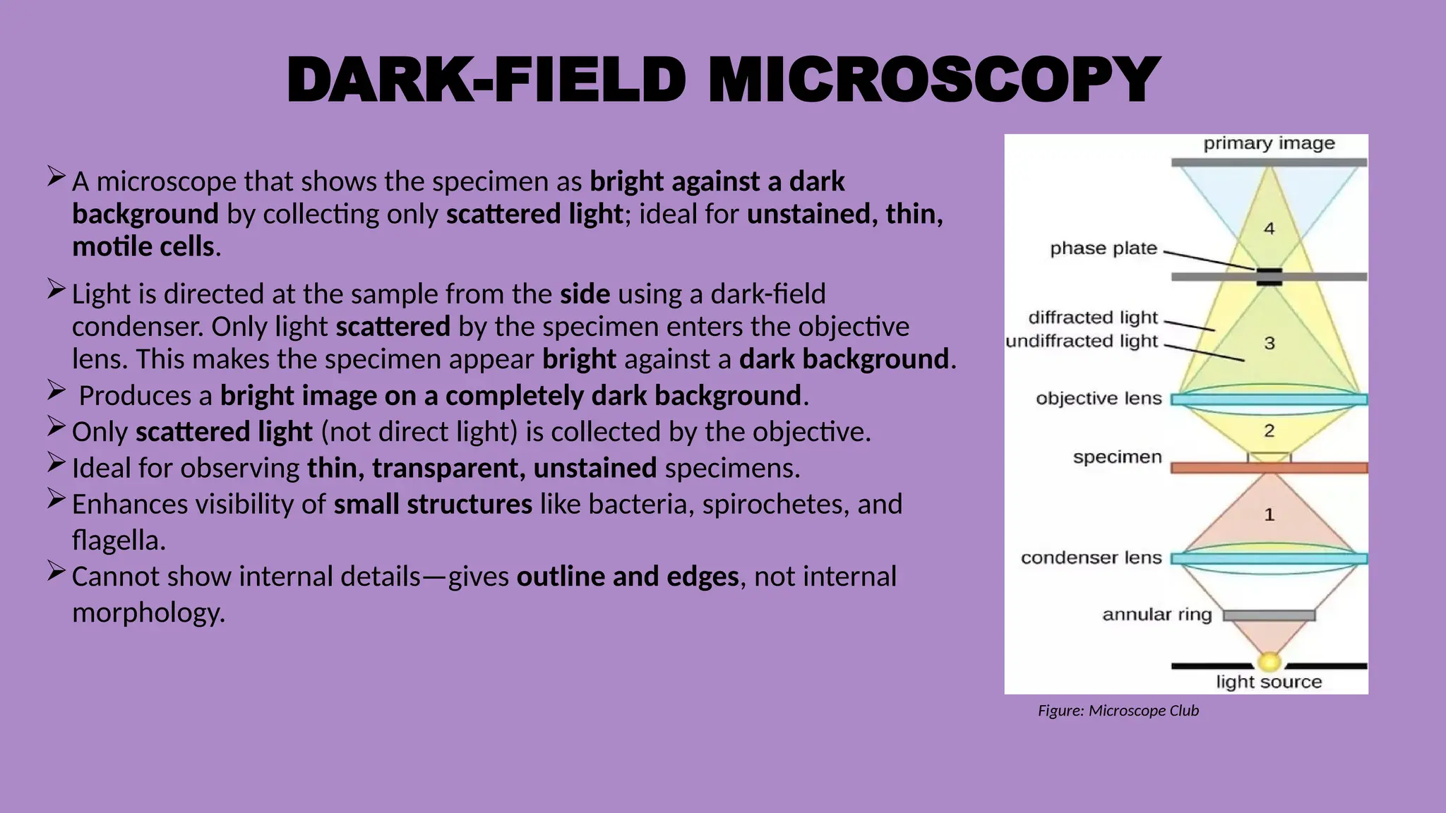
DARK-FIELD MICROSCOPY
A microscopethat shows the specimen as bright against a dark
background by collecting only scattered light; ideal for unstained, thin,
motile cells.
Light is directed at the sample from the side using a dark-field
condenser. Only light scattered by the specimen enters the objective
lens. This makes the specimen appear bright against a dark background.
Produces a bright image on a completely dark background.
Only scattered light (not direct light) is collected by the objective.
Ideal for observing thin, transparent, unstained specimens.
Enhances visibility of small structures like bacteria, spirochetes, and
flagella.
Cannot show internal details—gives outline and edges, not internal
morphology.
Figure: Microscope Club
6.
PHASE CONTRAST MICROSCOPY
Amicroscope that converts light phase differences into
contrast, allowing clear viewing of live, transparent,
unstained cells.
Phase-contrast converts differences in refractive index
(phase shifts) into visible brightness differences.
Transparent cells that normally look invisible become high-
contrast images.
Converts phase differences → intensity differences using
phase plates and annular rings.
Enables clear viewing of live, unstained cells without killing
them.
Excellent for observing organelles, cytoplasmic
movements, and cell division.
Produces images with bright halo or shade-off around
structures (optical artifact).
Very useful in cell biology, microbiology, and tissue culture
labs.
7.
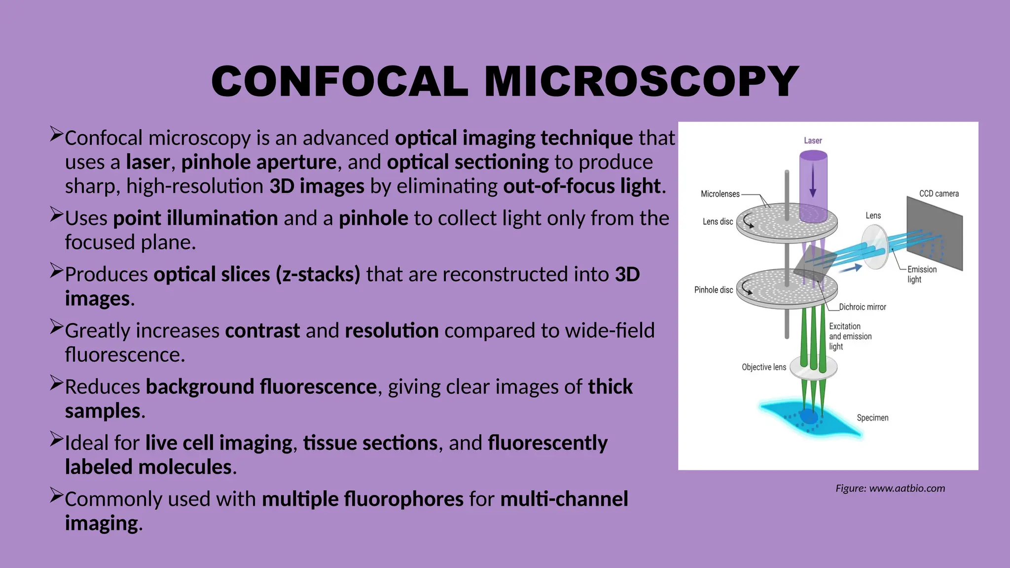
CONFOCAL MICROSCOPY
Confocal microscopyis an advanced optical imaging technique that
uses a laser, pinhole aperture, and optical sectioning to produce
sharp, high-resolution 3D images by eliminating out-of-focus light.
Uses point illumination and a pinhole to collect light only from the
focused plane.
Produces optical slices (z-stacks) that are reconstructed into 3D
images.
Greatly increases contrast and resolution compared to wide-field
fluorescence.
Reduces background fluorescence, giving clear images of thick
samples.
Ideal for live cell imaging, tissue sections, and fluorescently
labeled molecules.
Commonly used with multiple fluorophores for multi-channel
imaging.
Figure: www.aatbio.com
8.
Stimulated Emission DepletionMicroscopy
STED is a super-resolution fluorescence microscopy
technique that uses a depletion laser to shrink the
fluorescent spot beyond the diffraction limit, achieving
extremely high spatial resolution.
Uses two lasers: one for excitation, one for depleting
fluorescence around the focal point.
The depletion laser creates a donut-shaped beam that
leaves only the center fluorescing.
Achieves 20–50 nm resolution, far below the ~250 nm
light limit.
Provides real-time imaging of fine cellular structures like
synapses and the cytoskeleton.
Requires photostable fluorophores due to intense laser
power.
Useful in neurobiology, membrane studies, and
molecular architecture research.
Figure: www.Swinburne.edu.au
9.
Structured Illumination Microscopy
SIMis a super-resolution method that projects
striped/patterned light onto the specimen and uses
computational reconstruction of interference patterns to
achieve higher resolution.
Improves resolution to ~100 nm, about twice that of standard
fluorescence microscopes.
Uses Moiré patterns formed by patterned illumination
interacting with sample structures.
Produces super-resolution images with low phototoxicity,
suitable for live cells.
Allows fast imaging, ideal for dynamic cell processes.
Works well with standard fluorophores and normal sample
preparation.
Provides super-resolution over large fields of view.
Figure: www.slideserve.com
10.
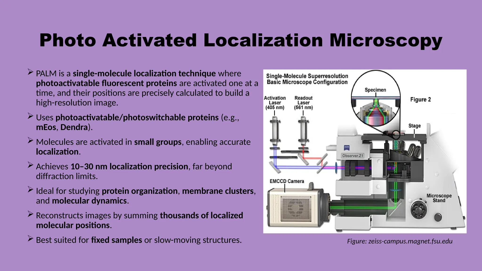
Photo Activated LocalizationMicroscopy
PALM is a single-molecule localization technique where
photoactivatable fluorescent proteins are activated one at a
time, and their positions are precisely calculated to build a
high-resolution image.
Uses photoactivatable/photoswitchable proteins (e.g.,
mEos, Dendra).
Molecules are activated in small groups, enabling accurate
localization.
Achieves 10–30 nm localization precision, far beyond
diffraction limits.
Ideal for studying protein organization, membrane clusters,
and molecular dynamics.
Reconstructs images by summing thousands of localized
molecular positions.
Best suited for fixed samples or slow-moving structures. Figure: zeiss-campus.magnet.fsu.edu
11.
Stochastic Optical ReconstructionMicroscopy
STORM is a single-molecule localization method that uses blinking
fluorophores which switch on and off randomly, allowing precise mapping of
each molecule to form a super-resolution image.
Uses dyes that stochastically blink, producing isolated signals for localization.
Achieves extremely high resolution (~20 nm) by fitting each blinking event.
Works well with organic dyes (e.g., Cy5), which give bright and stable signals.
Produces detailed images of protein networks, cytoskeleton, and membrane
nanostructures.
Requires thousands of frames, so imaging is slower than SIM or confocal.
Commonly used in cell biology, virology, and nanoscale molecular
organization studies.
12.
AI-ASSISTED IMAGE
RECONSTRUCTION
3D LIVE-CELL
IMAGING
Enhancesimage clarity and resolution by
reducing blur and noise using AI algorithms.
Allows low-light imaging, protecting live
cells from photodamage.
Reconstructs high-quality images from
limited or weak fluorescence signals.
Enables faster imaging by predicting and
restoring missing details.
Supports accurate 3D reconstruction,
especially in live-cell and super-resolution
microscopy.
Captures three-dimensional views of
living cells in real time.
Reveals dynamic processes like cell
division, migration, and organelle
movement.
Uses gentle illumination methods (e.g.,
light-sheet) to minimize phototoxicity.
Provides high temporal resolution,
allowing tracking of rapid cell changes.
Essential for studying cell behavior, drug
response, and intracellular interactions.
13.
SUMMARY
Optical microscopy providesthe foundation of cell imaging, with major types such
as bright-field, dark-field, and phase-contrast enabling contrast enhancement and
visualization of unstained or live cells.
Microscopes are broadly classified into light microscopy and electron microscopy,
with additional divisions based on illumination, contrast mechanisms, and resolution
capabilities.
Modern optical advancements—including confocal, STED, SIM, PALM, and STORM—
overcome the diffraction limit to deliver high-resolution and super-resolution
imaging of cellular structures.
Single-molecule localization techniques such as PALM and STORM provide
nanoscale visualization by precisely mapping individual fluorescent molecules.
Integration of AI-assisted image reconstruction and 3D live-cell imaging enables
faster, clearer, and dynamic visualization of cellular processes, transforming modern
biomedical research.
14.
REFERNCE
Karp, G. (2018).Cell and Molecular Biology: Concepts and Experiments. Wiley.
Alberts, B. et al. (2017). Molecular Biology of the Cell. Garland Science.
Chopra, A., & Panwar, H. (2021). Instrumentation and Techniques in Biotechnology.
PHI Learning, India.
Gupta, P. K. (2017). Elements of Biotechnology. Rastogi Publications, India.
Byju’s – Biology & Microscopy Articles https://byjus.com/biology/
Microbe Notes – Microscopy & Microbiology Notes https://microbenotes.com/
Khan Academy – Cell Biology & Microscopy Basics
https://www.khanacademy.org/science/biology