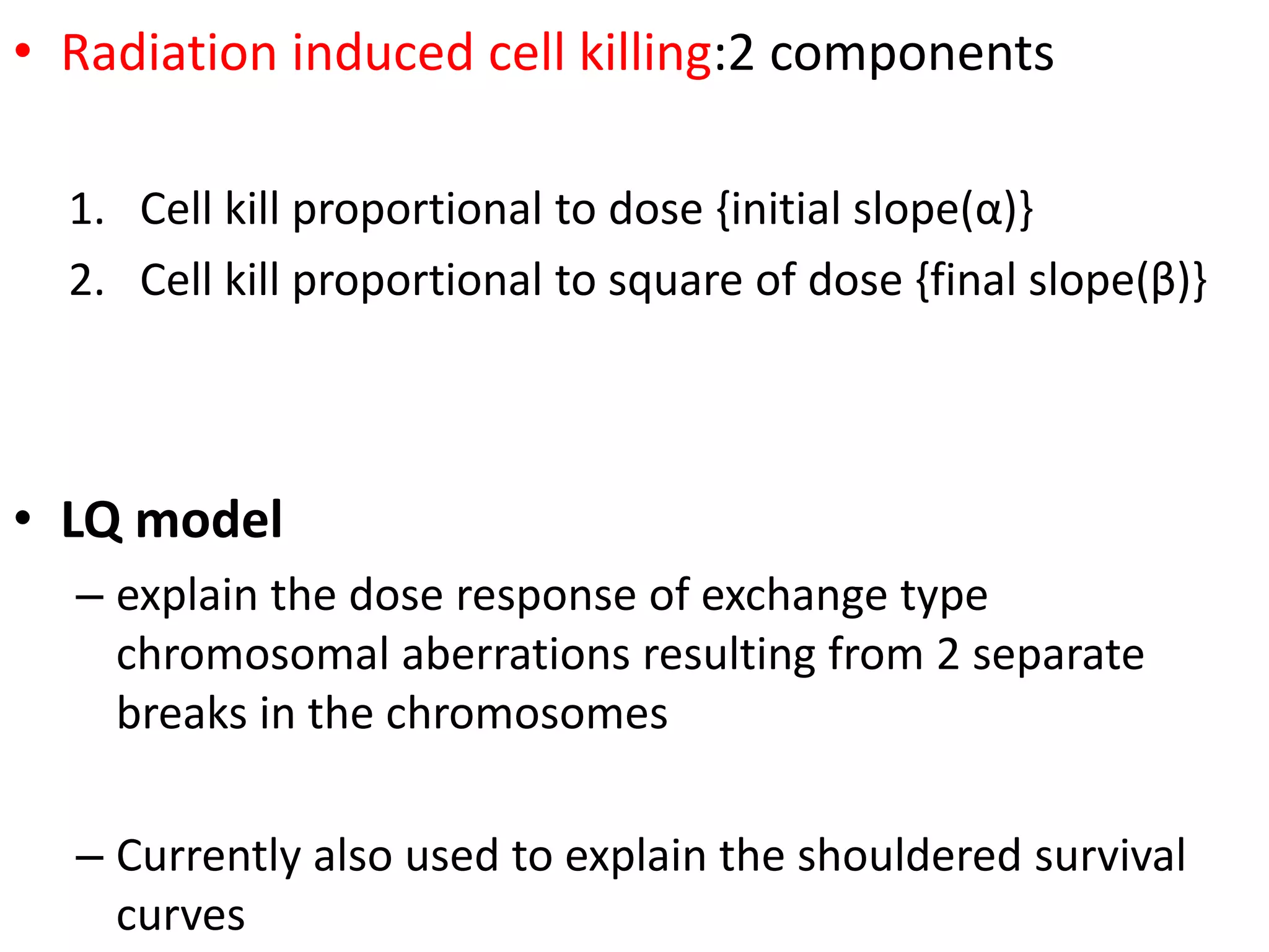
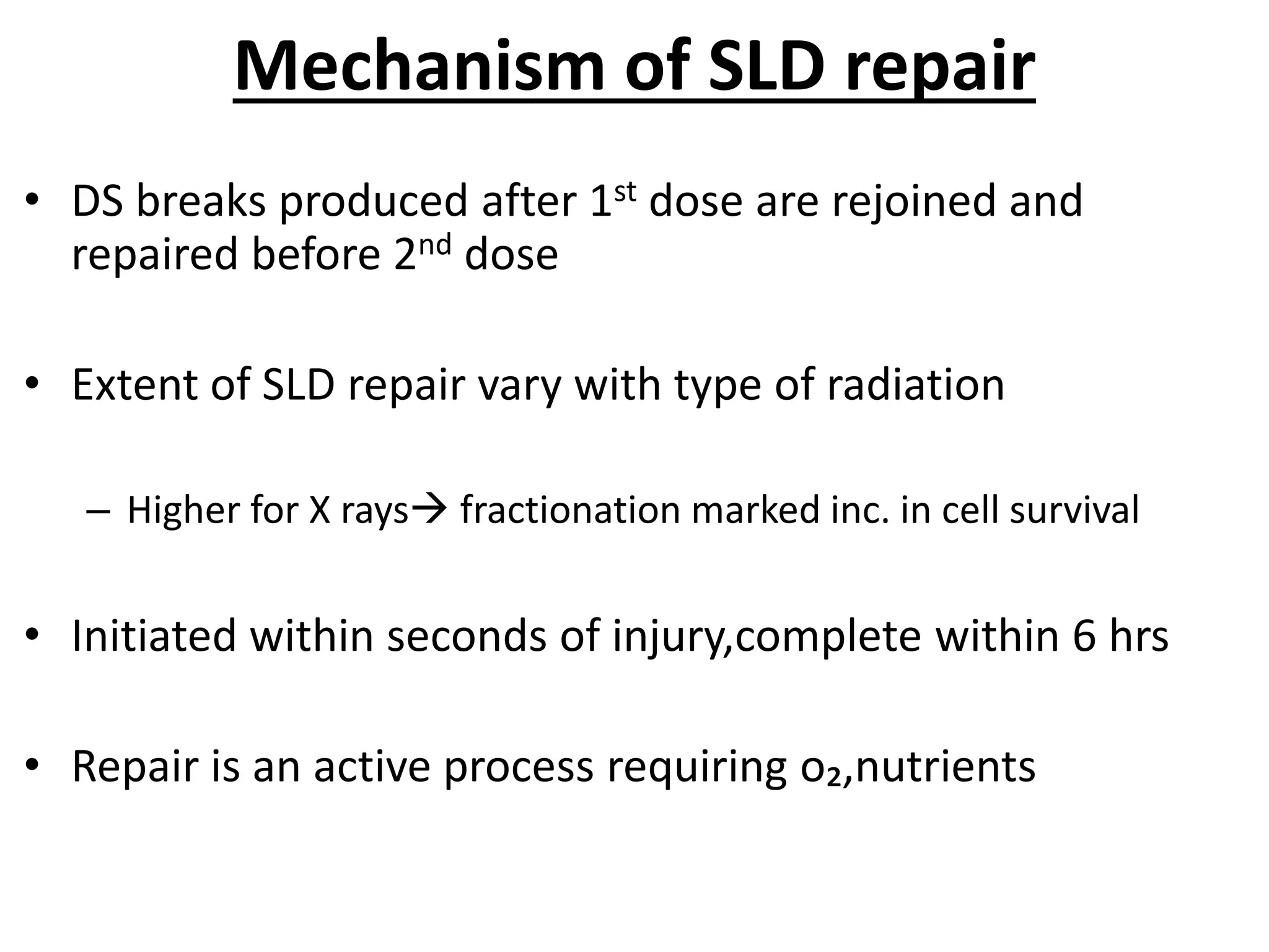
FRACTIONATION
-HISTORY
-RATIONALE
-CONCEPTS
Dr AMALU AUGUSTINE
Dept of Radiation oncology
GOVT MEDICAL COLLEGE
THIRUVANANTHAPURAM
• Definition
• History
– TDF models
• Stranquist
• Cohens
• Fowler
• Ellis
• Target model
– LQ model
• BED
• R’s of radiobiology
• Conventional fractionation
Fractionation
division of total dose of radiation into a no. of
separate fractions over total treatment time
– To deliver precisely measured dose of radiation
to a defined tumor volume
– with minimal damage to surrounding normal
tissue.
Historical review
• From the very beginning of RT , treatments
were fractionated
• X-ray
– just 1 month after its discovery
– Emil Grubbe(1896)
• Carcinoma breast
• 18 daily 1 hr #s
– Forced to use fractionated regime because of
low output from early Xray machines
• Single fraction radiotherapy -in
1914
• advent of Coolidge hot
cathode tube, with
– high output,
– adjustable tube currents &
–reproducible exposures
• The following ten years was a
period of uncertainty , about the
proper ways to fractionate.
ErIangen school ( Germany)
• Wintz
• Advocated single RT
• Fractionation allow tumor cells
time for recovery
• BERGONIE TRIBONDEAU LAW
– Rapidly growing tumor cells -
metabolically more active-
– Better able to recover from injury-
– will favor tumor cells if the
tumoricidal dose is not applied in the
first treatment.
Paris school ( France)
•Used radiobiological
principles of Regaud
• The Ram
experiment(1920’s)
Single dose of X-rays
• extensive scrotal skin
damage
Same dose in multiple #s
• sterilisation, no skin
damage
Tumour Normal
tissue
• HENRI COUTARD-1932
• Curie institute,Paris
• FRACTIONATION OF RADIATION PRODUCED
BETTER TUMOR CONTROL FOR A GIVEN LEVEL OF
NORMAL TISSUR TOXICITY THAN A SINGLE LARGE
DOSE.
Time ,Dose , Fractination Models
• With introduction of various fractionation schemes
– need for quantitative comparisons of treatments was felt
– to optimize treatment for particular tumor.
• Importance of tdf models
• 1. to calculate new total dose required to keep
biological effectiveness when conventional
fractionation is altered.
• 2. to compare diff treatment techniques that
differ in no of #, dpf, and overall treatment time.
• 3. To strive for optimal fractionation regimen.
• Strandquist -
• Cube root model- 1944
– first to device scientific
approach
– for correlating dose to
overall treatment time
– to produce an equivalent
biological isoeffect
– Isoeffect curves are a set of
curves which relate total
dose to overall treatment
time for definite effects of
radiation
• Stranquist plot / Cube root model
• relation between total dose & overall
treatment time
• He demonstrated that
– isoeffect curves (i.e. dose vs. no. of #s to
produce equal biological effect)
– on log-log graph
– for skin reactions (erythema & skin
tolerance)
– were straight lines with a slope of 0.33 i.e.
COHEN’S (1960)
• Cohen analyzed
– three diff. set of data
• erythema, skin damage and tumor control
• were documented for treatment times
from 1 to 40 days.
• Isoeffect curve for tumor control had a
smaller slope, m=0.22.
• Cohen found an exponent of
– 0.33for skin erythema / skin tolerance &
– 0.22 for skin cancers.
• According to Cohen’s results
– relationship b/w total dose & overall treatment time for normal tissue
tolerance & tumor can be written as
• Dn = K1 T.33
• D
t= K2 T.22
– K1& K2 are proportionality constants.
– Dn, Dt &T are normal tissue tolerance dose , tumor lethal dose & overall treatment
time respectively
• 0.33& 0.22 -> the repair capabilities
– of normal tissue & tumor cells respectively.
• This means as the treatment time is increased, tumor control
comes closer to the maximum tolerated skin dose.
• i.e. Tumor control can be achieved with , less normal tissue
damage.
Fowler
• Difference in exponents of time factor in Cohen’s formulations
– indicate that repair capacity of normal tissue is larger than that of tumor
• Fowler
– studies on pig skin
– showing normal tissue have two type of repair capabilities
• Intracellular
• having short repair half time of 0.5 to 3hrs &
• is complete within few hrs of irradiation.
• multiplicity of completion of recovery is equal to no. of #s.
• Homeostatic recovery - takes longer time to complete
• Hence no. of #s are more important than overall t/t
• This led Ellis to formulate NSD
NSD MODEL
Cube root law was the result of biological effect that were functions of
N and T
• time factor was a composite of
N (no. of #s) &
T (overall treatment time)
• Exponents for
– intracellular -0.22
– homeostatic recovery - 0.33-0.22=0.11
• Frank Ellis, British, 1969
• Fractionation is twice as important as time according to clinical
observations
• Hence dose is related to time & no. of #s as
D =NSD X T .11 X N.24
• This correlated well with Strandquist’s data.
– i.e. For treating once a day, everyday. T0.11 x T0.24 = T0.35.
• By not treating on weekends this will be reduced to T0.33
– The constant NSD is Nominal Standard Dose.
• NSD is a constant of proportionality
• which can be thought of as a bioeffective dose
• i.e. dose corrected for time and fractionation.
• NSD= D. T-0.11 .N-0.24
• Unit of NSD is RET ( Roentgen Equivalent Therapy)
• NSD can be used to compare two fractionation regimes.
Clinical use
• Enable clinicians to change from one
fractionation regimen to another,
– while maintaining equivalent biological effects on
both tumour and normal tissues
• Limitation:-
– omitted the importance of dose per fraction in
determining late effects in normal tissues
• Limitaitions of Elllis
• Was based on
• early Xray damage to skin &
• for trtmt upto 6 weeks.
• So cannot be applied for:
• 1.Late effects.
• 2.For other normal tissue effects that limit maximum dose.
• 3.For n<4
• 4.For high LET radiation.
• 5.Does not allow for explanation of important differences btw early and late
effects in fr. RT.
TDF
• Ortan & Ellis (1973)
• Basic formula of NSD is
– NSD = D x T-0.11 x N-0.24
– Replacing
• D= Nd (where N– no. of #s & d – dose/#)
• NSD = Nd x (T /N) -0.11 x N -0.24
• Raising both side of equation to power 1.538
• TDF = 1.19 Nd 1.54 (T/N) -0.17
• TDF contd.
• Allowance must be made for repopulation during rest period or
break
• According to Ellis,
– TDF before break should be reduced by decay factor to calculate
TDF after break
• Decay factor = {T/ (T+R)}.11
• T= time from beginning of RT to break.
• R= rest interval in days.
• TDF factor is derived from the basic NSD equation.
• TDF tables are available for rapid solution of NSD
problem.
• In split course regimes, overall effect= sum of TDF
factors
• TDF1 . [T/T+R]0.11 + TDF2
CRITICISMS OF NSD
• Do not take into account complex biological processes that take place
during or after irradiation
• Values of exponent of N in NSD eq. are not same for diff. tissue types.
• Validity of NSD w.r.t. different effects in same tissue is doubtful.
– For late effects in skin, the influence of no. of #s may be considerably
larger than for acute skin responses
• Uncertainty relates to no. of #s for which formula provides reasonable
approximation of tolerance dose of a given tissue.
– For effects in skin , approximation is obtained b/w 10 to 25 #s
SINGLE HIT SINGLE TARGET THEORY
• Crowther & expanded by Lea
• Single hit is sufficient to produce measured effect
or to inactivate a cell
• To express relationship b/w no. of cells killed &
dose delivered in mathematical terms
• The curve is exponential
– i.e. at low doses the relationship is linear & as
process continues larger doses are required to
inactivate same no. of organisms.
S = e –p
p= D / D0
S = e –(D/D0)
• Where 1/Do is constant of proportionality
• Where Do is the mean lethal dose that will produce avg. one
hit per cell
• such log survival curve is linear showing Do as dose that reduce
cell survival fraction to 37%
• Such curves are observed in mammalians cells only
– When cell are irradiated with
• high LET radn e.g. α - particles
• are synchronized in most sensitive phases of cell cycle (lateG2 or M)
MULTITARGET SINGLE HIT THEORY
• According to this theory some organisms
contain more than one target & to inactivate
organism each target should receive one hit
• Survival curves corresponding to this theory
start with less sensitive region at low doses &
show exponential behavior at large doses i.e.
curves show a shoulder region in the
beginning.
• Such curves are observed when mammalian
cells are irradiated with low LET radn e.g. x-
rays
• Shoulder represents cells in which fewer than
n targets have been damaged after receiving a
dose D i.e. cells have received SLD which can
be repaired.
• Radiation induced cell killing:2 components
1. Cell kill proportional to dose {initial slope(α)}
2. Cell kill proportional to square of dose {final slope(β)}
• LQ model
– explain the dose response of exchange type
chromosomal aberrations resulting from 2 separate
breaks in the chromosomes
– Currently also used to explain the shouldered survival
curves
LINEAR QUADRATIC MODEL
• Basis of LQ theory is that cell is damaged when both strands of
DNA are damaged.
• This can be produced either by single ionizing particle i.e.
E= αD
S= e –αd
– Where α is constant of proportionality
– Or it can be accomplished by independent interaction by
two separate ionizing particles such that
» E ∞ D2
» E= βD2
» S= e –(βD2)
• Overall LQ eq. for cell survival is therefore
• S= e (–αD – βD2)
• Linear component(α)
irreparable damage
• Quadratic component (β
reparable damage
• α/βcurviness of cell
survival
• Higher αstraighter curve
• At a particular
dose,D=α/β,both
components equal
• D= α / β
– is the dose at which log of surviving # for α-
damage equals that for β- damage.
• α / β represents curviness of cell survival curve.
– Higher the α / β,
• straighter is the curve &
• cells show little repair of SLD
– low α / β
• high capability of repair.
• Tumor
– high α / β values in range of 5-20Gy (mean
10Gy)
• late responding normal tissue
– α / β in range 1-4Gy (mean 2.5Gy)
Radiation response
• Depending on response,tissues are
either:
1)early responding-
fast proliferating
skin,mucosa,int. epithelium,colon,testis
2)late responding-
eg.spinal cord,bladder,lung,kidney
• Early responding tissues are
triggered to proliferate within 2-3
wks after start of fractionated RT
Dose response :early vs late
Early responding
• Curve –less curved
• Less level of SLD
repair
• Low β
• High α/β
• Highly sensitive to
dose delivered/#
Late responding
• More curved
• More repair
• Higher β
• Lower α/β
• Sensitive to total
dose,not dose/#
Linear and quadratic components of
cell killing are equal by about 2 Gy
Why diff in response?
Cells may be radioresistant in two diff situations:-
• 1)Popln proliferating so
slow that most cells are in
G1 phase/not
proliferating at all(G0
phase)
• Quadratic(β) cell killing
predominates
• Such resistance
disappears at high dose/#
• So,late responding tissues
are highly affected by
dose/# rather than total
dose
• Proliferation so fast-S
phase occupies a major
portn
• Linear(α) cell killing more
predominates
• Fraction size &overall
treatment time are
important
• NSD & TDF models are empirical models while LQ
model is derived from cell survival curves.
• LQ model is based on fundamental mechanism of
interaction of radiation with biological systems.
Biological effective dose(BED)
• Barendsen
• Jack Fowler
• quantity by which diff. fractionation regimens are
intercompared
• Where
• n - no. of #s
• d - dose/#
RADIOBIOLOGICAL RATIONALE FOR FRACTIONATION
• Delivery of tumorocidal dose in small dose fractions in conventional
multifraction regimen is based on 4R’s of radiobiology namely
• 4 R’s of radiobiology(Withers-1975)
Repair
Reassortment
Repopulation
Reoxygenation
5th R Radiosensitivity
6th R – Remote bystander effect
Repair of sublethal damage
• Most important ‘R’
• Most important rationale for fractionation
• Mammalian cells can repair radiation damage in b/w dose
fractions.
• complex process –
– repair of SLD by a variety of repair enzymes & pathways.
Radiation induced damage:
• Lethal-
• irreversible, irreparable & leads to cell death
• Potentially lethal-
• can be manipulated by repair when cells are allowed to remain
in non-dividing state.
• Sublethal-
• repaired in hrs, unless additional SLD is added to it
• Tumerocidal doses are very high as compared to NTT
– two ways to deliver such high doses:
1. to deliver much higher dose to tumor than to normal tissue –
basis of conformal radiotherapy
2. to fractionate the dose.
• sufficient time b/w consecutive fractions
• for complete repair of all cell that suffered SLD during 1st #
before 2nd #& so on
– small dose /#
• spares late reactions preferentially
– a reasonable schedule duration
• allows regeneration of early reacting tissues.
Split dose expt
• demonstrated by Elkind & Sutton
• Single dose-15.58 Gy SF 0.005
• 2 doses split by interval 30 min
• SF increases as time interval b/w
doses is incr until 2 hrs0.02
• Further increase in time
interval no advantage
Mechanism of SLD repair
• DS breaks produced after 1st dose are rejoined and
repaired before 2nd dose
• Extent of SLD repair vary with type of radiation
– Higher for X rays fractionation marked inc. in cell survival
• Initiated within seconds of injury,complete within 6 hrs
• Repair is an active process requiring o₂,nutrients
How it helps?
• Advantageous to normal tissues,
– as they are able to repair radiation
damage better than that of tumour
cells
Redistribution
• Increase in survival during 1st 2hrs in
split dose experiment results from
repair of SLD
• If interval b/w doses is 6hrs then
– resistant cells move to sensitive
phases
– increases cell kill in fractionated
treatment relative to a single session
treatment.
• Cells are
– sensitive during M & G2 phase
– resistant during S phase of cell cycle .
Reassortment
non-cycling cells recruited
into the cycling pool
Reassortment
replacement by cells from
less sensitive parts of cycle
(within two cycles )
Cells killed in sensitive
phases
leave a gap in the cell population
•‘movement of cells through
the cell cycle during the
interval between the split
doses’.
•Benefit : if tumour cells are
caught in the radiosensitive
phase of cell cycle after each
fraction.
• Asynchronous population of cells
irradiated
• more cells are killed in sensitive
phase
• surviving fraction of cells partly
synchronized(in radioresistant S
phase)
• In the next 6 hrs,the surviving cells
move through the cell cycle and
reach sensitive phase
• killed by2nd dose
Repopulation
• In b/w dose fractions normal cells as well as tumor cells
repopulate.
– difficult it becomes to control tumor & may be detrimental
• acutely responding normal tissue need to repopulate during
course of radiotherapy .
• fractionation must be controlled so as
– not to allow too much time for excessive repopulation of tumor
cells
– at the same time not treating so fast that acute tolerance is
exceeded
• If the total dose is delivered in 2 #s
separated by a time interval,
– there is increase in cell survival
• In a rapidly div cell,after the first 2 hrs,
– there is a dip in survival
(reassortment)
• If time interval b/w split doses exceed
cell cycle,
– there is an increase in
survival(repopulation)
Repopulation
•Tissues with large clonogenic populations do
better
• Rapid repopulation may reduce level of repair
ACCELERATED REPOPULATION
• Treatment with any cytotoxic agent , including
radn
– triggers surviving cells (clonogens) in a tumor
to divide faster than before
• tumour regressing -> clonogens rapidly
dividing
• Wither and colleagues-
– clonogen repopulation in human H&N cancer
accelerates at 28 days after start of
fractionated regime
• Implication
– 1)treatment to be completed as soon as it is
practicable
– 2)Better to delay start of treatment than to
introduce gaps in b/w
Reoxygenation
• center of tumor
– hypoxic
– resistant to low LET radiation.
• Hypoxic cells get reoxygenated
– occurs during a fractionated course of treatment,
– making them more radiosensitive to subsequent doses
of radiation.
Oxygen enhancement ratio(OER)
• The ratio of doses administered
under hypoxic to aerated
conditions needed to achieve the
same biological effect
• Sparsely ionising radiation(X-
rays)-OER 2.5-3.5 @high doses
• Slightly lower 2.5@ low doses
Oxygen effect is of great importance in sparsely ionising radiation
e.g,X-rays,intermediate value for fast neutrons and absent for
densely ionising radiation eg:α particle
HYPOXIA
1)Acute-
temporary closure of bv d/t
malformed vasculature
2)chronic-
d/t ltd diffusion distance of
oxygen through respiring tissue
Most tumors >1 cm have some
hypoxic cells in them
Some tumor types have larger %
Major contributor to tumor
radiation resistance.
Reoxygenation
• Mixed population of aerobic
& hypoxic cells
• Irradiated -preferential killing
of aerated cells
• Resulting popln-mainly
hypoxic cells
• If sufficient time allowed
before next dose,the process
of reoxygenation restores the
proportion of hypoxic cells
back to 15%
Mechanism of reoxygenation
• as cells killed by radiation are broken down
• Restructuring/revascularisation of tumors
• tmr shrinks in size
• cells beyond the range of oxygen diffusion previously
become closer to a bld supply(days)
• Radiosensitivity
• Radiosensitivity expresses the response of the tumor
to irradiation.
• Bergonie and Tribondeau:
– RS will be greater if the cell:
• Is highly mitotic.
• Is undifferentiated.
• Has a high carcinogenetic feature.
• Malignant cells have greater reproductive capacity
hence are more radiosensitivity
though tumors are
more sensitive,
therapeutic ratio is
low.
NTT exceeds TLD by
only small #.
e.g. sq. cell. ca. &
adenoca.
Skin, Mesoderm
organs (liver, heart,
lungs…)
• Therapeutic ratio is
high
• Normal tissue tolerates
doses several times
magnitude of TLD.
• e.g. lymphoma,
leukemia,seminoma
• ,dysgerminoma ,
• Bone Marrow, Spleen,
Thymus ,Lymphatic
nodes,Gonads,Eye lens,
Lymphocytes
(exception to the RS
laws)
• Dose required to
produce lethal
effect is more
than NTT.
• Hence therapeutic
index is very low.
• e.g. soft tissue &
bone sarcoma,
melanoma etc.
• Muscle, Bones,
Nervous system
Highly radiosensitive Moderately sensitive Resistant
• 6 th R - remote bystander effect
• Non irradiated cells that are located near the
irradiated cells undergo damage similar to that of
irradiated cells.
• due to cellular communication through gap
junctions
• Occurs both in tumour cells and normal cells
Summary
fractionation
Spares normal tissues
• Repair of sublethal damage
in between dose fractions
• Repopulation of cells,if
sufficiently long time
Increases damage to tumour
• Reoxygenation
• Reassortment of cells into
radiosensitive phases of cell
cycle
ADV. OF FRACTIONATION
• Acute effects of single dose of radiation can be decreased
• Pt.’s tolerance improves
• Exploits diff. in recovery rate b/w normal tissues & tumors.
• Radn induced redistribution & sensitization of rapidly
proliferating cells.
• Reduction in hypoxic cells leads to –
– Reoxygenation
– Reduction in no. of tumor cells with each dose #
Conventional fractionation
• 1.8-2 Gy/day
• 5 days/week
• Total dose b/w 60-70 Gy for gross solid
tumour
• Evolved as conventional regimen because it is
• Convenient (no weekend treatment)
• Efficient (treatment every weekday)
• Effective (high doses can be delivered without exceeding
either acute or chronic normal tissue tolerance)
• Rationale for using conventional fractionation
• Most tried & trusted method
• Both tumorocidal & tolerance doses are well documented
THANK YOU

Fractionation in radiotherapy

  • 1.
    FRACTIONATION -HISTORY -RATIONALE -CONCEPTS Dr AMALU AUGUSTINE Deptof Radiation oncology GOVT MEDICAL COLLEGE THIRUVANANTHAPURAM
  • 2.
    • Definition • History –TDF models • Stranquist • Cohens • Fowler • Ellis • Target model – LQ model • BED • R’s of radiobiology • Conventional fractionation
  • 3.
    Fractionation division of totaldose of radiation into a no. of separate fractions over total treatment time – To deliver precisely measured dose of radiation to a defined tumor volume – with minimal damage to surrounding normal tissue.
  • 4.
    Historical review • Fromthe very beginning of RT , treatments were fractionated • X-ray – just 1 month after its discovery – Emil Grubbe(1896) • Carcinoma breast • 18 daily 1 hr #s – Forced to use fractionated regime because of low output from early Xray machines
  • 5.
    • Single fractionradiotherapy -in 1914 • advent of Coolidge hot cathode tube, with – high output, – adjustable tube currents & –reproducible exposures • The following ten years was a period of uncertainty , about the proper ways to fractionate.
  • 6.
    ErIangen school (Germany) • Wintz • Advocated single RT • Fractionation allow tumor cells time for recovery • BERGONIE TRIBONDEAU LAW – Rapidly growing tumor cells - metabolically more active- – Better able to recover from injury- – will favor tumor cells if the tumoricidal dose is not applied in the first treatment. Paris school ( France) •Used radiobiological principles of Regaud • The Ram experiment(1920’s)
  • 8.
    Single dose ofX-rays • extensive scrotal skin damage Same dose in multiple #s • sterilisation, no skin damage Tumour Normal tissue
  • 9.
    • HENRI COUTARD-1932 •Curie institute,Paris • FRACTIONATION OF RADIATION PRODUCED BETTER TUMOR CONTROL FOR A GIVEN LEVEL OF NORMAL TISSUR TOXICITY THAN A SINGLE LARGE DOSE.
  • 10.
    Time ,Dose ,Fractination Models • With introduction of various fractionation schemes – need for quantitative comparisons of treatments was felt – to optimize treatment for particular tumor.
  • 11.
    • Importance oftdf models • 1. to calculate new total dose required to keep biological effectiveness when conventional fractionation is altered. • 2. to compare diff treatment techniques that differ in no of #, dpf, and overall treatment time. • 3. To strive for optimal fractionation regimen.
  • 12.
    • Strandquist - •Cube root model- 1944 – first to device scientific approach – for correlating dose to overall treatment time – to produce an equivalent biological isoeffect – Isoeffect curves are a set of curves which relate total dose to overall treatment time for definite effects of radiation
  • 13.
    • Stranquist plot/ Cube root model • relation between total dose & overall treatment time • He demonstrated that – isoeffect curves (i.e. dose vs. no. of #s to produce equal biological effect) – on log-log graph – for skin reactions (erythema & skin tolerance) – were straight lines with a slope of 0.33 i.e.
  • 14.
    COHEN’S (1960) • Cohenanalyzed – three diff. set of data • erythema, skin damage and tumor control • were documented for treatment times from 1 to 40 days. • Isoeffect curve for tumor control had a smaller slope, m=0.22. • Cohen found an exponent of – 0.33for skin erythema / skin tolerance & – 0.22 for skin cancers.
  • 15.
    • According toCohen’s results – relationship b/w total dose & overall treatment time for normal tissue tolerance & tumor can be written as • Dn = K1 T.33 • D t= K2 T.22 – K1& K2 are proportionality constants. – Dn, Dt &T are normal tissue tolerance dose , tumor lethal dose & overall treatment time respectively • 0.33& 0.22 -> the repair capabilities – of normal tissue & tumor cells respectively. • This means as the treatment time is increased, tumor control comes closer to the maximum tolerated skin dose. • i.e. Tumor control can be achieved with , less normal tissue damage.
  • 16.
    Fowler • Difference inexponents of time factor in Cohen’s formulations – indicate that repair capacity of normal tissue is larger than that of tumor • Fowler – studies on pig skin – showing normal tissue have two type of repair capabilities • Intracellular • having short repair half time of 0.5 to 3hrs & • is complete within few hrs of irradiation. • multiplicity of completion of recovery is equal to no. of #s. • Homeostatic recovery - takes longer time to complete • Hence no. of #s are more important than overall t/t • This led Ellis to formulate NSD
  • 17.
    NSD MODEL Cube rootlaw was the result of biological effect that were functions of N and T • time factor was a composite of N (no. of #s) & T (overall treatment time) • Exponents for – intracellular -0.22 – homeostatic recovery - 0.33-0.22=0.11 • Frank Ellis, British, 1969 • Fractionation is twice as important as time according to clinical observations
  • 18.
    • Hence doseis related to time & no. of #s as D =NSD X T .11 X N.24 • This correlated well with Strandquist’s data. – i.e. For treating once a day, everyday. T0.11 x T0.24 = T0.35. • By not treating on weekends this will be reduced to T0.33 – The constant NSD is Nominal Standard Dose. • NSD is a constant of proportionality • which can be thought of as a bioeffective dose • i.e. dose corrected for time and fractionation. • NSD= D. T-0.11 .N-0.24 • Unit of NSD is RET ( Roentgen Equivalent Therapy) • NSD can be used to compare two fractionation regimes.
  • 19.
    Clinical use • Enableclinicians to change from one fractionation regimen to another, – while maintaining equivalent biological effects on both tumour and normal tissues • Limitation:- – omitted the importance of dose per fraction in determining late effects in normal tissues
  • 20.
    • Limitaitions ofElllis • Was based on • early Xray damage to skin & • for trtmt upto 6 weeks. • So cannot be applied for: • 1.Late effects. • 2.For other normal tissue effects that limit maximum dose. • 3.For n<4 • 4.For high LET radiation. • 5.Does not allow for explanation of important differences btw early and late effects in fr. RT.
  • 21.
    TDF • Ortan &Ellis (1973) • Basic formula of NSD is – NSD = D x T-0.11 x N-0.24 – Replacing • D= Nd (where N– no. of #s & d – dose/#) • NSD = Nd x (T /N) -0.11 x N -0.24 • Raising both side of equation to power 1.538 • TDF = 1.19 Nd 1.54 (T/N) -0.17
  • 22.
    • TDF contd. •Allowance must be made for repopulation during rest period or break • According to Ellis, – TDF before break should be reduced by decay factor to calculate TDF after break • Decay factor = {T/ (T+R)}.11 • T= time from beginning of RT to break. • R= rest interval in days.
  • 23.
    • TDF factoris derived from the basic NSD equation. • TDF tables are available for rapid solution of NSD problem. • In split course regimes, overall effect= sum of TDF factors • TDF1 . [T/T+R]0.11 + TDF2
  • 24.
    CRITICISMS OF NSD •Do not take into account complex biological processes that take place during or after irradiation • Values of exponent of N in NSD eq. are not same for diff. tissue types. • Validity of NSD w.r.t. different effects in same tissue is doubtful. – For late effects in skin, the influence of no. of #s may be considerably larger than for acute skin responses • Uncertainty relates to no. of #s for which formula provides reasonable approximation of tolerance dose of a given tissue. – For effects in skin , approximation is obtained b/w 10 to 25 #s
  • 25.
    SINGLE HIT SINGLETARGET THEORY • Crowther & expanded by Lea • Single hit is sufficient to produce measured effect or to inactivate a cell • To express relationship b/w no. of cells killed & dose delivered in mathematical terms • The curve is exponential – i.e. at low doses the relationship is linear & as process continues larger doses are required to inactivate same no. of organisms. S = e –p p= D / D0 S = e –(D/D0) • Where 1/Do is constant of proportionality
  • 26.
    • Where Dois the mean lethal dose that will produce avg. one hit per cell • such log survival curve is linear showing Do as dose that reduce cell survival fraction to 37% • Such curves are observed in mammalians cells only – When cell are irradiated with • high LET radn e.g. α - particles • are synchronized in most sensitive phases of cell cycle (lateG2 or M)
  • 27.
    MULTITARGET SINGLE HITTHEORY • According to this theory some organisms contain more than one target & to inactivate organism each target should receive one hit • Survival curves corresponding to this theory start with less sensitive region at low doses & show exponential behavior at large doses i.e. curves show a shoulder region in the beginning. • Such curves are observed when mammalian cells are irradiated with low LET radn e.g. x- rays • Shoulder represents cells in which fewer than n targets have been damaged after receiving a dose D i.e. cells have received SLD which can be repaired.
  • 28.
    • Radiation inducedcell killing:2 components 1. Cell kill proportional to dose {initial slope(α)} 2. Cell kill proportional to square of dose {final slope(β)} • LQ model – explain the dose response of exchange type chromosomal aberrations resulting from 2 separate breaks in the chromosomes – Currently also used to explain the shouldered survival curves
  • 29.
    LINEAR QUADRATIC MODEL •Basis of LQ theory is that cell is damaged when both strands of DNA are damaged. • This can be produced either by single ionizing particle i.e. E= αD S= e –αd – Where α is constant of proportionality – Or it can be accomplished by independent interaction by two separate ionizing particles such that » E ∞ D2 » E= βD2 » S= e –(βD2) • Overall LQ eq. for cell survival is therefore • S= e (–αD – βD2)
  • 30.
    • Linear component(α) irreparabledamage • Quadratic component (β reparable damage • α/βcurviness of cell survival • Higher αstraighter curve • At a particular dose,D=α/β,both components equal
  • 31.
    • D= α/ β – is the dose at which log of surviving # for α- damage equals that for β- damage. • α / β represents curviness of cell survival curve. – Higher the α / β, • straighter is the curve & • cells show little repair of SLD – low α / β • high capability of repair. • Tumor – high α / β values in range of 5-20Gy (mean 10Gy) • late responding normal tissue – α / β in range 1-4Gy (mean 2.5Gy)
  • 32.
    Radiation response • Dependingon response,tissues are either: 1)early responding- fast proliferating skin,mucosa,int. epithelium,colon,testis 2)late responding- eg.spinal cord,bladder,lung,kidney • Early responding tissues are triggered to proliferate within 2-3 wks after start of fractionated RT
  • 33.
    Dose response :earlyvs late Early responding • Curve –less curved • Less level of SLD repair • Low β • High α/β • Highly sensitive to dose delivered/# Late responding • More curved • More repair • Higher β • Lower α/β • Sensitive to total dose,not dose/# Linear and quadratic components of cell killing are equal by about 2 Gy
  • 34.
    Why diff inresponse? Cells may be radioresistant in two diff situations:- • 1)Popln proliferating so slow that most cells are in G1 phase/not proliferating at all(G0 phase) • Quadratic(β) cell killing predominates • Such resistance disappears at high dose/# • So,late responding tissues are highly affected by dose/# rather than total dose • Proliferation so fast-S phase occupies a major portn • Linear(α) cell killing more predominates • Fraction size &overall treatment time are important
  • 35.
    • NSD &TDF models are empirical models while LQ model is derived from cell survival curves. • LQ model is based on fundamental mechanism of interaction of radiation with biological systems.
  • 36.
    Biological effective dose(BED) •Barendsen • Jack Fowler • quantity by which diff. fractionation regimens are intercompared • Where • n - no. of #s • d - dose/#
  • 37.
    RADIOBIOLOGICAL RATIONALE FORFRACTIONATION • Delivery of tumorocidal dose in small dose fractions in conventional multifraction regimen is based on 4R’s of radiobiology namely • 4 R’s of radiobiology(Withers-1975) Repair Reassortment Repopulation Reoxygenation 5th R Radiosensitivity 6th R – Remote bystander effect
  • 38.
    Repair of sublethaldamage • Most important ‘R’ • Most important rationale for fractionation • Mammalian cells can repair radiation damage in b/w dose fractions. • complex process – – repair of SLD by a variety of repair enzymes & pathways.
  • 39.
    Radiation induced damage: •Lethal- • irreversible, irreparable & leads to cell death • Potentially lethal- • can be manipulated by repair when cells are allowed to remain in non-dividing state. • Sublethal- • repaired in hrs, unless additional SLD is added to it
  • 40.
    • Tumerocidal dosesare very high as compared to NTT – two ways to deliver such high doses: 1. to deliver much higher dose to tumor than to normal tissue – basis of conformal radiotherapy 2. to fractionate the dose. • sufficient time b/w consecutive fractions • for complete repair of all cell that suffered SLD during 1st # before 2nd #& so on – small dose /# • spares late reactions preferentially – a reasonable schedule duration • allows regeneration of early reacting tissues.
  • 41.
    Split dose expt •demonstrated by Elkind & Sutton • Single dose-15.58 Gy SF 0.005 • 2 doses split by interval 30 min • SF increases as time interval b/w doses is incr until 2 hrs0.02 • Further increase in time interval no advantage
  • 42.
    Mechanism of SLDrepair • DS breaks produced after 1st dose are rejoined and repaired before 2nd dose • Extent of SLD repair vary with type of radiation – Higher for X rays fractionation marked inc. in cell survival • Initiated within seconds of injury,complete within 6 hrs • Repair is an active process requiring o₂,nutrients
  • 43.
    How it helps? •Advantageous to normal tissues, – as they are able to repair radiation damage better than that of tumour cells
  • 44.
    Redistribution • Increase insurvival during 1st 2hrs in split dose experiment results from repair of SLD • If interval b/w doses is 6hrs then – resistant cells move to sensitive phases – increases cell kill in fractionated treatment relative to a single session treatment. • Cells are – sensitive during M & G2 phase – resistant during S phase of cell cycle .
  • 45.
    Reassortment non-cycling cells recruited intothe cycling pool Reassortment replacement by cells from less sensitive parts of cycle (within two cycles ) Cells killed in sensitive phases leave a gap in the cell population •‘movement of cells through the cell cycle during the interval between the split doses’. •Benefit : if tumour cells are caught in the radiosensitive phase of cell cycle after each fraction.
  • 46.
    • Asynchronous populationof cells irradiated • more cells are killed in sensitive phase • surviving fraction of cells partly synchronized(in radioresistant S phase) • In the next 6 hrs,the surviving cells move through the cell cycle and reach sensitive phase • killed by2nd dose
  • 47.
    Repopulation • In b/wdose fractions normal cells as well as tumor cells repopulate. – difficult it becomes to control tumor & may be detrimental • acutely responding normal tissue need to repopulate during course of radiotherapy . • fractionation must be controlled so as – not to allow too much time for excessive repopulation of tumor cells – at the same time not treating so fast that acute tolerance is exceeded
  • 48.
    • If thetotal dose is delivered in 2 #s separated by a time interval, – there is increase in cell survival • In a rapidly div cell,after the first 2 hrs, – there is a dip in survival (reassortment) • If time interval b/w split doses exceed cell cycle, – there is an increase in survival(repopulation)
  • 49.
    Repopulation •Tissues with largeclonogenic populations do better • Rapid repopulation may reduce level of repair
  • 50.
    ACCELERATED REPOPULATION • Treatmentwith any cytotoxic agent , including radn – triggers surviving cells (clonogens) in a tumor to divide faster than before • tumour regressing -> clonogens rapidly dividing • Wither and colleagues- – clonogen repopulation in human H&N cancer accelerates at 28 days after start of fractionated regime • Implication – 1)treatment to be completed as soon as it is practicable – 2)Better to delay start of treatment than to introduce gaps in b/w
  • 51.
    Reoxygenation • center oftumor – hypoxic – resistant to low LET radiation. • Hypoxic cells get reoxygenated – occurs during a fractionated course of treatment, – making them more radiosensitive to subsequent doses of radiation.
  • 52.
    Oxygen enhancement ratio(OER) •The ratio of doses administered under hypoxic to aerated conditions needed to achieve the same biological effect • Sparsely ionising radiation(X- rays)-OER 2.5-3.5 @high doses • Slightly lower 2.5@ low doses
  • 53.
    Oxygen effect isof great importance in sparsely ionising radiation e.g,X-rays,intermediate value for fast neutrons and absent for densely ionising radiation eg:α particle
  • 54.
    HYPOXIA 1)Acute- temporary closure ofbv d/t malformed vasculature 2)chronic- d/t ltd diffusion distance of oxygen through respiring tissue Most tumors >1 cm have some hypoxic cells in them Some tumor types have larger % Major contributor to tumor radiation resistance.
  • 55.
    Reoxygenation • Mixed populationof aerobic & hypoxic cells • Irradiated -preferential killing of aerated cells • Resulting popln-mainly hypoxic cells • If sufficient time allowed before next dose,the process of reoxygenation restores the proportion of hypoxic cells back to 15%
  • 56.
    Mechanism of reoxygenation •as cells killed by radiation are broken down • Restructuring/revascularisation of tumors • tmr shrinks in size • cells beyond the range of oxygen diffusion previously become closer to a bld supply(days)
  • 57.
    • Radiosensitivity • Radiosensitivityexpresses the response of the tumor to irradiation. • Bergonie and Tribondeau: – RS will be greater if the cell: • Is highly mitotic. • Is undifferentiated. • Has a high carcinogenetic feature. • Malignant cells have greater reproductive capacity hence are more radiosensitivity
  • 58.
    though tumors are moresensitive, therapeutic ratio is low. NTT exceeds TLD by only small #. e.g. sq. cell. ca. & adenoca. Skin, Mesoderm organs (liver, heart, lungs…) • Therapeutic ratio is high • Normal tissue tolerates doses several times magnitude of TLD. • e.g. lymphoma, leukemia,seminoma • ,dysgerminoma , • Bone Marrow, Spleen, Thymus ,Lymphatic nodes,Gonads,Eye lens, Lymphocytes (exception to the RS laws) • Dose required to produce lethal effect is more than NTT. • Hence therapeutic index is very low. • e.g. soft tissue & bone sarcoma, melanoma etc. • Muscle, Bones, Nervous system Highly radiosensitive Moderately sensitive Resistant
  • 59.
    • 6 thR - remote bystander effect • Non irradiated cells that are located near the irradiated cells undergo damage similar to that of irradiated cells. • due to cellular communication through gap junctions • Occurs both in tumour cells and normal cells
  • 60.
    Summary fractionation Spares normal tissues •Repair of sublethal damage in between dose fractions • Repopulation of cells,if sufficiently long time Increases damage to tumour • Reoxygenation • Reassortment of cells into radiosensitive phases of cell cycle
  • 61.
    ADV. OF FRACTIONATION •Acute effects of single dose of radiation can be decreased • Pt.’s tolerance improves • Exploits diff. in recovery rate b/w normal tissues & tumors. • Radn induced redistribution & sensitization of rapidly proliferating cells. • Reduction in hypoxic cells leads to – – Reoxygenation – Reduction in no. of tumor cells with each dose #
  • 62.
    Conventional fractionation • 1.8-2Gy/day • 5 days/week • Total dose b/w 60-70 Gy for gross solid tumour
  • 63.
    • Evolved asconventional regimen because it is • Convenient (no weekend treatment) • Efficient (treatment every weekday) • Effective (high doses can be delivered without exceeding either acute or chronic normal tissue tolerance) • Rationale for using conventional fractionation • Most tried & trusted method • Both tumorocidal & tolerance doses are well documented
  • 64.

Editor's Notes

  • #39 Single dose-15.58 Gy SF 0.005 In 2 doses split by interval 30 mts SF increases as time interval b/w doses is incr until 2 hrs0.02 Further increase in time interval no advantage 24 degree Cno cell cycle movt taking place