![2
Body Fluid Compartments
• 2/3 (65%) of TBW is intracellular fluid (ICF)
• 1/3 extracellular fluid (ECF)
– 25 % interstitial fluid (ISF)
– 5-8 % in plasma [(IVF) intravascular fluid]
– 1-2 % in trans-cellular fluids: CSF, intraocular
fluids, serous membranes, and in GI, respiratory
and urinary tracts (third space)](data:image/gif;base64,R0lGODlhAQABAIAAAAAAAP///yH5BAEAAAAALAAAAAABAAEAAAIBRAA7)
Recommended
More Related Content
What's hot
What's hot (20)
Similar to Fluids & electrolytes imbalances
Similar to Fluids & electrolytes imbalances (20)
Recently uploaded
Recently uploaded (20)
Fluids & electrolytes imbalances
- 2. 2 Body Fluid Compartments • 2/3 (65%) of TBW is intracellular fluid (ICF) • 1/3 extracellular fluid (ECF) – 25 % interstitial fluid (ISF) – 5-8 % in plasma [(IVF) intravascular fluid] – 1-2 % in trans-cellular fluids: CSF, intraocular fluids, serous membranes, and in GI, respiratory and urinary tracts (third space)
- 3. 3
- 4. 4 • Fluid compartments are separated by membranes that are freely permeable to water. • Movement of fluids due to: – Diffusion – Osmotic pressure – Active transport – Hydrostatic pressure – Reabsorption Movement of Fluids
- 5. DIFFUSION •Solutes shift from an area of greater concentration to an area of higher concentration •Passive process
- 6. OSMOSIS •Movement of fluid across membrane from a lower solute concentration to a higher solute concentration •Passive process
- 7. ACTIVE TRANSPORT •Solutes move from an area of lower concentration to an area of higher concentration •Process requires energy
- 8. Hydrostatic Pressure •Capillary filtration •Movement of fluid through capillaries results from blood pushing against walls of the capillary. It forces fluids and solutes through the capillary wall
- 9. REABSORPTION •Prevents too much fluid from leaving capillaries no matter how much hydrostatic static pressure is inside them
- 10. 10 Homeostasis Maintained by: – Ion transport – Water movement – Kidney function
- 11. 11 TONICITY: Isotonic – A solution that has the same solute concentration as another solution to which it’s being compared • i.e. sodium in blood vs. 0.9% NSS
- 12. 12 • Hypertonic - A solution that has a higher solute concentration than another solution to which it’s being compared • Dextrose 5% in NSS TONICITY:
- 13. 13 • Hypotonic - A solution that has a lower solute concentration than another solution to which it’s being compared • 0.45%NSS TONICITY:
- 14. 14 Balance Fluid and electrolyte homeostasis is maintained in the body • Neutral balance: input = output • Positive balance: input > output • Negative balance: input < output
- 15. Fluid Gain & Loss Routes of Gain and Loss: Kidneys (urine) Skin (perspiration) Lungs (respiration) GI Tract (feces)
- 16. Fluid Gain & Loss Average Intake of Body H2O = 2600 ml/day Liquid = 1500 ml Solid Foods = 800 ml Oxidation = 300 ml
- 17. Fluid Gain & Loss Sensible Loss • Fluid loss that can be measured – Urination – Defecation – Bleeding – Wound drainage – Gastric drainage – Vomiting
- 18. Fluid Gain & Loss Insensible Loss • Fluid loss that cannot be measured – Perspiration – Respiration – Changes in humidity levels, respiratory rate and depth, and fever affect insensible loss
- 19. Fluid Gain & Loss Average Output of Body H2O = 2600 ml/day Urine = 1500 ml Feces = 100 ml Lungs = 400 ml Skin = 600 ml
- 20. Balancing Systems Renal System (kidneys) –RF = difficulty maintaining fluid balance –Na+ & K+ are either filtered or reabsorbed via the renal system
- 21. Balancing Systems Antidiuretic Hormone (ADH) –Water-retaining hormone –Hypothalamus senses low blood volume & increased serum osmolality; triggers its release from the pituitary gland –Prompts kidneys to retain H2O –Increases concentration of urine
- 22. Balancing Systems Renin-Angiotensin-Aldoseterone System (RAAS) –Release of renin triggered by low pressures –Angiotensin II potent vasoconstrictor and triggers the release of aldosterone from the adrenal cortex –Aldosterone = fluid retention and secretion of K+; triggers the thirst center
- 23. Balancing Systems Atrial Natriuretic Peptide (ANP) – Released when atrial pressures increase – Opposes the RAAS (shuts it off) – Key Functions of ANP: • Suppresses serum renin levels • Decreases aldosterone release • Increases glomerular filtration rate (excretion of Na+ and H2O) • Decreases ADH release • Decreases vascular resistance by causing vasodilation
- 24. Balancing Systems Thirst Mechanism – Simplest mechanism in maintaining fluid balance – Increases after even small fluid loss – Increase in salty foods dries mucous membranes, which stimulates the thirst center in the hypothalamus
- 25. Hypovolemia blood volume caused by internal/external bleeding, fluid losses, or inadequate fluid intake. (AKA: Fluid Volume Deficit (FVD) or Extracellular Fluid Volume Deficit (ECFVD))
- 26. Hypovolemia FVD occurs when loss of ECF exceeds intake of fluid. Hypovolemia or FVD ≠ dehydration Dehydration is loss of H2O only!! FVD → Fluid Loss = Electrolyte Loss Ratio Remains the Same (usually)
- 27. Hypovolemia Signs & Symptoms Weight Loss Skin Turgor Oliguria Concentrated Urine Postural Hypotension Weak, rapid pulse Flattened Neck Veins Temp Cool, clammy skin Thirst Anorexia Nausea Muscle Weakness Muscle Cramps
- 28. Hypovolemia Treatment: Infusion of Isotonic IV solutions: Hypotensive patients Infusion of Hypotonic IV solutions: Normotensive patients Hypovolemia d/t blood loss: Blood transfusion
- 29. Hypervolemia ECF → H2O gain is balanced 𝒄 retention of sodium. • Usually 2 retention of Na+ • Concentration of sodium to H2O is balanced. • serum sodium levels WNL (usually) (A.K.A. Extracellular Fluid Volume Excess (ECFVE))
- 30. Hypervolemia Hormonal Imbalances - ADH • Can occur 2 heart failure, renal failure, or cirrhosis. • Fluid overload r/t administration of excessive IV fluids • Dietary: Excessive sodium intake
- 31. Hypervolemia Signs & Symptoms JVD Edema Crackles Tachycardia B/P Weight Gain Urine Output SOB/Wheezing
- 32. Hypervolemia Treatment: Treat the underlying cause!!! • Renal Failure: dialysis • Heart Failure: diuretics, etc. • Dietary: low-salt diet and/or fluid restriction • Discontinuation of IV infusions
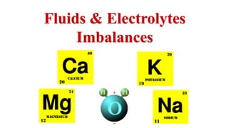
- 33. Sodium Reference Range: 135 – 145 mEq/L
- 34. Sodium • Accounts for 90% of ECF cations. • Almost all Na+ is found in ECF; 10% in ICF. • Na+ attracts fluid + helps preserve ECF volume/fluid distribution. • Na+ helps transmit impulses in nerve/muscle fibers, & combines w/ Cl- & HCO3 to regulate acid-base balance
- 35. Sodium • Excreted mainly via the kidneys (GU) – Also via the GI tract and perspiration • Increased Na+ levels trigger thirst & ADH • Sodium-Potassium pump helps maintain normal Na+ levels – Pump also creates an electrical charge for both cardiac & neuromuscular function
- 36. Sodium Hyponatremia is Na+ < 135 Hypernatremia is Na+ > 145
- 37. Hyponatremia Causes an osmotic fluid shift from plasma into brain cells
- 38. Hyponatremia Signs & Symptoms: Nausea/Vomiting Headache Malaise Confusion Diminished Reflexes Confusion Convulsions Stupor or Coma
- 39. Hyponatremia Causes: • ↑ Vasopressin/ADH • SIADH • Adrenal Insufficiency • Diuretics • Hypervolemia • Liver Failure • Heart Failure
- 40. Hyponatremia Treatment: • Administration of oral or IV Na+ (3%) Supplements • Encourage foods high in Na+ • Fluid restriction • Monitor Neuro Status • Monitor for Arrhythmias • Normovolemic hyponatremia – Vaprisol (conivaptan) – IV infusion – Samsca (tolvaptan) - PO
- 41. Hypernatremia Causes: • Dehydration/Hypovolemia • Diabetes Insipidus • Ingestion of Hypertonic Solutions • IV Infusion of Hypertonic Solutions • Cushing’s Syndrome • Hyperaldosteronism • Loss of pure water (excessive sweating or respiratory infections)
- 42. Hypernatremia Signs & symptoms • Thirst • Lethargy • Neurologic Dysfunction (d/t dehydration of brain cells) – Irritability – Weakness – Seizures – Coma • Edema • Decreased vascular volume
- 43. Hypernatremia Treatment: • Administration of IV Fluids – (Isotonic Salt-Free) • Encourage foods low in Na+ • Push P.O. Fluids • Monitor Neuro Status • Monitor for Arrhythmias
- 44. Potassium Reference Range: 3.5 – 5.0 mEq/L
- 45. Potassium Potassium is gained by intake and lost by excretion. If either is altered, hyperkalemia or hypokalemia may result! Regulated by aldosterone and insulin
- 46. Potassium Potassium levels directly affect cell, nerve, & muscle function: – Maintains electrical neutrality and osmolality of cells – Aids in neuromuscular transmission of nerve impulses – Assists skeletal & cardiac muscle contraction and electrical conductivity – Affects acid-base balance in relationship to H+ (another cation)
- 47. Potassium Hypokalemia is K+ < 3.5 Hyperkalemia is K+ > 5.o
- 48. Hypokalemia Levels < 3.5 Mildly Low Levels usually asymptomatic If level < 3.2, usually accompanied by symptoms
- 49. Hypokalemia Causes of Hypokalemia: Increased Urine Output Malnutrition Vomiting and/or Diarrhea Hypomagnesemia DKA
- 50. Hypokalemia May be a result of acid-base imbalances = alkalosis • In alkalosis, K+ moves into cell to maintain balance, -may lead to hypokalemia
- 52. Treatment • Oral or IV Potassium Chloride Replacement • D/C or adjust medications that may cause hypokalemia • Reverse alkalosis, if cause • Monitor closely for arrhythmias • Monitor Respiratory Status • Monitor LOC • Monitor GI symptoms
- 53. Hyperkalemia Levels > 5.0 Mildly elevated levels usually asymptomatic Levels > 8.0 Disturbances in cardiac conduction occur
- 54. Hyperkalemia Causes: • Renal Failure • Meds (ACEIs, ARBs, K+ sparing diuretics, NSAIDs) • Addison’s Disease • Aldosterone Insufficiencies • Dig Overdose • Beta-Blocker Therapy
- 55. Hyperkalemia May be a result of acid-base imbalances = acidosis In acidosis, excess [H+] move into cells & push K+ into ECF, - may lead to hyperkalemia as K+ moves out of cell to maintain balance.
- 57. Hyperkalemia Treatment: Medications: – Cation-exchange resins (bind with K+ and excreted via feces) – IVP insulin & glucose (K+ binds to insulin) – IV Ca++ (protect the heart from the effects of hyperkalemia) – Sodium bicarbonate (to reverse acidosis) – Diuretics (non-K+ sparing) – Beta2 Adrenergic agonists (epinephrine, albuterol) D/C meds that may cause hyperkalemia Restrict foods with K+ Dialysis for renal failure Monitor closely for arrhythmias Monitor Blood Pressure Monitor GI symptoms
- 58. Calcium Reference Range: 8.5 – 10.5 mg/dl
- 59. Calcium • 99% Ca++ in bones; 1% in serum/soft tissue (measured in blood serum levels) • Found in both ECF & ICF • Can be measured in 2 ways: – Total serum calcium (total Ca++in blood) – Ionized calcium level (various forms of Ca++ in ECF) • 41% ECF Ca++ is bound to protein; 9% bound to citrate or other organic ions
- 60. Calcium • Ca++ functions in the following ways: – Responsible for formation of teeth & bones – Helps maintain cell structure & function – Plays a role in cell membrane permeability & impulse transmission – Affects contraction of cardiac, smooth, and skeletal muscle – Participates in blood-clotting process
- 61. Calcium Ca++ helps K+ & Na+ move into and out of cells in the sodium-potassium pump mechanism
- 62. Hypocalcemia Causes: • Vitamin D Deficiency – Vitamin D promotes Ca++ absorption in intestines, resorption from bones, and kidney resorption all of which raise Ca++ levels • Deficiency of parathyroid hormone – Calcitonin, secreted by PTH, helps regulate Ca++ – s absorption of Ca++/enhances excretion by kidneys • Inefficient parathyroid hormone
- 63. Hypocalcemia Manifestations • Tetany • Laryngospasm • Cardiac Arrhythmias • EKG Δ’s → prolonged QT interval
- 64. Hypocalcemia Management… • PO or IV calcium replacement (depends on severity of symptoms or deficiency) • Vitamin D supplement • Encourage foods high in calcium
- 65. Hypercalcemia Causes: • Excessive calcium release • Increased intestinal calcium absorption ** Decreased renal calcium excretion **
- 66. Hypercalcemia Manifestations: • Cardiac Arrhythmias • EKG Δ’s → shortened QT interval
- 67. Hypercalcemia Severe Hypercalcemia (> 15mg/dl) is a… Medical Emergency May result in Coma or Cardiac Arrest
- 68. Hypercalcemia Signs & Symptoms Fatigue Depression Confusion Anorexia N/V Constipation Pancreatitis Increased Urination
- 69. Hypercalcemia Treatment… • Hydration • Increased Salt Intake • Diuretics • Dialysis (renal failure) • Glucocorticoids
- 70. Magnesium Reference Range: 1.3 – 2.3 mEq/L
- 71. Magnesium • 2nd most abundant ICF cation (K+ #1) • 60% Mg+ found in bones, < 1% ECF • Mg+ performs the following functions: – Promotes enzyme reactions in carbohydrate metabolism – Helps produce ADP (adenosine triphosphate) – Helps with protein synthesis – Influences vasodilation (normal CV function) – Helps Na+ and K+ ions cross cell membranes
- 72. Magnesium • Mg+ performs the following functions: – Regulates muscle contractions – Affects irritability and contractility of cardiac and skeletal muscle – Influences Ca++ levels • maintain Ca++ levels in ECF
- 73. Magnesium Hypomagnesemia is Mg+ < 1.8 Hypermagnesemia is Mg+ > 2.4
- 74. Hypomagnesemia Results in cardiac dysrhythmias and irritates the nervous system (tetany)
- 75. Hypomagnesemia Causes: • ETOH Abuse (#1) • Malnutrition • Chronic Diarrhea • Malabsorption • Diuretics • AMI • Pancreatitis
- 76. Hypomagnesemia • Does not produce specific EKG changes • May contribute to arrhythmias caused by digoxin toxicity, ischemia, or K+ imbalances • Monitor: – EKG for Arrhythmias – Muscle cramps
- 77. Hypomagnesemia Replacement of Mg: PO or IV • PO = Mg Oxide 400mg tabs • MgSo4 IV administration is usually given at a rate of 1 gram/hr (1 gram/100 ml) • Encourage foods high in magnesium
- 78. Hypermagnesemia Severe hypermagnesemia is associated with: – AV blocks – Intraventricular conduction disturbances