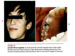
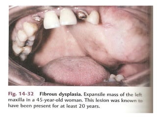
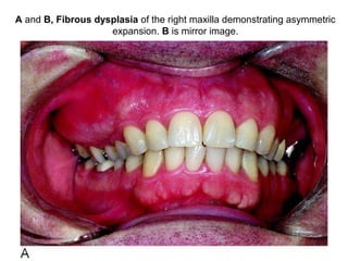
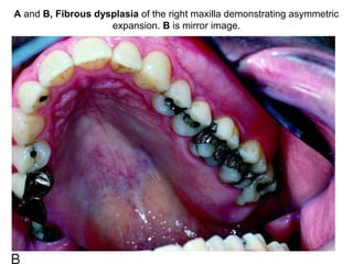
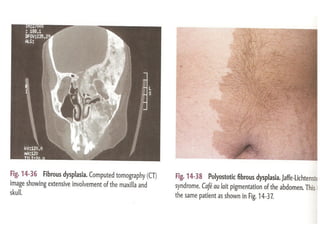
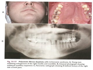
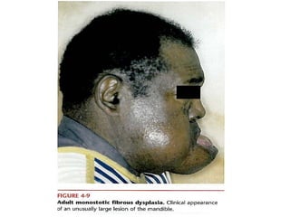
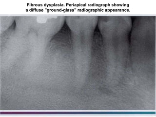
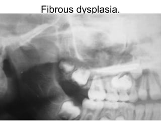
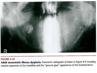
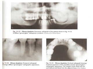
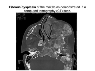
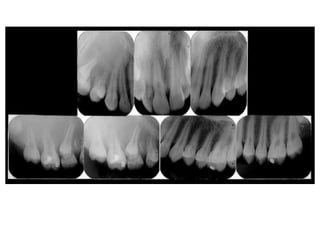
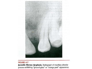
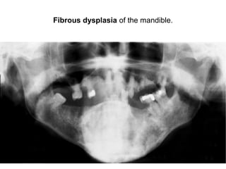
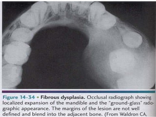
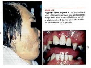
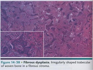
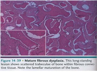
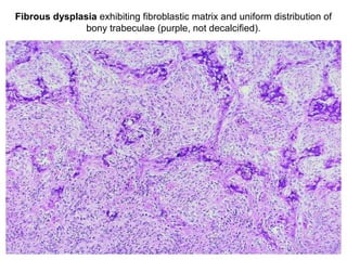
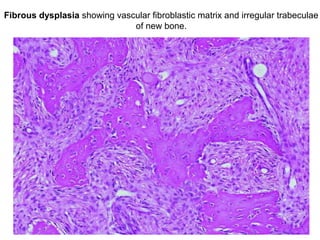
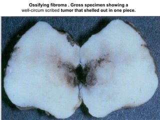
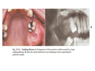
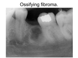
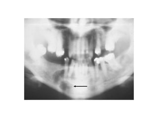
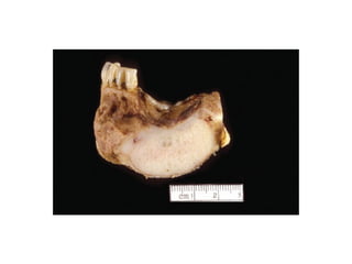
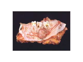
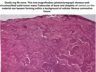
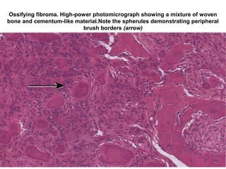
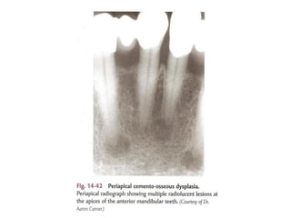
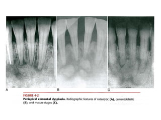
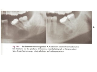
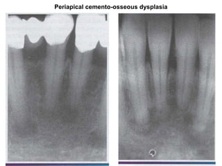
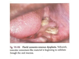
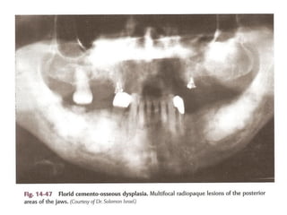
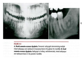
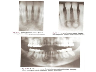
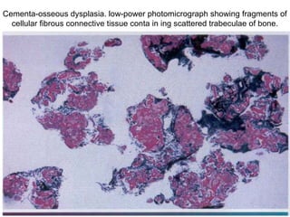
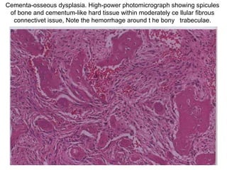
A and B, Fibrous dysplasia of the right maxilla demonstrating asymmetric
expansion. B is mirror image.
A and B, Fibrous dysplasia of the right maxilla demonstrating asymmetric
expansion. B is mirror image.
Fibrous dysplasia. Periapical radiograph showing
a diffuse "ground-glass" radiographic appearance.
Fibrous dysplasia.
Fibrous dysplasia of the maxilla as demonstrated in a
computed tomography (CT) scan.
Fibrous dysplasia of the mandible.
Fibrous dysplasia exhibiting fibroblastic matrix and uniform distribution of
bony trabeculae (purple, not decalcified).
Fibrous dysplasia showing vascular fibroblastic matrix and irregular trabeculae
of new bone.
Ossifying fibroma . Gross specimen showing a
well-circum scribed tumor that shelled out in one piece.
Ossifying fibroma.
Ossify ing fib roma. This low-magnification photomicrograph showsa well-
circumscribed solid tumor mass.Trabeculae of bone and droplets of cement um-like
material can beseen forming within a background of cellular fibrous connective
tissue.
Ossifying fibroma. High-power photomicrograph showing a mixture of woven
bone and cementum-like material.Note the spherules demonstrating peripheral
brush borders (arrow)
Periapical cemento-osseous dysplasia
Cementa-osseous dysplasia. low-power photomicrograph showing fragments of
cellular fibrous connective tissue conta in ing scattered trabeculae of bone.
Cementa-osseous dysplasia. High-power photomicrograph showing spicules
of bone and cementum-like hard tissue within moderately ce llular fibrous
connectivet issue, Note the hemorrhage around t he bony trabeculae.

Fibro osseous lesion dina patho