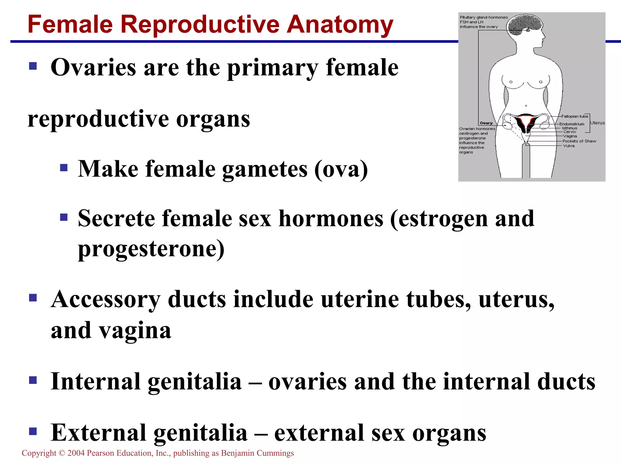
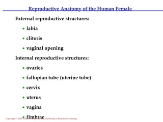
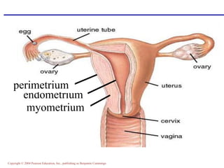
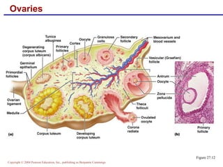
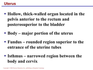
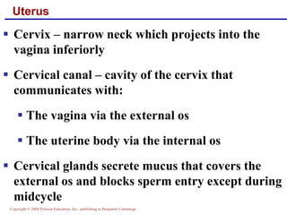
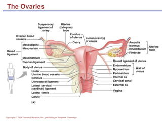
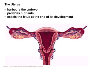
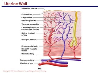
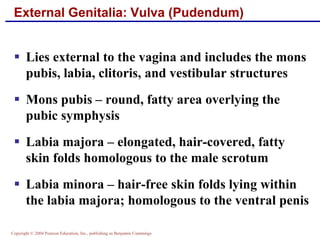
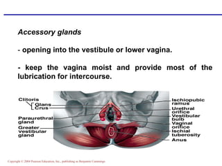
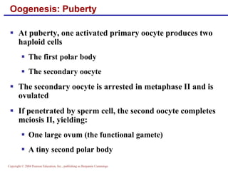
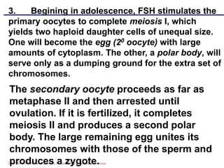
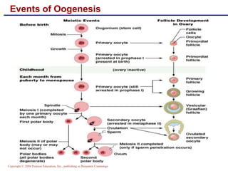
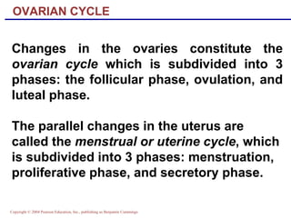
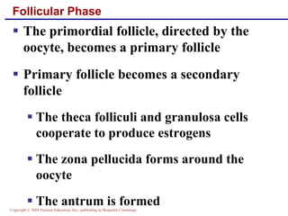
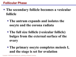
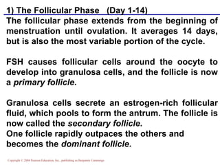
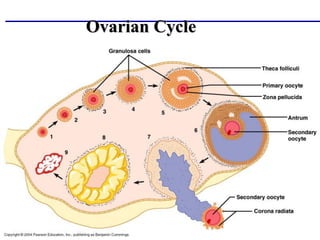
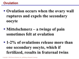
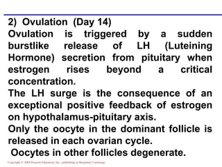
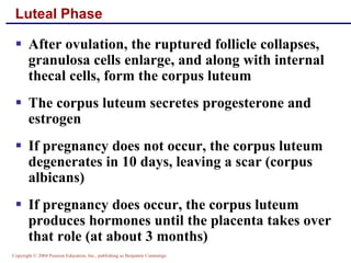
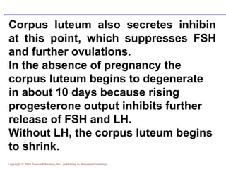
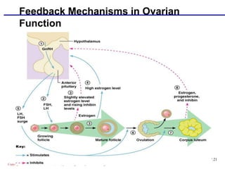
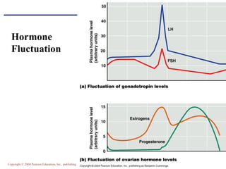
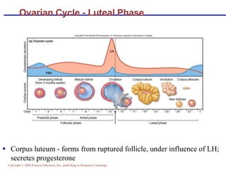
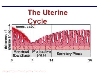
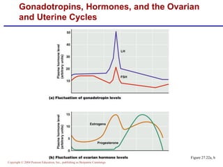
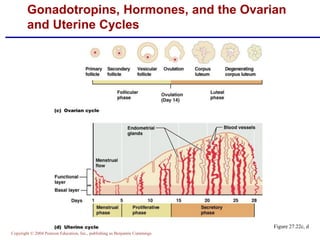
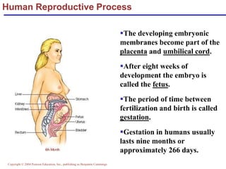
Copyright © 2004 Pearson Education, Inc., publishing as Benjamin Cummings
Female Reproductive Anatomy
▪ Ovaries are the primary female
reproductive organs
▪ Make female gametes (ova)
▪ Secrete female sex hormones (estrogen and
progesterone)
▪ Accessory ducts include uterine tubes, uterus,
and vagina
▪ Internal genitalia – ovaries and the internal ducts
▪ External genitalia – external sex organs
Copyright © 2004 Pearson Education, Inc., publishing as Benjamin Cummings
Reproductive Anatomy of the Human Female
External reproductive structures:
• labia
• clitoris
• vaginal opening
Internal reproductive structures:
• ovaries
• fallopian tube (uterine tube)
• cervix
• uterus
• vagina
• fimbrae
Copyright © 2004 Pearson Education, Inc., publishing as Benjamin Cummings
endometrium
myometrium
perimetrium
Copyright © 2004 Pearson Education, Inc., publishing as Benjamin Cummings
The Ovaries
▪ Paired organs on each side of the uterus held in
place by several ligaments
▪ Ovarian – anchors the ovary medially to the
uterus
▪ Suspensory – anchors the ovary laterally to the
pelvic wall
▪ Mesovarium – suspends the ovary in between
▪ Broad ligament – contains the suspensory
ligament and the mesovarium
Copyright © 2004 Pearson Education, Inc., publishing as Benjamin Cummings
The Ovaries
Figure 27.14a
Copyright © 2004 Pearson Education, Inc., publishing as Benjamin Cummings
Ovaries
▪ Blood supply – ovarian arteries and the
ovarian branch of the uterine artery
▪ They are surrounded by a fibrous tunica
albuginea, which is covered by a layer of
epithelial cells called the germinal
epithelium
▪ Embedded in the ovary cortex are ovarian
follicles
Copyright © 2004 Pearson Education, Inc., publishing as Benjamin Cummings
Ovaries
▪ Each follicle consists of an immature
germ cell called an oocyte
▪ Cells around the oocyte are called:
▪ Follicle cells (one cell layer thick)
▪ Granulosa cells (when more than one
layer is present)
Copyright © 2004 Pearson Education, Inc., publishing as Benjamin Cummings
Ovaries
▪ Primordial follicle – one layer of
squamouslike follicle cells surrounds
the oocyte
▪ Primary follicle – two or more layers
of cuboidal granulosa cells enclose the
oocyte
▪ Secondary follicle – has a fluid-filled
space between granulosa cells that
coalesces to form a central antrum
Copyright © 2004 Pearson Education, Inc., publishing as Benjamin Cummings
Ovaries
▪ Graafian follicle – secondary follicle at
its most mature stage that bulges from
the surface of the ovary
▪ Ovulation – ejection of the oocyte from
the ripening follicle
▪ Corpus luteum – ruptured follicle after
ovulation
Copyright © 2004 Pearson Education, Inc., publishing as Benjamin Cummings
Ovaries
Figure 27.12
Copyright © 2004 Pearson Education, Inc., publishing as Benjamin Cummings
Uterine Tubes (Fallopian Tubes) and Oviducts
▪ Receive the ovulated oocyte and provide a
site for fertilization
▪ Empty into the superolateral region of the
uterus via the isthmus
▪ Expand distally around the ovary forming
the ampulla
▪ The ampulla ends in the funnel-shaped,
ciliated infundibulum containing fingerlike
projections called fimbriae
Copyright © 2004 Pearson Education, Inc., publishing as Benjamin Cummings
Uterine Tubes
▪ The uterine tubes have no contact with
the ovaries and the ovulated oocyte is
cast into the peritoneal cavity
▪ Beating cilia on the fimbriae create
currents to carry the oocyte into the
uterine tube
▪ The oocyte is carried toward the
uterus by peristalsis and ciliary action
Copyright © 2004 Pearson Education, Inc., publishing as Benjamin Cummings
Uterine Tubes
▪ Nonciliated cells keep the oocyte and
the sperm nourished and moist
▪ Mesosalpinx – visceral peritoneum
that supports the uterine tubes
Copyright © 2004 Pearson Education, Inc., publishing as Benjamin Cummings
Uterus
▪ Hollow, thick-walled organ located in the
pelvis anterior to the rectum and
posterosuperior to the bladder
▪ Body – major portion of the uterus
▪ Fundus – rounded region superior to the
entrance of the uterine tubes
▪ Isthmus – narrowed region between the
body and cervix
Copyright © 2004 Pearson Education, Inc., publishing as Benjamin Cummings
Uterus
▪ Cervix – narrow neck which projects into the
vagina inferiorly
▪ Cervical canal – cavity of the cervix that
communicates with:
▪ The vagina via the external os
▪ The uterine body via the internal os
▪ Cervical glands secrete mucus that covers the
external os and blocks sperm entry except during
midcycle
Copyright © 2004 Pearson Education, Inc., publishing as Benjamin Cummings
The Ovaries
Copyright © 2004 Pearson Education, Inc., publishing as Benjamin Cummings
The Uterus
• harbours the embryo
• provides nutrients
• expels the fetus at the end of its development
Copyright © 2004 Pearson Education, Inc., publishing as Benjamin Cummings
Uterine Wall
▪ Composed of three layers
▪ Perimetrium – outermost serous
layer; the visceral peritoneum
▪ Myometrium – middle layer;
interlacing layers of smooth muscle
▪ Endometrium – mucosal lining of the
uterine cavity
Copyright © 2004 Pearson Education, Inc., publishing as Benjamin Cummings
Endometrium
▪ Has numerous uterine glands that change in length
as the endometrial thickness changes
▪ Stratum functionalis:
▪ Undergoes cyclic changes in response to ovarian
hormones
▪ Is shed during menstruation
▪ Stratum basalis:
▪ Forms a new functionalis after menstruation ends
▪ Does not respond to ovarian hormones
Copyright © 2004 Pearson Education, Inc., publishing as Benjamin Cummings
Uterine Vascular Supply
▪ Uterine arteries – arise from the internal iliacs,
ascend the sides of the uterus and send branches
into the uterine wall
▪ Arcuate arteries – branches of the uterine
arteries in the myometrium that give rise to
radial branches
▪ Radial branches – descend into the endometrium
and give off:
▪ Spiral arteries to the stratum functionalis
▪ Straight arteries to the stratum basalis
Copyright © 2004 Pearson Education, Inc., publishing as Benjamin Cummings
Uterine Vascular Supply
▪ Degeneration and regeneration of
spiral arteries causes the functionalis
to shed during menstruation
▪ Veins of the endometrium are thin-
walled with occasional sinusoidal
enlargements
Copyright © 2004 Pearson Education, Inc., publishing as Benjamin Cummings
Uterine Wall
Copyright © 2004 Pearson Education, Inc., publishing as Benjamin Cummings
Vagina
▪ Thin-walled tube lying between the bladder
and the rectum, extending from the cervix
to the exterior of the body
▪ The urethra is embedded in the anterior
wall
▪ Provides a passageway for birth, menstrual
flow, and is the organ of copulation
Copyright © 2004 Pearson Education, Inc., publishing as Benjamin Cummings
Vagina
▪ Wall consists of three coats: fibroelastic
adventitia, smooth muscle muscularis, and
a stratified squamous mucosa
▪ Mucosa near the vaginal orifice forms an
incomplete partition called the hymen
▪ Vaginal fornix – upper end of the vagina
surrounding the cervix
Copyright © 2004 Pearson Education, Inc., publishing as Benjamin Cummings
Vagina
Copyright © 2004 Pearson Education, Inc., publishing as Benjamin Cummings
External Genitalia: Vulva (Pudendum)
▪ Lies external to the vagina and includes the mons
pubis, labia, clitoris, and vestibular structures
▪ Mons pubis – round, fatty area overlying the
pubic symphysis
▪ Labia majora – elongated, hair-covered, fatty
skin folds homologous to the male scrotum
▪ Labia minora – hair-free skin folds lying within
the labia majora; homologous to the ventral penis
Copyright © 2004 Pearson Education, Inc., publishing as Benjamin Cummings
Accessory glands
- opening into the vestibule or lower vagina.
- keep the vagina moist and provide most of the
lubrication for intercourse.
Copyright © 2004 Pearson Education, Inc., publishing as Benjamin Cummings
A.Oogenesis
1. Egg production is called oogenesis,
which is a distinctly cyclic event.
2. Most primary oocytes undergo a
process of degeneration called atresia.
Only 2 million remain at the time of birth,
and by puberty, only 400,000 remain.
OOGENESIS
Copyright © 2004 Pearson Education, Inc., publishing as Benjamin Cummings
Oogenesis
▪ Production of female sex cells by meiosis
▪ In the fetal period, oogonia (2n ovarian stem
cells) multiply by mitosis and store nutrients
▪ Primordial follicles appear as oogonia are
transformed into primary oocytes
▪ Primary oocytes begin meiosis but stall in
prophase I
Copyright © 2004 Pearson Education, Inc., publishing as Benjamin Cummings
Oogenesis: Puberty
▪ At puberty, one activated primary oocyte produces two
haploid cells
▪ The first polar body
▪ The secondary oocyte
▪ The secondary oocyte is arrested in metaphase II and is
ovulated
▪ If penetrated by sperm cell, the second oocyte completes
meiosis II, yielding:
▪ One large ovum (the functional gamete)
▪ A tiny second polar body
Copyright © 2004 Pearson Education, Inc., publishing as Benjamin Cummings
3. Begining in adolescence, FSH stimulates the
primary oocytes to complete meiosis I, which
yields two haploid daughter cells of unequal size.
One will become the egg (20 oocyte) with large
amounts of cytoplasm. The other, a polar body, will
serve only as a dumping ground for the extra set of
chromosomes.
The secondary oocyte proceeds as far as
metaphase II and then arrested until
ovulation. If it is fertilized, it completes
meiosis II and produces a second polar
body. The large remaining egg unites its
chromosomes with those of the sperm and
produces a zygote.
Copyright © 2004 Pearson Education, Inc., publishing as Benjamin Cummings
Events of Oogenesis
Copyright © 2004 Pearson Education, Inc., publishing as Benjamin Cummings
Changes in the ovaries constitute the
ovarian cycle which is subdivided into 3
phases: the follicular phase, ovulation, and
luteal phase.
The parallel changes in the uterus are
called the menstrual or uterine cycle, which
is subdivided into 3 phases: menstruation,
proliferative phase, and secretory phase.
OVARIAN CYCLE
Copyright © 2004 Pearson Education, Inc., publishing as Benjamin Cummings
Follicular Phase
▪ The primordial follicle, directed by the
oocyte, becomes a primary follicle
▪ Primary follicle becomes a secondary
follicle
▪ The theca folliculi and granulosa cells
cooperate to produce estrogens
▪ The zona pellucida forms around the
oocyte
▪ The antrum is formed
Copyright © 2004 Pearson Education, Inc., publishing as Benjamin Cummings
Follicular Phase
▪ The secondary follicle becomes a vesicular
follicle
▪ The antrum expands and isolates the
oocyte and the corona radiata
▪ The full size follicle (vesicular follicle)
bulges from the external surface of the
ovary
▪ The primary oocyte completes meiosis I,
and the stage is set for ovulation
Copyright © 2004 Pearson Education, Inc., publishing as Benjamin Cummings
1) The Follicular Phase (Day 1-14)
The follicular phase extends from the beginning of
menstruation until ovulation. It averages 14 days,
but is also the most variable portion of the cycle.
FSH causes follicular cells around the oocyte to
develop into granulosa cells, and the follicle is now
a primary follicle.
Granulosa cells secrete an estrogen-rich follicular
fluid, which pools to form the antrum. The follicle is
now called the secondary follicle.
One follicle rapidly outpaces the others and
becomes the dominant follicle.
Copyright © 2004 Pearson Education, Inc., publishing as Benjamin Cummings
Ovarian Cycle
Copyright © 2004 Pearson Education, Inc., publishing as Benjamin Cummings
Ovulation
▪ Ovulation occurs when the ovary wall
ruptures and expels the secondary
oocyte
▪ Mittelschmerz – a twinge of pain
sometimes felt at ovulation
▪ 1-2% of ovulations release more than
one secondary oocyte, which if
fertilized, results in fraternal twins
Copyright © 2004 Pearson Education, Inc., publishing as Benjamin Cummings
2) Ovulation (Day 14)
Ovulation is triggered by a sudden
burstlike release of LH (Luteining
Hormone) secretion from pituitary when
estrogen rises beyond a critical
concentration.
The LH surge is the consequence of an
exceptional positive feedback of estrogen
on hypothalamus-pituitary axis.
Only the oocyte in the dominant follicle is
released in each ovarian cycle.
Oocytes in other follicles degenerate.
Copyright © 2004 Pearson Education, Inc., publishing as Benjamin Cummings
Luteal Phase
▪ After ovulation, the ruptured follicle collapses,
granulosa cells enlarge, and along with internal
thecal cells, form the corpus luteum
▪ The corpus luteum secretes progesterone and
estrogen
▪ If pregnancy does not occur, the corpus luteum
degenerates in 10 days, leaving a scar (corpus
albicans)
▪ If pregnancy does occur, the corpus luteum
produces hormones until the placenta takes over
that role (at about 3 months)
Copyright © 2004 Pearson Education, Inc., publishing as Benjamin Cummings
Corpus luteum also secretes inhibin
at this point, which suppresses FSH
and further ovulations.
In the absence of pregnancy the
corpus luteum begins to degenerate
in about 10 days because rising
progesterone output inhibits further
release of FSH and LH.
Without LH, the corpus luteum begins
to shrink.
Copyright © 2004 Pearson Education, Inc., publishing as Benjamin Cummings
If pregnancy occurs, the corpus luteum
continues to secrete progesterone and
estrogen for about 3 months under the
stimulation of LH-like hormone
released by the developing embryo.
The secretion by corpus luteum does
not stop until the placenta is ready to
take over its homone-producing duties.
This is known as corpus luteum
rescue.
Copyright © 2004 Pearson Education, Inc., publishing as Benjamin Cummings
Establishing the Ovarian Cycle
▪ During childhood, ovaries grow and secrete
small amounts of estrogens that inhibit the
hypothalamic release of GnRH
▪ As puberty nears, GnRH is released; FSH
and LH are released by the pituitary, which
act on the ovaries
▪ These events continue until an adult cyclic
pattern is achieved and menarche occurs
Copyright © 2004 Pearson Education, Inc., publishing as Benjamin Cummings
Feedback Mechanisms in Ovarian
Function
Figure 27.21
Copyright © 2004 Pearson Education, Inc., publishing as Benjamin Cummings
Hormone
Fluctuation
Copyright © 2004 Pearson Education, Inc., publishing as Benjamin Cummings
Ovarian Cycle - Luteal Phase
▪ Corpus luteum - forms from ruptured follicle, under influence of LH;
secretes progesterone
Copyright © 2004 Pearson Education, Inc., publishing as Benjamin Cummings
The Uterine
Cycle
Copyright © 2004 Pearson Education, Inc., publishing as Benjamin Cummings
Menstrual (Uterine) Cycle
1) Menstruation (Day 1-5)
The superficial layer stratum
functionalis of the uterus detaches
from the uterine wall, accompanied by
bleeding for 3-5 days.
Sex hormones are at their lowest
normal levels at Day 1
Menstrual fluid contains fibrolysin,
therefore it normally does not clot.
Copyright © 2004 Pearson Education, Inc., publishing as Benjamin Cummings
2) Proliferative Phase (Day 6-14)
Estrogen stimulates mitosis, the prolific growth of
blood vessels, and the formation of a new stratum
functionalis.
Estrogen also stimulates the endometrium to
develop progesterone receptors.
As ovulation approaches, the uterine tube becomes
edematous, its fimbriae develop and caress the
ovary, and its cilia create a gentle current in the
nearby peritoneal fluid.
Copyright © 2004 Pearson Education, Inc., publishing as Benjamin Cummings
The ovulated egg is usually caught up in this current and
swept into the tube.
An oocyte has only 24 hours to be fertilized.
The chance of fertilization is enhanced by changes in the
cervical mucus at the time of ovulation.
It becomes thinner and more stringy.
Copyright © 2004 Pearson Education, Inc., publishing as Benjamin Cummings
Menstrual Cycle - Proliferative
Phase
▪ Day 6-14 rebuild endometrial tissue
▪ mitosis occurs in stratum basalis
▪ result of estrogen from developing follicles
Copyright © 2004 Pearson Education, Inc., publishing as Benjamin Cummings
3) Secretory Phase (Day 15-28)
a. In response to rising level of
progesterone, the endometrium of the
uterus proliferates further in preparation for
possible pregnancy.
Spiral arteries elaborate and coil more
tightly
Uterine glands enlarge, coil, and begin
secreting nutritients into the uterine cavity
to sustain the embryo until implantation.
The cervical mucus becomes viscous,
forming the cervical plug, which prevents
sperm entry.
Copyright © 2004 Pearson Education, Inc., publishing as Benjamin Cummings
▪ Further thickening of endometrium due to secretion and fluid
accumulation -- not mitosis
▪ Due to progesterone stimulation of glands
Menstrual Cycle - Secretory Phase
Copyright © 2004 Pearson Education, Inc., publishing as Benjamin Cummings
If pregnancy does not occur towards
the end of the secretory phase, LH
level drops due to negative feedback
of high level of progesterone.
Progesterone level decline following
the drop of LH. Without the support
of progesterone, the endometrium
undergoes degeneration in the
following sequence.
Copyright © 2004 Pearson Education, Inc., publishing as Benjamin Cummings
The spiral arteries close due to continuous
and intensive constriction (spasm).
The superficial layer stratum functionalis of
the uterus is deprived of blood supply.
The endometrial cells die of ischemia.
The spiral arteries suddenly relax and open
wide.
Blood gushes into the weakened capillary
beds, causing the capillaries to fragment
and the stratum functionalis to slough off.
Copyright © 2004 Pearson Education, Inc., publishing as Benjamin Cummings
Menstrual Cycle Premenstrual
Phase
▪ Involution of corpus luteum, progesterone falls
▪ spiral arteries constrict causes endometrial ischemia
▪ stratum functionalis sloughs
Copyright © 2004 Pearson Education, Inc., publishing as Benjamin Cummings
Menstrual Cycle - Menstrual
Phase
▪ Blood, serous fluid and endometrial tissue are discharged
Copyright © 2004 Pearson Education, Inc., publishing as Benjamin Cummings
Menses
▪ If fertilization does not occur, progesterone levels fall,
depriving the endometrium of hormonal support
▪ Spiral arteries kink and go into spasms and endometrial
cells begin to die
▪ The functional layer begins to digest itself
▪ Spiral arteries constrict one final time then suddenly
relax and open wide
▪ The rush of blood fragments weakened capillary beds
and the functional layer sloughs
Copyright © 2004 Pearson Education, Inc., publishing as Benjamin Cummings
▪ c. The menstrual cycle starts over again on this
first day of vaginal discharge.
Copyright © 2004 Pearson Education, Inc., publishing as Benjamin Cummings
Figure 27.22a, b
Gonadotropins, Hormones, and the Ovarian
and Uterine Cycles
Copyright © 2004 Pearson Education, Inc., publishing as Benjamin Cummings
Figure 27.22c, d
Gonadotropins, Hormones, and the Ovarian
and Uterine Cycles
Copyright © 2004 Pearson Education, Inc., publishing as Benjamin Cummings
▪ Estrogen levels rise during puberty
▪ Promote oogenesis and follicle growth in the ovary
▪ Exert anabolic effects on the female reproductive
tract
▪ Uterine tubes, uterus, and vagina grow larger and
become functional
▪ Uterine tubes and uterus exhibit enhanced motility
▪ Vaginal mucosa thickens and external genitalia
mature
Extrauterine Effects of
Estrogens and Progesterone
Copyright © 2004 Pearson Education, Inc., publishing as Benjamin Cummings
▪ Growth of the breasts
▪ Increased deposition of subcutaneous fat,
especially in the hips and breasts
▪ Widening and lightening of the pelvis
▪ Growth of axillary and pubic hair
▪ Stimulate bone growth
▪ Increases HDL and lowers LDL
Estrogen-Induced Secondary Sex
Characteristics
Copyright © 2004 Pearson Education, Inc., publishing as Benjamin Cummings
FERTILIZATION
▪The fusion of sperm cell
nucleus and an egg cell
nucleus is known as
fertilization. This process
takes place in the distal
portion of the uterine tube
called the ampulla.
▪ The fertilized egg is now
refered to as a zygote.
▪The appropriate number
of chromosomes has also
been restored.
This process takes place
within 48hrs of ovulation .
Once the sperm cell
penetrate the ovum, the
2nd polar body is extruded
and the fertilized ovum
begins to divide.
Copyright © 2004 Pearson Education, Inc., publishing as Benjamin Cummings
Accomplishing Fertilization
▪ The oocyte is viable for 12 to 24 hours
▪ Sperm is viable 24 to 72 hours
▪ For fertilization to occur, coitus must occur no
more than:
▪ Three days before ovulation
▪ 24 hours after ovulation
▪ Fertilization – when a sperm fuses with an egg
to form a zygote
Copyright © 2004 Pearson Education, Inc., publishing as Benjamin Cummings
Sperm Transport and Capacitation
▪ Fates of ejaculated sperm
▪ Leak out of the vagina immediately after
deposition
▪ Destroyed by the acidic vaginal environment
▪ Fail to make it through the cervix
▪ Dispersed in the uterine cavity or destroyed by
phagocytic leukocytes
▪ Reach the uterine tubes
▪ Sperm must undergo capacitation before they
can penetrate the oocyte
Copyright © 2004 Pearson Education, Inc., publishing as Benjamin Cummings
Acrosomal Reaction and Sperm Penetration
▪ An ovulated oocyte is encapsulated by:
▪ The corona radiata and zona pellucida
▪ Extracellular matrix
▪ Sperm binds to the zona pellucida and
undergoes the acrosomal reaction
▪ Enzymes are released near the oocyte
▪ Hundreds of acrosomes release their
enzymes to digest the zona pellucida
Copyright © 2004 Pearson Education, Inc., publishing as Benjamin Cummings
Acrosomal Reaction and Sperm Penetration
▪Once a sperm makes contact
with the oocyte’s membrane:
▪Beta protein finds and binds to
receptors on the oocyte
membrane
▪Alpha protein causes it to insert
into the membrane
Copyright © 2004 Pearson Education, Inc., publishing as Benjamin Cummings
Acrosomal Reaction and Sperm Penetration
Figure 28.2a
Copyright © 2004 Pearson Education, Inc., publishing as Benjamin Cummings
Blocks to Polyspermy
▪ Only one sperm is allowed to penetrate the oocyte
▪ Two mechanisms ensure monospermy
▪ Fast block to polyspermy – membrane
depolarization prevents sperm from fusing with
the oocyte membrane
▪ Slow block to polyspermy – zonal inhibiting
proteins (ZIPs):
▪ Destroy sperm receptors
▪ Cause sperm already bound to receptors to
detach
Copyright © 2004 Pearson Education, Inc., publishing as Benjamin Cummings
IMPLANTATION
▪ The zygote passes down the fallopian tube
and enters into the uterus.
▪ Continuous cell divisions result in the
formation of a hollow ball of cells called a
blastocyst.
▪ The zygote obtains nutrients from fluids
secreted by the mother.
▪ On the sixth day, the zygote/ball of cells
implants in the uterine wall.
Copyright © 2004 Pearson Education, Inc., publishing as Benjamin Cummings
Implantation
▪ Viability of the corpus luteum is maintained by
human chorionic gonadotropin (hCG) secreted by
the trophoblasts.
▪ HCG prompts the corpus luteum to continue to
secrete progesterone and estrogen
▪ Chorion – developed from trophoblasts after
implantation, continues this hormonal stimulus
▪ Between the second and third month, the placenta:
▪ Assumes the role of progesterone and estrogen
production
▪ Is providing nutrients and removing wastes
Copyright © 2004 Pearson Education, Inc., publishing as Benjamin Cummings
PREGNANCY
▪ Pregnancy – events that occur from fertilization until
the infant is born
▪ Conceptus – the developing offspring
▪ Gestation period – from the last menstrual period
until birth
▪ Preembryo – conceptus from fertilization until it is
two weeks old
▪ Embryo – conceptus during the third through the
eighth week
▪ Fetus – conceptus from the 9th wk till birth
Copyright © 2004 Pearson Education, Inc., publishing as Benjamin Cummings
EVENTS OF EARLY PREGNANCY
EVENT DAYS AFTER
OVULATION
OVULATION 0
FERTILIZATION 1-2
BLASTOCYSTS enters uterine
cavity
4
IMPLANTATION 5
TROPHOBLAST forms and
attaches to endometrium
6
TROPHOBLAST begins to
secreteHCG
8
HCG rescues corpus luteum 10
Copyright © 2004 Pearson Education, Inc., publishing as Benjamin Cummings
EMBRYONIC DEVELOPMENT
▪The zygote undergoes a series of rapid mitotic cell
divisions called cleavage.
▪The process begins by the zygote dividing into two
identical cells, followed by four, then eight, ect.
▪Three cell layers are formed and are called primary
germ layers.
▪This begin the process of forming all the tissues,
organs and systems in an animal and is known as
differentiation.
Copyright © 2004 Pearson Education, Inc., publishing as Benjamin Cummings
Human Reproductive Process
▪The developing embryonic
membranes become part of the
placenta and umbilical cord.
▪After eight weeks of
development the embryo is
called the fetus.
▪The period of time between
fertilization and birth is called
gestation.
▪Gestation in humans usually
lasts nine months or
approximately 266 days.
Copyright © 2004 Pearson Education, Inc., publishing as Benjamin Cummings
PREGNANCY HORMONES
▪ 1ST TRIMESTER – HCG,
PROGESTERONE,OESTROGEN, RELAXIN.
▪ 2ND TRIMESTER – HPL, STEROID
HORMONES
▪ 3RD TRIMESTER – CROSSOVER OF
OESTROGEN, HPL, PROLACTIN, RELAXIN
Copyright © 2004 Pearson Education, Inc., publishing as Benjamin Cummings
Hormonal Changes During Pregnancy
Figure 28.6
Copyright © 2004 Pearson Education, Inc., publishing as Benjamin Cummings
PARTURITION
▪ The delivery of the foetus – 40weeks after the onset
of the last menstrual period.
▪ Once the foetus reaches a critical size, ditention of
the uterus increases contractility
▪ Uncoordinated contractions known as Braxton
Hicks contractions begins- about a month before
parturition
▪ Oestrogen/progesterone ratio increases which
increases the sensitivity of of the uterus to
contracting stimuli
Copyright © 2004 Pearson Education, Inc., publishing as Benjamin Cummings
Parturition..........
▪ Oestrogen stimulates local
production of prostaglandins
which increases the intracellular
Ca++ concentration of uterine
smooth muscle. Also, increases the
sensitivity of oxytocin receptors.
▪ Oxytocin stimulates powerful
uterine contraction
Copyright © 2004 Pearson Education, Inc., publishing as Benjamin Cummings
Parturition: Initiation of Labor
Figure 28.16
Copyright © 2004 Pearson Education, Inc., publishing as Benjamin Cummings
3 Stages of labour
▪ Ist stage: uterine contractions originating
at the fundus and sweeping downwards
move the head of the foetus toward the
cervix and progressively widens the cervix
▪ 2nd stage: the foetus is forced through the
cervix and delivered per vagina
▪ 3rd stage: the placenta separates from the
uterine decidual tissue and its delivered.
Copyright © 2004 Pearson Education, Inc., publishing as Benjamin Cummings
83
Lactation
▪ Physiology of lactation
▪ Changes during pregnancy
▪ Increased breast tissue
▪ Maturation of structure
▪ Hormonal controls
▪ Prolactin: stimulates milk production
▪ Oxytocin: stimulates milk release
▪ “let-down” reflex
Copyright © 2004 Pearson Education, Inc., publishing as Benjamin Cummings
84
Copyright © 2004 Pearson Education, Inc., publishing as Benjamin Cummings

FEMALE REPRODUCTIVE PHYSIOLOGY .pdf

  • 1.
    Copyright © 2004Pearson Education, Inc., publishing as Benjamin Cummings Female Reproductive Anatomy ▪ Ovaries are the primary female reproductive organs ▪ Make female gametes (ova) ▪ Secrete female sex hormones (estrogen and progesterone) ▪ Accessory ducts include uterine tubes, uterus, and vagina ▪ Internal genitalia – ovaries and the internal ducts ▪ External genitalia – external sex organs
  • 2.
    Copyright © 2004Pearson Education, Inc., publishing as Benjamin Cummings Reproductive Anatomy of the Human Female External reproductive structures: • labia • clitoris • vaginal opening Internal reproductive structures: • ovaries • fallopian tube (uterine tube) • cervix • uterus • vagina • fimbrae
  • 3.
    Copyright © 2004Pearson Education, Inc., publishing as Benjamin Cummings endometrium myometrium perimetrium
  • 4.
    Copyright © 2004Pearson Education, Inc., publishing as Benjamin Cummings The Ovaries ▪ Paired organs on each side of the uterus held in place by several ligaments ▪ Ovarian – anchors the ovary medially to the uterus ▪ Suspensory – anchors the ovary laterally to the pelvic wall ▪ Mesovarium – suspends the ovary in between ▪ Broad ligament – contains the suspensory ligament and the mesovarium
  • 5.
    Copyright © 2004Pearson Education, Inc., publishing as Benjamin Cummings The Ovaries Figure 27.14a
  • 6.
    Copyright © 2004Pearson Education, Inc., publishing as Benjamin Cummings Ovaries ▪ Blood supply – ovarian arteries and the ovarian branch of the uterine artery ▪ They are surrounded by a fibrous tunica albuginea, which is covered by a layer of epithelial cells called the germinal epithelium ▪ Embedded in the ovary cortex are ovarian follicles
  • 7.
    Copyright © 2004Pearson Education, Inc., publishing as Benjamin Cummings Ovaries ▪ Each follicle consists of an immature germ cell called an oocyte ▪ Cells around the oocyte are called: ▪ Follicle cells (one cell layer thick) ▪ Granulosa cells (when more than one layer is present)
  • 8.
    Copyright © 2004Pearson Education, Inc., publishing as Benjamin Cummings Ovaries ▪ Primordial follicle – one layer of squamouslike follicle cells surrounds the oocyte ▪ Primary follicle – two or more layers of cuboidal granulosa cells enclose the oocyte ▪ Secondary follicle – has a fluid-filled space between granulosa cells that coalesces to form a central antrum
  • 9.
    Copyright © 2004Pearson Education, Inc., publishing as Benjamin Cummings Ovaries ▪ Graafian follicle – secondary follicle at its most mature stage that bulges from the surface of the ovary ▪ Ovulation – ejection of the oocyte from the ripening follicle ▪ Corpus luteum – ruptured follicle after ovulation
  • 10.
    Copyright © 2004Pearson Education, Inc., publishing as Benjamin Cummings Ovaries Figure 27.12
  • 11.
    Copyright © 2004Pearson Education, Inc., publishing as Benjamin Cummings Uterine Tubes (Fallopian Tubes) and Oviducts ▪ Receive the ovulated oocyte and provide a site for fertilization ▪ Empty into the superolateral region of the uterus via the isthmus ▪ Expand distally around the ovary forming the ampulla ▪ The ampulla ends in the funnel-shaped, ciliated infundibulum containing fingerlike projections called fimbriae
  • 12.
    Copyright © 2004Pearson Education, Inc., publishing as Benjamin Cummings Uterine Tubes ▪ The uterine tubes have no contact with the ovaries and the ovulated oocyte is cast into the peritoneal cavity ▪ Beating cilia on the fimbriae create currents to carry the oocyte into the uterine tube ▪ The oocyte is carried toward the uterus by peristalsis and ciliary action
  • 13.
    Copyright © 2004Pearson Education, Inc., publishing as Benjamin Cummings Uterine Tubes ▪ Nonciliated cells keep the oocyte and the sperm nourished and moist ▪ Mesosalpinx – visceral peritoneum that supports the uterine tubes
  • 14.
    Copyright © 2004Pearson Education, Inc., publishing as Benjamin Cummings Uterus ▪ Hollow, thick-walled organ located in the pelvis anterior to the rectum and posterosuperior to the bladder ▪ Body – major portion of the uterus ▪ Fundus – rounded region superior to the entrance of the uterine tubes ▪ Isthmus – narrowed region between the body and cervix
  • 15.
    Copyright © 2004Pearson Education, Inc., publishing as Benjamin Cummings Uterus ▪ Cervix – narrow neck which projects into the vagina inferiorly ▪ Cervical canal – cavity of the cervix that communicates with: ▪ The vagina via the external os ▪ The uterine body via the internal os ▪ Cervical glands secrete mucus that covers the external os and blocks sperm entry except during midcycle
  • 16.
    Copyright © 2004Pearson Education, Inc., publishing as Benjamin Cummings The Ovaries
  • 17.
    Copyright © 2004Pearson Education, Inc., publishing as Benjamin Cummings The Uterus • harbours the embryo • provides nutrients • expels the fetus at the end of its development
  • 18.
    Copyright © 2004Pearson Education, Inc., publishing as Benjamin Cummings Uterine Wall ▪ Composed of three layers ▪ Perimetrium – outermost serous layer; the visceral peritoneum ▪ Myometrium – middle layer; interlacing layers of smooth muscle ▪ Endometrium – mucosal lining of the uterine cavity
  • 19.
    Copyright © 2004Pearson Education, Inc., publishing as Benjamin Cummings Endometrium ▪ Has numerous uterine glands that change in length as the endometrial thickness changes ▪ Stratum functionalis: ▪ Undergoes cyclic changes in response to ovarian hormones ▪ Is shed during menstruation ▪ Stratum basalis: ▪ Forms a new functionalis after menstruation ends ▪ Does not respond to ovarian hormones
  • 20.
    Copyright © 2004Pearson Education, Inc., publishing as Benjamin Cummings Uterine Vascular Supply ▪ Uterine arteries – arise from the internal iliacs, ascend the sides of the uterus and send branches into the uterine wall ▪ Arcuate arteries – branches of the uterine arteries in the myometrium that give rise to radial branches ▪ Radial branches – descend into the endometrium and give off: ▪ Spiral arteries to the stratum functionalis ▪ Straight arteries to the stratum basalis
  • 21.
    Copyright © 2004Pearson Education, Inc., publishing as Benjamin Cummings Uterine Vascular Supply ▪ Degeneration and regeneration of spiral arteries causes the functionalis to shed during menstruation ▪ Veins of the endometrium are thin- walled with occasional sinusoidal enlargements
  • 22.
    Copyright © 2004Pearson Education, Inc., publishing as Benjamin Cummings Uterine Wall
  • 23.
    Copyright © 2004Pearson Education, Inc., publishing as Benjamin Cummings Vagina ▪ Thin-walled tube lying between the bladder and the rectum, extending from the cervix to the exterior of the body ▪ The urethra is embedded in the anterior wall ▪ Provides a passageway for birth, menstrual flow, and is the organ of copulation
  • 24.
    Copyright © 2004Pearson Education, Inc., publishing as Benjamin Cummings Vagina ▪ Wall consists of three coats: fibroelastic adventitia, smooth muscle muscularis, and a stratified squamous mucosa ▪ Mucosa near the vaginal orifice forms an incomplete partition called the hymen ▪ Vaginal fornix – upper end of the vagina surrounding the cervix
  • 25.
    Copyright © 2004Pearson Education, Inc., publishing as Benjamin Cummings Vagina
  • 26.
    Copyright © 2004Pearson Education, Inc., publishing as Benjamin Cummings External Genitalia: Vulva (Pudendum) ▪ Lies external to the vagina and includes the mons pubis, labia, clitoris, and vestibular structures ▪ Mons pubis – round, fatty area overlying the pubic symphysis ▪ Labia majora – elongated, hair-covered, fatty skin folds homologous to the male scrotum ▪ Labia minora – hair-free skin folds lying within the labia majora; homologous to the ventral penis
  • 27.
    Copyright © 2004Pearson Education, Inc., publishing as Benjamin Cummings Accessory glands - opening into the vestibule or lower vagina. - keep the vagina moist and provide most of the lubrication for intercourse.
  • 28.
    Copyright © 2004Pearson Education, Inc., publishing as Benjamin Cummings A.Oogenesis 1. Egg production is called oogenesis, which is a distinctly cyclic event. 2. Most primary oocytes undergo a process of degeneration called atresia. Only 2 million remain at the time of birth, and by puberty, only 400,000 remain. OOGENESIS
  • 29.
    Copyright © 2004Pearson Education, Inc., publishing as Benjamin Cummings Oogenesis ▪ Production of female sex cells by meiosis ▪ In the fetal period, oogonia (2n ovarian stem cells) multiply by mitosis and store nutrients ▪ Primordial follicles appear as oogonia are transformed into primary oocytes ▪ Primary oocytes begin meiosis but stall in prophase I
  • 30.
    Copyright © 2004Pearson Education, Inc., publishing as Benjamin Cummings Oogenesis: Puberty ▪ At puberty, one activated primary oocyte produces two haploid cells ▪ The first polar body ▪ The secondary oocyte ▪ The secondary oocyte is arrested in metaphase II and is ovulated ▪ If penetrated by sperm cell, the second oocyte completes meiosis II, yielding: ▪ One large ovum (the functional gamete) ▪ A tiny second polar body
  • 31.
    Copyright © 2004Pearson Education, Inc., publishing as Benjamin Cummings 3. Begining in adolescence, FSH stimulates the primary oocytes to complete meiosis I, which yields two haploid daughter cells of unequal size. One will become the egg (20 oocyte) with large amounts of cytoplasm. The other, a polar body, will serve only as a dumping ground for the extra set of chromosomes. The secondary oocyte proceeds as far as metaphase II and then arrested until ovulation. If it is fertilized, it completes meiosis II and produces a second polar body. The large remaining egg unites its chromosomes with those of the sperm and produces a zygote.
  • 32.
    Copyright © 2004Pearson Education, Inc., publishing as Benjamin Cummings Events of Oogenesis
  • 33.
    Copyright © 2004Pearson Education, Inc., publishing as Benjamin Cummings Changes in the ovaries constitute the ovarian cycle which is subdivided into 3 phases: the follicular phase, ovulation, and luteal phase. The parallel changes in the uterus are called the menstrual or uterine cycle, which is subdivided into 3 phases: menstruation, proliferative phase, and secretory phase. OVARIAN CYCLE
  • 34.
    Copyright © 2004Pearson Education, Inc., publishing as Benjamin Cummings Follicular Phase ▪ The primordial follicle, directed by the oocyte, becomes a primary follicle ▪ Primary follicle becomes a secondary follicle ▪ The theca folliculi and granulosa cells cooperate to produce estrogens ▪ The zona pellucida forms around the oocyte ▪ The antrum is formed
  • 35.
    Copyright © 2004Pearson Education, Inc., publishing as Benjamin Cummings Follicular Phase ▪ The secondary follicle becomes a vesicular follicle ▪ The antrum expands and isolates the oocyte and the corona radiata ▪ The full size follicle (vesicular follicle) bulges from the external surface of the ovary ▪ The primary oocyte completes meiosis I, and the stage is set for ovulation
  • 36.
    Copyright © 2004Pearson Education, Inc., publishing as Benjamin Cummings 1) The Follicular Phase (Day 1-14) The follicular phase extends from the beginning of menstruation until ovulation. It averages 14 days, but is also the most variable portion of the cycle. FSH causes follicular cells around the oocyte to develop into granulosa cells, and the follicle is now a primary follicle. Granulosa cells secrete an estrogen-rich follicular fluid, which pools to form the antrum. The follicle is now called the secondary follicle. One follicle rapidly outpaces the others and becomes the dominant follicle.
  • 37.
    Copyright © 2004Pearson Education, Inc., publishing as Benjamin Cummings Ovarian Cycle
  • 38.
    Copyright © 2004Pearson Education, Inc., publishing as Benjamin Cummings Ovulation ▪ Ovulation occurs when the ovary wall ruptures and expels the secondary oocyte ▪ Mittelschmerz – a twinge of pain sometimes felt at ovulation ▪ 1-2% of ovulations release more than one secondary oocyte, which if fertilized, results in fraternal twins
  • 39.
    Copyright © 2004Pearson Education, Inc., publishing as Benjamin Cummings 2) Ovulation (Day 14) Ovulation is triggered by a sudden burstlike release of LH (Luteining Hormone) secretion from pituitary when estrogen rises beyond a critical concentration. The LH surge is the consequence of an exceptional positive feedback of estrogen on hypothalamus-pituitary axis. Only the oocyte in the dominant follicle is released in each ovarian cycle. Oocytes in other follicles degenerate.
  • 40.
    Copyright © 2004Pearson Education, Inc., publishing as Benjamin Cummings Luteal Phase ▪ After ovulation, the ruptured follicle collapses, granulosa cells enlarge, and along with internal thecal cells, form the corpus luteum ▪ The corpus luteum secretes progesterone and estrogen ▪ If pregnancy does not occur, the corpus luteum degenerates in 10 days, leaving a scar (corpus albicans) ▪ If pregnancy does occur, the corpus luteum produces hormones until the placenta takes over that role (at about 3 months)
  • 41.
    Copyright © 2004Pearson Education, Inc., publishing as Benjamin Cummings Corpus luteum also secretes inhibin at this point, which suppresses FSH and further ovulations. In the absence of pregnancy the corpus luteum begins to degenerate in about 10 days because rising progesterone output inhibits further release of FSH and LH. Without LH, the corpus luteum begins to shrink.
  • 42.
    Copyright © 2004Pearson Education, Inc., publishing as Benjamin Cummings If pregnancy occurs, the corpus luteum continues to secrete progesterone and estrogen for about 3 months under the stimulation of LH-like hormone released by the developing embryo. The secretion by corpus luteum does not stop until the placenta is ready to take over its homone-producing duties. This is known as corpus luteum rescue.
  • 43.
    Copyright © 2004Pearson Education, Inc., publishing as Benjamin Cummings Establishing the Ovarian Cycle ▪ During childhood, ovaries grow and secrete small amounts of estrogens that inhibit the hypothalamic release of GnRH ▪ As puberty nears, GnRH is released; FSH and LH are released by the pituitary, which act on the ovaries ▪ These events continue until an adult cyclic pattern is achieved and menarche occurs
  • 44.
    Copyright © 2004Pearson Education, Inc., publishing as Benjamin Cummings Feedback Mechanisms in Ovarian Function Figure 27.21
  • 45.
    Copyright © 2004Pearson Education, Inc., publishing as Benjamin Cummings Hormone Fluctuation
  • 46.
    Copyright © 2004Pearson Education, Inc., publishing as Benjamin Cummings Ovarian Cycle - Luteal Phase ▪ Corpus luteum - forms from ruptured follicle, under influence of LH; secretes progesterone
  • 47.
    Copyright © 2004Pearson Education, Inc., publishing as Benjamin Cummings The Uterine Cycle
  • 48.
    Copyright © 2004Pearson Education, Inc., publishing as Benjamin Cummings Menstrual (Uterine) Cycle 1) Menstruation (Day 1-5) The superficial layer stratum functionalis of the uterus detaches from the uterine wall, accompanied by bleeding for 3-5 days. Sex hormones are at their lowest normal levels at Day 1 Menstrual fluid contains fibrolysin, therefore it normally does not clot.
  • 49.
    Copyright © 2004Pearson Education, Inc., publishing as Benjamin Cummings 2) Proliferative Phase (Day 6-14) Estrogen stimulates mitosis, the prolific growth of blood vessels, and the formation of a new stratum functionalis. Estrogen also stimulates the endometrium to develop progesterone receptors. As ovulation approaches, the uterine tube becomes edematous, its fimbriae develop and caress the ovary, and its cilia create a gentle current in the nearby peritoneal fluid.
  • 50.
    Copyright © 2004Pearson Education, Inc., publishing as Benjamin Cummings The ovulated egg is usually caught up in this current and swept into the tube. An oocyte has only 24 hours to be fertilized. The chance of fertilization is enhanced by changes in the cervical mucus at the time of ovulation. It becomes thinner and more stringy.
  • 51.
    Copyright © 2004Pearson Education, Inc., publishing as Benjamin Cummings Menstrual Cycle - Proliferative Phase ▪ Day 6-14 rebuild endometrial tissue ▪ mitosis occurs in stratum basalis ▪ result of estrogen from developing follicles
  • 52.
    Copyright © 2004Pearson Education, Inc., publishing as Benjamin Cummings 3) Secretory Phase (Day 15-28) a. In response to rising level of progesterone, the endometrium of the uterus proliferates further in preparation for possible pregnancy. Spiral arteries elaborate and coil more tightly Uterine glands enlarge, coil, and begin secreting nutritients into the uterine cavity to sustain the embryo until implantation. The cervical mucus becomes viscous, forming the cervical plug, which prevents sperm entry.
  • 53.
    Copyright © 2004Pearson Education, Inc., publishing as Benjamin Cummings ▪ Further thickening of endometrium due to secretion and fluid accumulation -- not mitosis ▪ Due to progesterone stimulation of glands Menstrual Cycle - Secretory Phase
  • 54.
    Copyright © 2004Pearson Education, Inc., publishing as Benjamin Cummings If pregnancy does not occur towards the end of the secretory phase, LH level drops due to negative feedback of high level of progesterone. Progesterone level decline following the drop of LH. Without the support of progesterone, the endometrium undergoes degeneration in the following sequence.
  • 55.
    Copyright © 2004Pearson Education, Inc., publishing as Benjamin Cummings The spiral arteries close due to continuous and intensive constriction (spasm). The superficial layer stratum functionalis of the uterus is deprived of blood supply. The endometrial cells die of ischemia. The spiral arteries suddenly relax and open wide. Blood gushes into the weakened capillary beds, causing the capillaries to fragment and the stratum functionalis to slough off.
  • 56.
    Copyright © 2004Pearson Education, Inc., publishing as Benjamin Cummings Menstrual Cycle Premenstrual Phase ▪ Involution of corpus luteum, progesterone falls ▪ spiral arteries constrict causes endometrial ischemia ▪ stratum functionalis sloughs
  • 57.
    Copyright © 2004Pearson Education, Inc., publishing as Benjamin Cummings Menstrual Cycle - Menstrual Phase ▪ Blood, serous fluid and endometrial tissue are discharged
  • 58.
    Copyright © 2004Pearson Education, Inc., publishing as Benjamin Cummings Menses ▪ If fertilization does not occur, progesterone levels fall, depriving the endometrium of hormonal support ▪ Spiral arteries kink and go into spasms and endometrial cells begin to die ▪ The functional layer begins to digest itself ▪ Spiral arteries constrict one final time then suddenly relax and open wide ▪ The rush of blood fragments weakened capillary beds and the functional layer sloughs
  • 59.
    Copyright © 2004Pearson Education, Inc., publishing as Benjamin Cummings ▪ c. The menstrual cycle starts over again on this first day of vaginal discharge.
  • 60.
    Copyright © 2004Pearson Education, Inc., publishing as Benjamin Cummings Figure 27.22a, b Gonadotropins, Hormones, and the Ovarian and Uterine Cycles
  • 61.
    Copyright © 2004Pearson Education, Inc., publishing as Benjamin Cummings Figure 27.22c, d Gonadotropins, Hormones, and the Ovarian and Uterine Cycles
  • 62.
    Copyright © 2004Pearson Education, Inc., publishing as Benjamin Cummings ▪ Estrogen levels rise during puberty ▪ Promote oogenesis and follicle growth in the ovary ▪ Exert anabolic effects on the female reproductive tract ▪ Uterine tubes, uterus, and vagina grow larger and become functional ▪ Uterine tubes and uterus exhibit enhanced motility ▪ Vaginal mucosa thickens and external genitalia mature Extrauterine Effects of Estrogens and Progesterone
  • 63.
    Copyright © 2004Pearson Education, Inc., publishing as Benjamin Cummings ▪ Growth of the breasts ▪ Increased deposition of subcutaneous fat, especially in the hips and breasts ▪ Widening and lightening of the pelvis ▪ Growth of axillary and pubic hair ▪ Stimulate bone growth ▪ Increases HDL and lowers LDL Estrogen-Induced Secondary Sex Characteristics
  • 64.
    Copyright © 2004Pearson Education, Inc., publishing as Benjamin Cummings FERTILIZATION ▪The fusion of sperm cell nucleus and an egg cell nucleus is known as fertilization. This process takes place in the distal portion of the uterine tube called the ampulla. ▪ The fertilized egg is now refered to as a zygote. ▪The appropriate number of chromosomes has also been restored. This process takes place within 48hrs of ovulation . Once the sperm cell penetrate the ovum, the 2nd polar body is extruded and the fertilized ovum begins to divide.
  • 65.
    Copyright © 2004Pearson Education, Inc., publishing as Benjamin Cummings Accomplishing Fertilization ▪ The oocyte is viable for 12 to 24 hours ▪ Sperm is viable 24 to 72 hours ▪ For fertilization to occur, coitus must occur no more than: ▪ Three days before ovulation ▪ 24 hours after ovulation ▪ Fertilization – when a sperm fuses with an egg to form a zygote
  • 66.
    Copyright © 2004Pearson Education, Inc., publishing as Benjamin Cummings Sperm Transport and Capacitation ▪ Fates of ejaculated sperm ▪ Leak out of the vagina immediately after deposition ▪ Destroyed by the acidic vaginal environment ▪ Fail to make it through the cervix ▪ Dispersed in the uterine cavity or destroyed by phagocytic leukocytes ▪ Reach the uterine tubes ▪ Sperm must undergo capacitation before they can penetrate the oocyte
  • 67.
    Copyright © 2004Pearson Education, Inc., publishing as Benjamin Cummings Acrosomal Reaction and Sperm Penetration ▪ An ovulated oocyte is encapsulated by: ▪ The corona radiata and zona pellucida ▪ Extracellular matrix ▪ Sperm binds to the zona pellucida and undergoes the acrosomal reaction ▪ Enzymes are released near the oocyte ▪ Hundreds of acrosomes release their enzymes to digest the zona pellucida
  • 68.
    Copyright © 2004Pearson Education, Inc., publishing as Benjamin Cummings Acrosomal Reaction and Sperm Penetration ▪Once a sperm makes contact with the oocyte’s membrane: ▪Beta protein finds and binds to receptors on the oocyte membrane ▪Alpha protein causes it to insert into the membrane
  • 69.
    Copyright © 2004Pearson Education, Inc., publishing as Benjamin Cummings Acrosomal Reaction and Sperm Penetration Figure 28.2a
  • 70.
    Copyright © 2004Pearson Education, Inc., publishing as Benjamin Cummings Blocks to Polyspermy ▪ Only one sperm is allowed to penetrate the oocyte ▪ Two mechanisms ensure monospermy ▪ Fast block to polyspermy – membrane depolarization prevents sperm from fusing with the oocyte membrane ▪ Slow block to polyspermy – zonal inhibiting proteins (ZIPs): ▪ Destroy sperm receptors ▪ Cause sperm already bound to receptors to detach
  • 71.
    Copyright © 2004Pearson Education, Inc., publishing as Benjamin Cummings IMPLANTATION ▪ The zygote passes down the fallopian tube and enters into the uterus. ▪ Continuous cell divisions result in the formation of a hollow ball of cells called a blastocyst. ▪ The zygote obtains nutrients from fluids secreted by the mother. ▪ On the sixth day, the zygote/ball of cells implants in the uterine wall.
  • 72.
    Copyright © 2004Pearson Education, Inc., publishing as Benjamin Cummings Implantation ▪ Viability of the corpus luteum is maintained by human chorionic gonadotropin (hCG) secreted by the trophoblasts. ▪ HCG prompts the corpus luteum to continue to secrete progesterone and estrogen ▪ Chorion – developed from trophoblasts after implantation, continues this hormonal stimulus ▪ Between the second and third month, the placenta: ▪ Assumes the role of progesterone and estrogen production ▪ Is providing nutrients and removing wastes
  • 73.
    Copyright © 2004Pearson Education, Inc., publishing as Benjamin Cummings PREGNANCY ▪ Pregnancy – events that occur from fertilization until the infant is born ▪ Conceptus – the developing offspring ▪ Gestation period – from the last menstrual period until birth ▪ Preembryo – conceptus from fertilization until it is two weeks old ▪ Embryo – conceptus during the third through the eighth week ▪ Fetus – conceptus from the 9th wk till birth
  • 74.
    Copyright © 2004Pearson Education, Inc., publishing as Benjamin Cummings EVENTS OF EARLY PREGNANCY EVENT DAYS AFTER OVULATION OVULATION 0 FERTILIZATION 1-2 BLASTOCYSTS enters uterine cavity 4 IMPLANTATION 5 TROPHOBLAST forms and attaches to endometrium 6 TROPHOBLAST begins to secreteHCG 8 HCG rescues corpus luteum 10
  • 75.
    Copyright © 2004Pearson Education, Inc., publishing as Benjamin Cummings EMBRYONIC DEVELOPMENT ▪The zygote undergoes a series of rapid mitotic cell divisions called cleavage. ▪The process begins by the zygote dividing into two identical cells, followed by four, then eight, ect. ▪Three cell layers are formed and are called primary germ layers. ▪This begin the process of forming all the tissues, organs and systems in an animal and is known as differentiation.
  • 76.
    Copyright © 2004Pearson Education, Inc., publishing as Benjamin Cummings Human Reproductive Process ▪The developing embryonic membranes become part of the placenta and umbilical cord. ▪After eight weeks of development the embryo is called the fetus. ▪The period of time between fertilization and birth is called gestation. ▪Gestation in humans usually lasts nine months or approximately 266 days.
  • 77.
    Copyright © 2004Pearson Education, Inc., publishing as Benjamin Cummings PREGNANCY HORMONES ▪ 1ST TRIMESTER – HCG, PROGESTERONE,OESTROGEN, RELAXIN. ▪ 2ND TRIMESTER – HPL, STEROID HORMONES ▪ 3RD TRIMESTER – CROSSOVER OF OESTROGEN, HPL, PROLACTIN, RELAXIN
  • 78.
    Copyright © 2004Pearson Education, Inc., publishing as Benjamin Cummings Hormonal Changes During Pregnancy Figure 28.6
  • 79.
    Copyright © 2004Pearson Education, Inc., publishing as Benjamin Cummings PARTURITION ▪ The delivery of the foetus – 40weeks after the onset of the last menstrual period. ▪ Once the foetus reaches a critical size, ditention of the uterus increases contractility ▪ Uncoordinated contractions known as Braxton Hicks contractions begins- about a month before parturition ▪ Oestrogen/progesterone ratio increases which increases the sensitivity of of the uterus to contracting stimuli
  • 80.
    Copyright © 2004Pearson Education, Inc., publishing as Benjamin Cummings Parturition.......... ▪ Oestrogen stimulates local production of prostaglandins which increases the intracellular Ca++ concentration of uterine smooth muscle. Also, increases the sensitivity of oxytocin receptors. ▪ Oxytocin stimulates powerful uterine contraction
  • 81.
    Copyright © 2004Pearson Education, Inc., publishing as Benjamin Cummings Parturition: Initiation of Labor Figure 28.16
  • 82.
    Copyright © 2004Pearson Education, Inc., publishing as Benjamin Cummings 3 Stages of labour ▪ Ist stage: uterine contractions originating at the fundus and sweeping downwards move the head of the foetus toward the cervix and progressively widens the cervix ▪ 2nd stage: the foetus is forced through the cervix and delivered per vagina ▪ 3rd stage: the placenta separates from the uterine decidual tissue and its delivered.
  • 83.
    Copyright © 2004Pearson Education, Inc., publishing as Benjamin Cummings 83 Lactation ▪ Physiology of lactation ▪ Changes during pregnancy ▪ Increased breast tissue ▪ Maturation of structure ▪ Hormonal controls ▪ Prolactin: stimulates milk production ▪ Oxytocin: stimulates milk release ▪ “let-down” reflex
  • 84.
    Copyright © 2004Pearson Education, Inc., publishing as Benjamin Cummings 84
  • 85.
    Copyright © 2004Pearson Education, Inc., publishing as Benjamin Cummings