Recommended

Recommended
More Related Content
What's hot
What's hot (19)
Viewers also liked
Viewers also liked (17)
Similar to Fall+2015+Research+Symposium+Presentation+Draft+2.compressed
Similar to Fall+2015+Research+Symposium+Presentation+Draft+2.compressed (20)
Fall+2015+Research+Symposium+Presentation+Draft+2.compressed
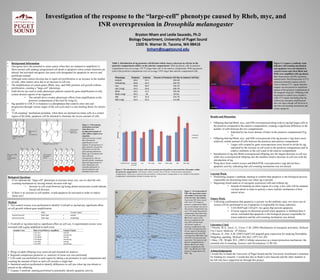
- 1. Investigation of the response to the “large-cell” phenotype caused by Rheb, myc, and INR overexpression in Drosophila melanogaster • Background Information • Oncogenes have the potential to cause cancer when they are mutated or amplified (1). Most normal cells undergo programmed cell death or apoptosis when certain functions are altered, but activated oncogenes can cause cells designated for apoptosis to survive and proliferate instead. • Although some tumors develop due to rapid cell proliferation or an increase in the number of cells, other tumors arise due to an increase in cell size. • The amplification of certain genes (Rheb, myc, and INR) promote cell growth without proliferation, creating a “large-cell” phenotype. • Gal4 drivers are used to study phenotypic patterns caused by gene amplification in only certain desired regions of an organism. • The enGal4 driver creates phenotypic effects from amplification in the posterior compartment of the fruit fly wing (2). • Stg (parallel to CDC25 in humans) is a phosphatase that controls entry into and progression through various stages of the cell cycle and is a rate-limiting factor for mitosis (3). • “Cell-counting” mechanism postulate: when there are deemed too many cells in a certain region of the body, apoptosis will be initiated to eliminate the excess amount of cells. Biological Questions • If we replicate the “large-cell” phenotype to increase tissue size, can we alert the cell- counting mechanism by forcing mitotic division with stg? • Increase in cell count between stg being absent and present would indicate forced cell division. • If there is an increase in cell number, would apoptosis be activated in order to reduce tissue size? Figure 2. The introduction of stg to cells overexpressing growth pathways increased the cell number within the posterior compartment. Cell density within a surface area of 40 µm2 in the posterior and anterior compartments. When stg is expressed the cell number in the posterior compartment is very similar to the anterior compartment, indicating an increase in mitotic activity. Table 1. Introduction of stg promotes cell division which causes a decrease in cell size in the posterior compartment relative to the anterior compartment. With stg absent, cells in posterior compartment were on average 159.7% larger than cells in the anterior compartment. With stg present, cells in the posterior compartment were on average 110% larger than anterior compartment cells. Figure 3. Overexpression of INR with stg present causes cell number to increase due to forced entry into mitosis. Expressing INR alone caused cells to be 155% larger in posterior compartment. When stg was introduced, cell size in the posterior compartment was only 115% larger than anterior compartment. (A) Adult wing with only INR overexpressed. (B) 40 µm2 region of the anterior compartment with only INR overexpressed. (C) 40 µm2 region of posterior compartment with only INR overexpressed. (C) 40 µm2 region of the anterior compartment with only INR overexpressed. (D) Adult wing with INR overexpressed and stg present. The posterior and anterior compartments are labeled. (E) 40 µm2 of the anterior compartment with INR + stg. (F) 40 µm2 of the posterior compartment with INR + stg. Figure 1. Cell-counting mechanism activated when there are an abnormal amount of cells within a region of an organism. Each cell secretes a single hair, allowing for quantification of cell quantity. Overexpression of genes promote cell growth, which does not alert the cell-counting mechanism. When stg is introduced into lines with gene overexpression, cells are forced to divide, creating an abnormal large amount of cells, alerting the cell- counting mechanism to activate apoptosis and eliminate excess cells. Results and Discussion • Offspring that had Rheb, myc, and INR overexpressed along with no stg had larger cells in the posterior compared to the anterior compartment, creating a significant difference in the number of cells between the two compartments. • Indicated by the lower density of hairs in the posterior compartment.(Fig. 3) • Offspring that had Rheb, myc, and INR overexpressed with stg present (+stg) had a more relatively similar amount of cells between the posterior and anterior compartment. • Larger cells created by gene overexpression were forced to divide by stg, indicated by the increase in cell count in the posterior compartment and its relative similarity to the cell count in the anterior compartment. • Introduction of stg into Rheb-overexpressed offspring saw the largest decrease in cell size while myc-overexpressed offspring saw the smallest relative decrease in cell size with the introduction of stg. • Wing discs from control crosses and Rheb,PI3K overexpression (-stg) did not have apoptotic activity, indicating that cell-counting mechanism was not alerted. Current Work • Performing caspase-3 antibody staining to confirm that apoptosis is the biological process responsible for decreasing tissue size when stg is present. • Beginning clonal analysis of oncogene expression with and without stg. • Instead of mutating an entire region of a wing, a few cells will be mutated via heat shock in order to portray a more realistic mechanism of how cancer arises. Future Work • Following confirmation that apoptosis is present via the antibody stain, two more sets of crosses will be performed to see if apoptosis is responsible for tissue reduction. • UAS DIAP and UAS p35: two genes that prevent apoptosis. • If tissue regains its abnormal growth when apoptosis is inhibited then it can be concluded that apoptosis is the biological process responsible for tissue reduction and the cell-counting mechanism was alerted. Literature Cited 1 Pierotti, M.A., Sozzi, G., Croce, C.M. (2003) Mechanisms of oncogenic activation. Holland- Frei Cancer Medicine. 6th Edition. 2 Busson, D., Pret, A.M. (2007) Gal4/UAS targeted gene expression for studying Drosophila Hedgehog signaling. Methods Mol Biol. (397) 161-201. 3 Shay, J.W., Wright, W.E. (2001) Cellular senescence as a tumor-protection mechanism: the essential role of counting. Genetics and Development 11:98-103. Acknowledgements I would like to thank the University of Puget Sound and the University Enrichment Committee for funding my research. I would also like to thank Leslie Saucedo and the other students in her lab who have supported me through this project. Method 1. Two control crosses were performed to identify if enGal4 or stg had any significant effect on cell growth without gene amplification. 2. If enGal4 or stg alone had no significant effect on cell size, 6 experimental crosses were executed with a gene amplified in each cross. 3. Wings of adult offspring were removed and mounted for analysis. 4. Regional comparison (posterior vs. anterior) of tissue size was performed. 5. Cell count was performed in each region by taking a set perimeter in each compartment and counting the amount of hairs as each cell secretes a single hair. 6. Statistical analysis performed to identify differences in cell size when stg was absent or present in the offspring. 7. Caspase-3 antibody staining performed to potentially identify apoptotic activity. Male Female (Virgin) Control Cross #1 Wild-‐Type x enGal4 Control Cross #2 Wild-‐Type x enGal4/stg Variable Cross Male (Gene/Pathway Amplified) Female (Virgin) 1 UAS myc x enGal4 2 UAS myc x enGal4/stg 3 UAS Rheb x enGal4 4 UAS Rheb x enGal4/stg 5 UAS INR x enGal4 6 UAS INR x enGal4/stg Phenotype Posterior Anterior Percent of Posterior Cell Size to Anterior Cell Size enGal4 49.4 51.1 103.4% enGal4/stg 50.8 52.2 102.7% myc 39.8 50.2 126.1% myc (+stg) 54.2 54.6 100.7% Rheb 26.2 51.2 195.4% Rheb (+stg) 44.2 44 99.5% INR 49.7 76.9 154.7% INR (+stg) 56.9 65.9 115.8% Figure 4. Caspase-3 antibody stain indicates cell-counting mechanism and apoptosis were not initiated in control crosses and when Rheb and PI3K were amplified with stg absent. Blue fluorescence (DAPI) represents stained nuclei. Red flourescence (CY3) represents potential caspase activity. Lack of intense CY3- signal indicates caspase was not present in significant regions of the posterior compartment of the imaginal wing disc. Offspring with stg present in control cross created a weak CY3 signal concluding that stg without any oncogene amplification does not cause enough cell division to alert the cell-counting mechanism and initiate apoptosis. ! Normal Tissue and Cells Enlarged Tissue and Cells Rheb, myc, or INR overexpression Cell-Counting Mechanism NOT alerted Rheb, myc, PI3K, or INR overexpression (+stg) Cell-Counting Mechanism alerted 0 10 20 30 40 50 60 70 80 90 enG4 (control) enG4/stg (control) myc myc (+stg) Rheb Rheb (+stg) INR INR (+stg) Cell Count Posterior Anterior A. D. B. C. E. F.