1. 30th Congress of the International Society for Advancement of Cytometry,
June 26-30, 2015 Glasgow, Scotland
373/B242
Negative Cell Sorting for Removal of Undifferentiated Cells
for Regenerative Medicine
Takahide Ino1*, Haixin Zhang2, Masayuki Ishige1, Kazuhiro Aiba2, Yuu Fujimura1, Norio Nakatsuji2, Kazuo Takeda1
1 On-chip Biotechnologies Co., Ltd., Tokyo, Japan
2 Institute for Integrated Cell-Material Sciences (WPI iCeMS), Kyoto University, Kyoto, Japan
In the field of regenerative medicine involving embryonic stem (ES) cells or induced pluripotent stem (iPS) cells, differentiated cells are often cultured to tissues and implanted
to a patient. However, there is a serious problem that some remaining undifferentiated cells in the implanted tissue may cause tumors, and thus undifferentiated cells must be
completely removed prior to implantation.
Conventional cell sorters are typically used for purification of cells, but cell sorting for regenerative medicine must be much gentler to cells and operated in sterile condition. In
2012, we have launched a disposable microfluidic chip-based cell sorting system (On-chip Sort) suitable for such applications (Fig.1). The channel size of the sorting chip is 80 μm
in height and width, and cells are deflected into a sorting chamber using pulse flow created by pneumatic actuation1,2.
Herein, we report the following: 1. improved sorting speed of On-chip Sort to 300 targets per second; 2. proposal of “Negative Cell Sorting”, a new purification method that
allows for 100 fold increase in collection speed of target cells for a sample containing about 1% of non-target cells; 3. application of this new method on undifferentiated cell removal
from neural cell population; and 4. realization of cell spheroid sorting using disposable chip.
Fig.1 Disposable microfluidic chip based cell sorter (On-chip Sort)
Sample reservoir
(1mL)
Sheath reservoir
(10mL)
Collection reservoir
(2mL)
Waste reservoir
(12mL)
Sorting region
Sorting reservoir
(2mL)
Disposable microfluidic chip (sterile)
Fig.2 Pulse flow sorting by air pressure in a microfluidic chip
Fig.3 Sorting speed and
sorting efficiency
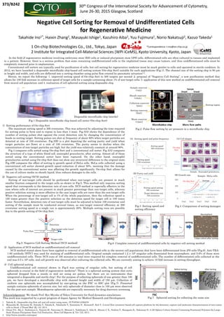
Fig.5 Negative Cell Sorting Method (NCS method)
Non-target cells (rare)
Detection
Target cells
Pulse flow
1) Sorting performance of On-chip Sort
The maximum sorting speed is 300 events/sec. This was achieved by adjusting the time required
for sorting pulse to form and to repose to less than 3 msec. Fig.3(A) shows the dependence of the
number of sorting pulses shot against the event detection rate by changing the concentration of
beads as sorting target. Sorting pulses are shot at frequency of about 80% when target particles are
detected at rate of 100 events/sec. Fig.3(B) is a plot depicting the sorting purity and yield when
target particles are flown at a rate of 100 events/sec. The purity seems to decline when the
concentration of non-target particles are high, but the yield was relatively constant at around 80%.
Fig.4 compares cells sorted using On-chip Sort and a conventional cell sorter. Fig.4(A) compares
the structure of a type of white blood cell, eosinophil granulocyte, after sorting. Almost all of those
sorted using the conventional sorter have been ruptured. On the other hand, eosinophil
granulocytes sorted using On-chip Sort does not show any structural difference to the original state.
Fig.4(B) compares the effect of sorting to growth speed of HeLa cells. HeLa cells collected using a
conventional cell sorter showed growth speed reduction of about 50%, meaning that the damage
caused by the conventional sorter was affecting the growth significantly. On-chip Sort allows for
the use of culture media as sheath liquid, thus reduces damages to the cells.
2) Negative cell sorting (NCS) method
Sorting of non-target cells should be performed when non-target cells are present in much
smaller fraction compared to the target cells as shown in Fig.5. This method only requires sorting
speed that corresponds to the detection rate of rare cells. NCS method is especially effective in the
case where cells of interest are present in much greater percentage than non-target cells, whereas
positive selection sorting is more effective in the case target cells are rarer than the non-target cells.
When the percentage of non-target cell is 1% of the whole population, then the collection speed is
100 times greater than the positive selection as the detection speed for target cell is 100 times
faster. Nevertheless, detection rate of non-target cells must be adjusted to below 100 events/sec and
sorting of the sample must be conducted several times, as non-target removal efficiency at 300
events/sec sorting speed in a single run is approximately 60%. Multiple sorting runs are possible
due to the gentle sorting of On-chip Sort.
Sorting speed
300 events/sec
Sorting speed
100 events/sec
Target detection rate
100 events/sec
Sorting speed setting
300 events/sec
(A) Sorting speed and pulse frequency
(B) Purity and yield
Fig.4 Comparison of sorting damages
Fig.6 Complete removal of undifferentiated cells by negative cell sorting method
3) Application of NCS method on undifferentiated cell removal
NCS method stated above have been applied to removal of undifferentiated cells in the neuron cell populations that have been differentiated from iPS cells (Fig.6). Anti-TRA-
1-60 antibody (FITC) was used for staining of undifferentiated cells. Original sample contained 1.2 x 10⁶ singular cells dissociated from neurosphere, and 5.6% of those were
undifferentiated cells. Three NCS runs of 100 minutes in total were required for complete removal of undifferentiated cells. The number of differentiated cells collected at the
end was 0.5 x 10⁶ cells, and cell growth was observed after culturing the collected cells. We are currently aiming to achieve 10 fold increase in sorting throughput.
FSC
SSC
150μm 150μm
Before Sorting After Sorting
FSC
(A) Cell shapes
0
100
200
300
Day1 Day2 Day3 Day5 Day8
On-chip Sort-1 On-chip Sort-2
Sorter X-1 Sorter X-2
damageOn-chip Sort
Other Sorter
On-chip
Sort
Other
Sorter
Sample : Eosinophil granulocyte
Sample : HeLa cells(B) Growth rate
Fig.7 Spheroid sorting for collecting the same size
Acknowledgement: We thank Prof. Ryo Miyake of Tokyo University for fluid dynamics simulations.
This work was supported by a grant program of Japan Agency for Medical Research and Development.
1. Takeda K.; Disposable chip flow cell and cell sorter using same.; PCT/JP2011/050270
2. Watanabe M., Serizawa M., Sawada T., Takeda K., Takahashi T., Yamamoto N., KoizumiF., Koh Y.; A novel flow cytometry-based cell capture platform for the detection, capture and molecular characterization of rare tumor
cells in blood. J Transl Med. 12, 143, 2014
3. Otsuji T.G., Bin J., Yoshimura A., Tomura M., Tateyama D., Minami I., Yoshikawa Y., Aiba K., Heuser J. E., Nishino T., Hasegawa K., Nakatsuji N.; A 3D Sphere Culture System Containing Functional Polymers for Large-
Scale Human Pluripotent Stem Cell Production. Stem Cell Reports, 2, 734–745, 2014
4. http://www.unionbio.com/copas/
4) Cell spheroid sorting
Undifferentiated cell removal shown in Fig.6 was sorting of singular cells, but sorting of cell
spheroids is crucial in the field of regenerative medicine3. There is a spheroid sorting system that sorts
spheroid dropped from a nozzle in mid air using air pulses, but there are no instruments that
integrated a disposable and sterile chip4. For the purpose of collecting spheroids of size greater than 100
μm, we have developed a microfluidic chip with channel height and width of 150 μm. Collection of
uniform size spheroids was accomplished by size-gating on the FSC vs SSC plot (Fig.7). Presorted
sample contains spheroids of uneven size, but only spheroids of diameter close to 100 μm were observed
for the post-sort sample. Monodisperse spheroids are especially necessary for drug discovery screening.
FSC
TRA-1-60(FITC)
sorting
3 days culture after sorting
500μm
Neurosphere
sortingsorting
*Correspondence: t-ino@on-chip.co.jp