NCCT OR CECT PELVIS BONE.pptx, INDICATIONS OF PELVIS BONE
1. NCCT/CECT PELVIS BONE
Rukmanee Yadav
Assistant Professor
Department of Radiology and
Imaging Technology
Mewar university
Rajasthan
2. INTRODUCTION
• Pelvis bone is also called bony pelvis or pelvis girdle.
• it is basin shaped and connect to trunk and legs
• It support and balance the trunk .
• It consist paired hipbone ,connected infront of pubic symphysis.
• Pelvis play many functions in human body-pelvis carries the entire weight
of upper body.
• Stabilize the body, sitting , standing.
• Bony pelvis provide the comfortable environmental for fetus during
pregnancy.
• CT scan is fast, non-invasive and give the 3D image.
3. Continue….
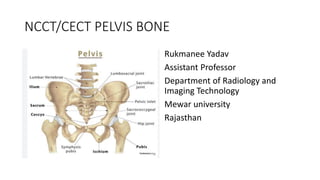
• Pelvic bone consist-
• Sacrum
• Iliac bone
• Ischium
• Pubic bone
• Acetabulum
• Abturator Foramen (opening in the hip bone b/t pubic and ischium)
passes of blood vessel and nerve.
• Coccyx bone
6. Patient preparation
Contrast media: nonionic, iodinated contrast media used.
Volume: 40-60ml.
Rate:1ml to 3ml/sec.
Contrast media administration:IV
7. Patient position
• Supine with head or feet first with the knee extension.
• After the position finish the laser light should be off.
• POSITION LANDMARK- Level of umbilicus
8. Protocol
Topogram- Anteroposterior Level of umbilicus.
Scan extent- Highest point of iliac crest to inter-trochantric region bilaterally.
Slice thickness —2-3 mm
Slice interval: 1-1.5 mm
Reconstruction algorithm - sharp for bone and medium for soft tissue
Scan direction : cranio-caudal (head to feet)
Scan delay-40-60 sec
Pre contrast series: Axial images are acquired covering from Highest point of iliac crest to inter-trochantric
region bilaterally.
Post contrast series: Axial images are acquired covering from the Highest point of iliac crest to inter-
trochantric region bilaterally.
10. After care
Patient is brought out from the gantry
Bring the patient down from the scanner table.
Ask the patient to dress up.
patient should be observed for possible CM reaction post procedure.
If non is observed , IV cannula should be removed from the patient’s hand gently.
Ensure that the patient is stable before leaving the department.
Inform patient time and where to collect the result.