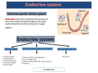
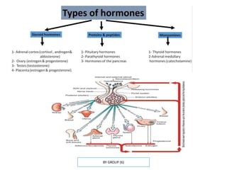
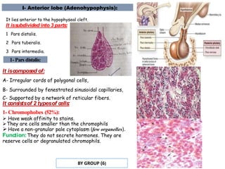
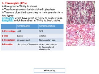
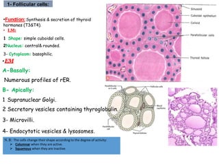
ENDOCRINE SYSTEM
BY GROUP (6)
BY GROUP (6)
Endocrine glands= ductless glands
Definition: the sites of synthesis & secretion of
hormones which spread throughout the body
by the bloodstream where they act on target
organs.
BY GROUP (6)
PITUITARY GLAND (HYPOPHYSIS CEREBRI):
Anatomically:
It is situated inside the hypophyseal fossa (sella
turcica) of the sphenoid bone, connected to the brain
by a stalk called the infundibulum& covered by the
diaphragma sellae.
Histologically:
It is divided into:
a) Anterior lobe (adenohypophysis):
dark& cellular.
b)Posterior lobe (neurohypophysis):
Pale&fibrous.
Development of Pituitary gland:
BY GROUP (6)
I- Anterior lobe (Adenohypophysis):
It lies anterior to the hypophyseal cleft.
It iss
ubdivided into 3 parts:
1 Pars distalis.
2 Pars tuberalis.
3 Pars intermedia.
1- Pars distalis:
It iscompos
ed of:
A- Irregular cords of polygonal cells,
B- Surrounded by fenestrated sinusoidal capillaries,
C- Supported by a network of reticular fibers.
It cons
is
tsof 2 typesof cells
:
1- Chromophobes (52%):
 Have weak affinity to stains.
They are cells smaller than the chromophils
 Have a non-granular pale cytoplasm (few organelles).
Function:They do not secrete hormones. They are
reserve cells or degranulated chromophils.
BY GROUP (6)
2- Chromophils (48%):
Have great affinity to stains.
They have granular darkly stained cytoplasm
They are classified according to their granules into
two types:
Acidophils: which have great affinity to acidic stains.
Basophils: which have great affinity to basic stains.
BY GROUP (6)
Chromophils Chromophobes
1- Percentage 48% 52%
2- size Larger Smaller
3- Cytoplasm Granular, dark Non granular, pale
4- Function Secretion of hormones A- Act as a reserve.
B- Degranulated
chromophils.
BY GROUP (6)
a- Acidophils (37%):
They secrete hormones of protein in nature. They are
periodic acid Schiff (PAS) negative.
They are smaller than the basophils but contain larger secretory
granules.
EM: protein secreting cells.
Acidophilsareof two types:
1 Somatotrophs: secret growth hormone.
2 Mammotrophs: secret prolactin.
Mammotrophs:
•The secretory granules in these cells are small, but in pregnant and
lactating females they become large and numerous& are called
pregnancy cells or Erdheim cells.
•When suckling is terminated, lysosomes eliminate the excess
secretory granules, a process known as
crinophagy.
BY GROUP (6)
b- Basophils (11%):
They secrete hormones of glycoprotein in nature.
They are PAS +ve (except the corticotrophs which secrete a polypeptide hormone (so, it is
PAS –ve).
They are larger than the acidophils but contain smaller secretory granules.
EM: protein secreting cells.
Bas
ophils are three types:
1 Thyrotrophs: secrete thyroid stimulating hormone (TSH).
2 Gonadotrophs: They secrete two hormones:
 Follicle stimulating hormone (FSH).
 Luteinizing hormone (LH)
3- Corticotrophs: secrete adrenocorticotrophic hormone (ACTH).
BY GROUP (6)
Acidophils Basophils
1- Percentage 37% 11%
2- Nature of
hormones
Protein Glycoprotein
3- PAS Negative Positive
4- size Smaller Larger
5- Granules Larger Smaller
6- Secretion Somatotrophs
(growth h.)
Mammotrophs
(prolactin)
Thyrotrophs (TSH)
Gonadotrophs
(FSH& LH)
Corticotrophs (ACTH)
BY GROUP (6)
2- Pars Tuberalis:
It is a superior extension that surrounds the
infundibular stalk. It is highly vascular.
It isformed of:cords of cells (show
immunoreactivity to ACTH,FSH&LH).
Function:nonspecific.
3- Pars Intermedia:
It lies between the pars distalis and pars nervosa.
It ismade up of: cords of faint
basophilic cells with few secretory
granules.
Function:nonspecific.
BY GROUP (6)
II- Posterior lobe (Neurohypophysis):
• It lies posterior to the hypophyseal cleft.
• It consists of 3 parts:
1The Median Eminence: a funnel shaped downward
extension of the hypothalamus.
2 The Infundibular s
talk.
3The Pars Nervosa: which is connected to the base
of the brain by the infundibular stalk and the median
eminence.
It cons
is
tsof:
1- Unmyelinated axons of neurosecretory cells:
Their cell bodies present in the supraoptic& paraventricular
nuclei of the hypothalamus.
 Their axons form the hypothalamo-hypophyseal tract
carrying the neurosecretion from the hypothalamic nuclei to
the pars nervosa.
2- Herring bodies:
 Acidophilic homogenous bodies.
Represent accumulation of neurosecretory granules at
the dilated terminal ends of axons.
3 Pituicytes: modified glial cells to support nutrition&
isolation.
4 Rich fenestrated blood capillaries.
ParsNervosa :
BY GROUP (6)
Neurosecretion:
Acts on smooth muscles of the uterus&mammary glands
2- Vasopressin (ADH)
↑ water reabsorption from collecting ducts → concentrated
urine.
1-Oxytocin
Blood supply of pituitary gland:
BY GROUP (6)
Anatomically:
It lies in the anterior aspect of the lower part of the neck.
It consists of two lobes connected by an isthmus.
STRUCTURE OF THYROID GLAND :
1- Stroma:
 A double capsule:
a. The outer: is the pretracheal fascia.
b. The inner: is the true CT capsule of the gland.
 CT septa: dividing the gland into incomplete lobes
& lobules.
Reticular fibers: form a
network supporting the
thyroid parenchyma.
2- Parenchyma:
 It is composed of follicles.
The follicles are surrounded by network of fenestrated
blood capillaries.
 They contain a gelatinous acidophilic material in their
lumina called colloid.
They are lined by cuboidal
cells of two types:
follicular cells & parafollicular
cells.
1- Follicular cells:
•Function:Synthesis & secretion of thyroid
hormones (T3&T4).
• LM:
1 Shape: simple cuboidal cells.
2Nucleus: central& rounded.
3- Cytoplasm: basophilic.
N.B: The cells change their shape according to the degree of activity:
 Columnar when they are active.
 Squamous when they are inactive.
•EM
A-Basally:
Numerous profiles of rER.
B- Apically:
1 Supranuclear Golgi.
2 Secretory vesicles containing thyroglobulin.
3- Microvilli.
4- Endocytotic vesicles & lysosomes.
BY GROUP (6)
2-Parafollicular (Clear cells or C cells):
• Function: synthesis & secretion of calcitonin.
•They are found as part of the follicular
epithelium, in between the follicular cells or as
isolated clusters between the follicles.
• Number: 0.1% of the follicular epithelium.
• LM:
 S
ize: larger than the follicular cells.
 Cytoplasm: pale (clear cells).
Their basal surfaces rest on the follicular
basement membrane but their apical surfaces never reach the lumen of the follicles.
• EM: Polypeptides secreting cells:
 Moderate amount of organelles.
 Infra nuclear Golgi (Why).
 The secretory granules are small & basal (Why).
BY GROUP (6)
Steps of synthesis of thyroid hormones:
1.Synthesis of thyroglobulin
(glycoprotein):
A- The protein part of the thyroglobulin are
synthesized from the amino acids tyrosine by the
rough endoplasmic reticulum.
B- The Golgi apparatus adds carbohydrate to the
protein to form the thyroglobulin& packed into
secretory vesicles.
C- The secretory vesicles are discharged into the
lumen of the follicle by exocytosis where
thyroglobulin contributes in the formation of the
colloid.
2 Uptake of iodide:
The follicular cells trap the iodide from the capillaries
released into the follicular lumen. oxidion into iodine
BY GROUP (6)
3 Iodination of thyroglobulin: takes place in the colloid to form
monoiodotyrosine& diiodotyrosine.
4 Formation of T3 & T4: by coupling reaction in the colloid, where they are
stored in the lumen of the follicle.
5 Release of T3 & T4:
 In response to TSH endocytosis of the colloid in endocytotic vesicles by the follicular cells
fusion of the vesicles with lysosomes hydrolysis release of T3&T4 blood stream.
n.
BY GROUP (6)
Follicular cells Parafollicular cells
1- Size Smaller Larger
2- Number Numerous Few
3- Extension Reach the lumen Never reach the lumen
4- Cytoplasm Basophilic Pale basophilic
5- rER Rich, basally Moderate
5- Golgi Supranuclear Infranuclear
5- Lysosomes Numerous Less
6- Secretion Stored extracellular Stored intracellular, inside secretory granules.
7-Function Secrete T3&T4 Secrete calcitonin
BY GROUP (6)
Anatomically: 4 glands situated behind thyroid gland.
I-Stroma:
 CT capsule: of that of the thyroid gland.
 CT septa.
Network of reticular fibers: support the
parenchymal cells.
II-Parenchyma:
•It consists of cords of polygonal cells
surrounded by a rich network of
fenestrated blood capillaries.
•The cells are of 2 types: Chief & oxyphil
cells.
STRUCTURE OF PARATHYROID :
BY GROUP (6)
A- The chief cells:
•Function:Secretion of parathyroid
hormone.
• Number:numerous.
• LM:
 Shape: small polygonal cells
 Nucleus: large vesicular nuclei
 Cytoplasm: pale (WHY).
• EM: Polypeptide secreting cells
The cytoplasm contains moderate amount of
organelles (rER, Golgi complex, mitochondria&
secretory granules.
Glycogen granules& lipid droplets: cause the
pale cytoplasm seen with the LM.
Parathyroid gland
BY GROUP (6)
B- Oxyphil cells •Function:Unknown.
•Number:few.
• LM:
 Shape: large polygonal.
 Nucleus: small& dense.
 Cytoplasm: deep eosinophilic (Why).
EM: The cytoplasm contains many
mitochondria.
• With increas
ing age:
 The chief cells are replaced by fat cells
which may constitute more than 50% of the
gland.
 The number of oxyphil cells increases.
Chief cell Oxyphil cell
1- Size Smaller Larger
2- Number Numerous Few
3- Cytoplasm Pale Deep acidophilic
4- Nucleus Large& vesicular Small & dense
5- rER More Less
6- Mitochondria Less Numerous
7- Function Secretion of
parathyroid
hormone
Unknown
BY GROUP (6)
•Anatomically:
Two glands, each one situated at the
upper pole of the kidney.
•Histologically:
Each gland consists of a cortex& medulla.
STRUC TURE OF SUPRARENAL GLANDS :
I.Cortex:
 Yellow in color (Why).
 It originates from the mesoderm.
 It constitutes 80- 90% of the gland.
II. Medulla:
 Reddish brown in color (Why).
 It originates from the neural crest (the same origin as the sympathetic nervous
system).
 It is considered as a modified sympathetic ganglion, where the neurons have
acquired an endocrine activity and secrete catecholamines.
STRUC TURE OF SUPRARENAL GLANDS :
A- Stroma:
 A connective tissue capsule.
 A CT septa.
 A network of reticular fibers.
BY GROUP (6)
B- Parenchyma: it is divided the cortex & the medulla.
I- Suprarenal Cortex:
• It is the steroid secreting portion of the gland.
• It is divided into 3 zones:
1Zona glomerulosa (15% of the cortex): Secretes
mineralocorticoids mainly aldosterone control
blood volume& blood pressure.
2Zona fasiculata (65-80%): Secretes
glucocorticoids control CHO, fat & protein
metabolism.
3 Zona reticularis (7%): Secretes androgen
development of axillary& pubic hair in females.
Structure of cells in adrenal cortex:
•Steroid secreting cells:
•LM:
 Cytoplasm: acidophilic& vacuolated.
•EM:
 Abundant sER.
 Mitochondria with tubular cristae.
Lipid droplets: they are numerous in zona
fasiculata & less in
 zona glomerulosa and reticularis.
BY GROUP (6)
A- Zona Glomerulosa:
• LM:
 It lies immediately under the capsule.
 Arrangement of the cells: in closely packed
arched clusters surrounded by fenestrated blood
capillaries.
 Shape of the cells: columnar.
 Nucleus: dense.
 Cytoplasm: acidophilic & slightly vacuolated.
•EM: The cells show the ultrastructural
features of steroid secreting cells with few lipid
droplets.
B- Zona Fsciculata:
•LM:
 It is the middle layer of the cortex
 Arrangement of the cells: in long straight
cords, perpendicular to the capsule.
The cords are one or two cells thick& separated
by sinusoidal blood capillaries.
 Shape of the cell: large polyhedral.
 Nucleus: large& pale.
Cytoplasm: pale acidophilic& highly vacuolated
so called spongiocytes.
EM: The cells show the ultrastructural features
of steroid secreting cells with
numerous lipid droplets.
BY GROUP (6)
C- Zona Reticularis:
•LM:
 It is the inner layer of the cortex.
 Arrangement of the cells: in irregular
cords enclosing fenestrated capillaries in
between.
 Shape of the cell: small polyhedral.
 Cytoplasm: acidophilic& slightly vacuolated.
 Dark cells: are seen near the medulla. They have
dark cytoplasm with pyknotic nuclei, suggesting that
they are degenerating cells.
 Lipofuscin pigments: are present in this layer.
EM: The cells show the ultrastructural features of
steroid secreting cells with few lipid droplets.
NB: Steroid secreting cells Never store their Hormones.
BY GROUP (6)
Zona glomerulosa Zona fasiculata Zona reticularis
1- Percentage 15% 65-80% 7%
2-Arrangement Closely packed arches Long straight
cords
Irregular cords
3- Shape of
the cells
Columnar Large polyhedral Small polyhedral
4- Nucleus Dense Large& pale
5- Cytoplasm Acidophilic& slightly
vacuolated
Acidophilic& highly
vacuolated
Acidophilic&
slightly
vacuolated
6-Lipid
droplets
Few Numerous Few
7- Function Secrets
mineralocorticoids
Secretes
glucocorticoids
Secretes
androgen
II- Suprarenal Medulla:
It contains two types of cells:
1 Chromaffin cells: secrete catecholamines
(epinephrine&norepinephrine).
2 Sympathetic ganglion cells.
BY GROUP (6)
1- Chromaffin cells = Pheochromocytes:
•They are called chromaffin cells due to
the affinity of their granules to react
with chromium salts and give brown color
(chromaffin reaction).
•LM:
 Arrangement of the cells: in irregular
cords enclosing the blood capillaries.
 Shape of the cell: large ovoid.
 Nucleus: large spherical.
 Cytoplasm: pale basophilic.
• EM: polypeptide secreting cells
Moderate amount of rER, prominent
Golgi and secretory granules.
BY GROUP (6)
-Adrenal medulla is considered as modified sympathetic ganglia
-Chromaffin cells are modified postganglionic neurons
2- Sympathetic ganglion cells:
Functions:
A-Modulate the secretory activity of the
cortex.
B- Innervate the blood vessels of the cortex.
BY GROUP (6)
B LOOD S UPPLY OF THE ADRENAL GLAND:
• The medulla hasa dual blood supply:
1 Arterial: from the medullary arterioles.
2 Venous: from the cortical sinusoids draining the
cortex (glucocorticoids reaching the medulla are
essential for production of epinephrine by
chromaffin cells).
BY GROUP (6)
Anatomically:
Pancreas is one of the largest glands of
the digestive tube. It lies in C- shaped
cavity of the duodenum. It is divided into
head, body & tail.
Histologically:
It is a mixed gland: composed of both
exocrine (described in GIT module) &
endocrine parts.
THE ENDOCRINE PANCREAS (THE ISLETS OF LANGERHANS)
• The islets of Langerhans are more numerous in
the tail than in the head & the body of the
pancreas.
• They appear in histological sections as lightly
stained areas (Why).
BY GROUP (6)
HISTOLOGIC AL STRUC TURE OF ISLETS OF LANGERHANS
A- Stroma:
 They are not encapsulated.
 Supported by network of reticular fibers.
B- Parenchyma:
• LM:
Arrangement of the cells: in short cords and separated
by network of fenestrated blood capillaries.
Shape of the cells: polygonal.
Cytoplasm: pale basophilic
•EM: Polypeptide secreting cells
Moderate amount of rER, Golgi complex & secretory
granules.
3 main typesof cellscan be identified:A cells, B
Cells& D cells.
1-A cells (alpha):
increases blood
•Function:secretion of glucagon
sugar level.
• S
ize: large.
• Number: 20%.
• S
ite: at the periphery of the islet.
•EM:the secretory granules are numerous and have a
homogeneous dense core.
BY GROUP (6)
2-B cells (Beta):
• Functions:
1- Secretion of insulin lower blood sugar level.
2- Secretion of C-peptide: as a consequence of insulin
production. It prevents neuropathy and other symptoms
of diabetic vascular deterioration. Measuring its level
can give an idea about the viable beta cell mass.
3- Secretion of GABA (γ amino butyric acid): suppress
glucagon secretion.
• S
ize of the cells:small.
• Number: 70% of the islet cells.
• Site: centrally located within the islet.
•EM:their secretory granules have a rectangular crystalline dense core surrounded
by an electron lucent halo.
3-D cells (Delta):
•Function: secretion of somatostatin
secretion of GH, insulin & glucagon.
• Number: 5 - 10% of the islet cells.
• S
ite: at the periphery of the islet.
inhibits
BY GROUP (6)
Alpha Beta Delta
Number 20% 70% 5-10%
Site Periphery Central Periphery
Size Large Small Small
Granules:
EM
Granuleswith
homogenous
dense core
Granules with rectangular
crystalline dense core
surrounded by an electro
lucent halo.
Function Glucagon Insulin, C peptide & GABA Somatostatin
BY GROUP (6)
•Definition: single or small groups of endocrine
cells with diffuse distribution in different tissues
and organs of the body.
• These cells secrete hormones, usually peptides.
•They synthesize monoamines by the uptake of
amine precursors and their decarboxylation(
APUD cells=Amine Precursor Uptake &
Decarboxylation).
BY GROUP (6)
Modes of action of their hormones:
1 Paracrine: act on the neighboring cells.
2 Autocrine: act on the cell that secrete the
hormone itself.
3 Endocrine: act on target cells in remote
tissue.
1 H&E: These cells are not well seen.
2 Special stain:
 chromium salts (chromaffin cells).
 silver salts (argentaffin cells).
3 Immunohistochemical stain: using antibodies against their peptide hormone.
Chromaffin cells Argentaffin cells
How to identify the DNES in histological sections?
BY GROUP (6)
Structure of DNES:
•EM:
1 Shape: pyramidal with narrow apex and a wide base.
2Cytoplasm: polypeptide secreting cells with moderate amount of organelles due
to slow rate & small amount of secretion:
 rER.
 Mitochondria.
 Golgi apparatus.
 Secretory granules.
Types of DNES:
1 Open type: the cell has long microvilli
extending into the lumen of the organ.
2Clos
ed type: where the cell apex is
covered by other epithelial cells of the
organs.
BY GROUP (6)
Distribution of DNES:
 Gastrointestinal tract (enteroendocrine cells).
 Respiratory system.
 Supraoptic and paraventricular hypothalamic nuclei.
 Parafollicular cells of thyroid gland.
 Chief cells of parathyroid gland.
 Chromaffin cells of the suprarenal medulla.
 Cells of islets of Langerhans.
 Pinealocytes.
BY GROUP (6)
Site: on the posterior part of third ventricle at the mid
line between two cerebral hemispheres.
Structure: It is covered by pia matter which sends CT
septa carrying the blood vessels.
It consists of 2 types of cells:
A- Pinealocytes (secretory cells).
B- Glial cells (resemble astrocytes): for support.
Pinelocytes
• Shape: branching with long tortuous processes end as
flat dilatations near blood capillaries (WHY).
• Nucleus: large, lobulated with prominent nucleoli.
• Cytoplasm: pale basophilic (WHY) with secretory
vesicles.
• Function: secretion of
melatonin.
BY GROUP (6)
• Definition: Formation of calcified
bodies which increase in number & size
by age.
• Importance: it can be used as a clinical landmark for the midline location of the
pineal gland during radiological examination of the brain.
Corpora arenacea (brain sand)
Functions of melatonin
1 Controls circadian rhythm.
2Induces feeling of sleepiness.
3- Decreases gonadotropin.
4- Protects CNS from oxidative stress.
BY GROUP (6)

endocrine-System-Group6.pptx

  • 1.
  • 2.
    BY GROUP (6) Endocrineglands= ductless glands Definition: the sites of synthesis & secretion of hormones which spread throughout the body by the bloodstream where they act on target organs.
  • 3.
  • 4.
    PITUITARY GLAND (HYPOPHYSISCEREBRI): Anatomically: It is situated inside the hypophyseal fossa (sella turcica) of the sphenoid bone, connected to the brain by a stalk called the infundibulum& covered by the diaphragma sellae. Histologically: It is divided into: a) Anterior lobe (adenohypophysis): dark& cellular. b)Posterior lobe (neurohypophysis): Pale&fibrous. Development of Pituitary gland: BY GROUP (6)
  • 5.
    I- Anterior lobe(Adenohypophysis): It lies anterior to the hypophyseal cleft. It iss ubdivided into 3 parts: 1 Pars distalis. 2 Pars tuberalis. 3 Pars intermedia. 1- Pars distalis: It iscompos ed of: A- Irregular cords of polygonal cells, B- Surrounded by fenestrated sinusoidal capillaries, C- Supported by a network of reticular fibers. It cons is tsof 2 typesof cells : 1- Chromophobes (52%):  Have weak affinity to stains. They are cells smaller than the chromophils  Have a non-granular pale cytoplasm (few organelles). Function:They do not secrete hormones. They are reserve cells or degranulated chromophils. BY GROUP (6)
  • 6.
    2- Chromophils (48%): Havegreat affinity to stains. They have granular darkly stained cytoplasm They are classified according to their granules into two types: Acidophils: which have great affinity to acidic stains. Basophils: which have great affinity to basic stains. BY GROUP (6) Chromophils Chromophobes 1- Percentage 48% 52% 2- size Larger Smaller 3- Cytoplasm Granular, dark Non granular, pale 4- Function Secretion of hormones A- Act as a reserve. B- Degranulated chromophils.
  • 7.
    BY GROUP (6) a-Acidophils (37%): They secrete hormones of protein in nature. They are periodic acid Schiff (PAS) negative. They are smaller than the basophils but contain larger secretory granules. EM: protein secreting cells. Acidophilsareof two types: 1 Somatotrophs: secret growth hormone. 2 Mammotrophs: secret prolactin. Mammotrophs: •The secretory granules in these cells are small, but in pregnant and lactating females they become large and numerous& are called pregnancy cells or Erdheim cells. •When suckling is terminated, lysosomes eliminate the excess secretory granules, a process known as crinophagy.
  • 8.
    BY GROUP (6) b-Basophils (11%): They secrete hormones of glycoprotein in nature. They are PAS +ve (except the corticotrophs which secrete a polypeptide hormone (so, it is PAS –ve). They are larger than the acidophils but contain smaller secretory granules. EM: protein secreting cells. Bas ophils are three types: 1 Thyrotrophs: secrete thyroid stimulating hormone (TSH). 2 Gonadotrophs: They secrete two hormones:  Follicle stimulating hormone (FSH).  Luteinizing hormone (LH) 3- Corticotrophs: secrete adrenocorticotrophic hormone (ACTH).
  • 9.
    BY GROUP (6) AcidophilsBasophils 1- Percentage 37% 11% 2- Nature of hormones Protein Glycoprotein 3- PAS Negative Positive 4- size Smaller Larger 5- Granules Larger Smaller 6- Secretion Somatotrophs (growth h.) Mammotrophs (prolactin) Thyrotrophs (TSH) Gonadotrophs (FSH& LH) Corticotrophs (ACTH)
  • 10.
    BY GROUP (6) 2-Pars Tuberalis: It is a superior extension that surrounds the infundibular stalk. It is highly vascular. It isformed of:cords of cells (show immunoreactivity to ACTH,FSH&LH). Function:nonspecific. 3- Pars Intermedia: It lies between the pars distalis and pars nervosa. It ismade up of: cords of faint basophilic cells with few secretory granules. Function:nonspecific.
  • 11.
    BY GROUP (6) II-Posterior lobe (Neurohypophysis): • It lies posterior to the hypophyseal cleft. • It consists of 3 parts: 1The Median Eminence: a funnel shaped downward extension of the hypothalamus. 2 The Infundibular s talk. 3The Pars Nervosa: which is connected to the base of the brain by the infundibular stalk and the median eminence. It cons is tsof: 1- Unmyelinated axons of neurosecretory cells: Their cell bodies present in the supraoptic& paraventricular nuclei of the hypothalamus.  Their axons form the hypothalamo-hypophyseal tract carrying the neurosecretion from the hypothalamic nuclei to the pars nervosa. 2- Herring bodies:  Acidophilic homogenous bodies. Represent accumulation of neurosecretory granules at the dilated terminal ends of axons. 3 Pituicytes: modified glial cells to support nutrition& isolation. 4 Rich fenestrated blood capillaries. ParsNervosa :
  • 12.
    BY GROUP (6) Neurosecretion: Actson smooth muscles of the uterus&mammary glands 2- Vasopressin (ADH) ↑ water reabsorption from collecting ducts → concentrated urine. 1-Oxytocin Blood supply of pituitary gland:
  • 13.
    BY GROUP (6) Anatomically: Itlies in the anterior aspect of the lower part of the neck. It consists of two lobes connected by an isthmus. STRUCTURE OF THYROID GLAND : 1- Stroma:  A double capsule: a. The outer: is the pretracheal fascia. b. The inner: is the true CT capsule of the gland.  CT septa: dividing the gland into incomplete lobes & lobules. Reticular fibers: form a network supporting the thyroid parenchyma. 2- Parenchyma:  It is composed of follicles. The follicles are surrounded by network of fenestrated blood capillaries.  They contain a gelatinous acidophilic material in their lumina called colloid. They are lined by cuboidal cells of two types: follicular cells & parafollicular cells.
  • 14.
    1- Follicular cells: •Function:Synthesis& secretion of thyroid hormones (T3&T4). • LM: 1 Shape: simple cuboidal cells. 2Nucleus: central& rounded. 3- Cytoplasm: basophilic. N.B: The cells change their shape according to the degree of activity:  Columnar when they are active.  Squamous when they are inactive. •EM A-Basally: Numerous profiles of rER. B- Apically: 1 Supranuclear Golgi. 2 Secretory vesicles containing thyroglobulin. 3- Microvilli. 4- Endocytotic vesicles & lysosomes.
  • 15.
    BY GROUP (6) 2-Parafollicular(Clear cells or C cells): • Function: synthesis & secretion of calcitonin. •They are found as part of the follicular epithelium, in between the follicular cells or as isolated clusters between the follicles. • Number: 0.1% of the follicular epithelium. • LM:  S ize: larger than the follicular cells.  Cytoplasm: pale (clear cells). Their basal surfaces rest on the follicular basement membrane but their apical surfaces never reach the lumen of the follicles. • EM: Polypeptides secreting cells:  Moderate amount of organelles.  Infra nuclear Golgi (Why).  The secretory granules are small & basal (Why).
  • 16.
    BY GROUP (6) Stepsof synthesis of thyroid hormones: 1.Synthesis of thyroglobulin (glycoprotein): A- The protein part of the thyroglobulin are synthesized from the amino acids tyrosine by the rough endoplasmic reticulum. B- The Golgi apparatus adds carbohydrate to the protein to form the thyroglobulin& packed into secretory vesicles. C- The secretory vesicles are discharged into the lumen of the follicle by exocytosis where thyroglobulin contributes in the formation of the colloid. 2 Uptake of iodide: The follicular cells trap the iodide from the capillaries released into the follicular lumen. oxidion into iodine
  • 17.
    BY GROUP (6) 3Iodination of thyroglobulin: takes place in the colloid to form monoiodotyrosine& diiodotyrosine. 4 Formation of T3 & T4: by coupling reaction in the colloid, where they are stored in the lumen of the follicle. 5 Release of T3 & T4:  In response to TSH endocytosis of the colloid in endocytotic vesicles by the follicular cells fusion of the vesicles with lysosomes hydrolysis release of T3&T4 blood stream. n.
  • 18.
    BY GROUP (6) Follicularcells Parafollicular cells 1- Size Smaller Larger 2- Number Numerous Few 3- Extension Reach the lumen Never reach the lumen 4- Cytoplasm Basophilic Pale basophilic 5- rER Rich, basally Moderate 5- Golgi Supranuclear Infranuclear 5- Lysosomes Numerous Less 6- Secretion Stored extracellular Stored intracellular, inside secretory granules. 7-Function Secrete T3&T4 Secrete calcitonin
  • 19.
    BY GROUP (6) Anatomically:4 glands situated behind thyroid gland. I-Stroma:  CT capsule: of that of the thyroid gland.  CT septa. Network of reticular fibers: support the parenchymal cells. II-Parenchyma: •It consists of cords of polygonal cells surrounded by a rich network of fenestrated blood capillaries. •The cells are of 2 types: Chief & oxyphil cells. STRUCTURE OF PARATHYROID :
  • 20.
    BY GROUP (6) A-The chief cells: •Function:Secretion of parathyroid hormone. • Number:numerous. • LM:  Shape: small polygonal cells  Nucleus: large vesicular nuclei  Cytoplasm: pale (WHY). • EM: Polypeptide secreting cells The cytoplasm contains moderate amount of organelles (rER, Golgi complex, mitochondria& secretory granules. Glycogen granules& lipid droplets: cause the pale cytoplasm seen with the LM. Parathyroid gland
  • 21.
    BY GROUP (6) B-Oxyphil cells •Function:Unknown. •Number:few. • LM:  Shape: large polygonal.  Nucleus: small& dense.  Cytoplasm: deep eosinophilic (Why). EM: The cytoplasm contains many mitochondria. • With increas ing age:  The chief cells are replaced by fat cells which may constitute more than 50% of the gland.  The number of oxyphil cells increases. Chief cell Oxyphil cell 1- Size Smaller Larger 2- Number Numerous Few 3- Cytoplasm Pale Deep acidophilic 4- Nucleus Large& vesicular Small & dense 5- rER More Less 6- Mitochondria Less Numerous 7- Function Secretion of parathyroid hormone Unknown
  • 22.
    BY GROUP (6) •Anatomically: Twoglands, each one situated at the upper pole of the kidney. •Histologically: Each gland consists of a cortex& medulla. STRUC TURE OF SUPRARENAL GLANDS : I.Cortex:  Yellow in color (Why).  It originates from the mesoderm.  It constitutes 80- 90% of the gland. II. Medulla:  Reddish brown in color (Why).  It originates from the neural crest (the same origin as the sympathetic nervous system).  It is considered as a modified sympathetic ganglion, where the neurons have acquired an endocrine activity and secrete catecholamines. STRUC TURE OF SUPRARENAL GLANDS : A- Stroma:  A connective tissue capsule.  A CT septa.  A network of reticular fibers.
  • 23.
    BY GROUP (6) B-Parenchyma: it is divided the cortex & the medulla. I- Suprarenal Cortex: • It is the steroid secreting portion of the gland. • It is divided into 3 zones: 1Zona glomerulosa (15% of the cortex): Secretes mineralocorticoids mainly aldosterone control blood volume& blood pressure. 2Zona fasiculata (65-80%): Secretes glucocorticoids control CHO, fat & protein metabolism. 3 Zona reticularis (7%): Secretes androgen development of axillary& pubic hair in females. Structure of cells in adrenal cortex: •Steroid secreting cells: •LM:  Cytoplasm: acidophilic& vacuolated. •EM:  Abundant sER.  Mitochondria with tubular cristae. Lipid droplets: they are numerous in zona fasiculata & less in  zona glomerulosa and reticularis.
  • 24.
    BY GROUP (6) A-Zona Glomerulosa: • LM:  It lies immediately under the capsule.  Arrangement of the cells: in closely packed arched clusters surrounded by fenestrated blood capillaries.  Shape of the cells: columnar.  Nucleus: dense.  Cytoplasm: acidophilic & slightly vacuolated. •EM: The cells show the ultrastructural features of steroid secreting cells with few lipid droplets. B- Zona Fsciculata: •LM:  It is the middle layer of the cortex  Arrangement of the cells: in long straight cords, perpendicular to the capsule. The cords are one or two cells thick& separated by sinusoidal blood capillaries.  Shape of the cell: large polyhedral.  Nucleus: large& pale. Cytoplasm: pale acidophilic& highly vacuolated so called spongiocytes. EM: The cells show the ultrastructural features of steroid secreting cells with numerous lipid droplets.
  • 25.
    BY GROUP (6) C-Zona Reticularis: •LM:  It is the inner layer of the cortex.  Arrangement of the cells: in irregular cords enclosing fenestrated capillaries in between.  Shape of the cell: small polyhedral.  Cytoplasm: acidophilic& slightly vacuolated.  Dark cells: are seen near the medulla. They have dark cytoplasm with pyknotic nuclei, suggesting that they are degenerating cells.  Lipofuscin pigments: are present in this layer. EM: The cells show the ultrastructural features of steroid secreting cells with few lipid droplets. NB: Steroid secreting cells Never store their Hormones.
  • 26.
    BY GROUP (6) Zonaglomerulosa Zona fasiculata Zona reticularis 1- Percentage 15% 65-80% 7% 2-Arrangement Closely packed arches Long straight cords Irregular cords 3- Shape of the cells Columnar Large polyhedral Small polyhedral 4- Nucleus Dense Large& pale 5- Cytoplasm Acidophilic& slightly vacuolated Acidophilic& highly vacuolated Acidophilic& slightly vacuolated 6-Lipid droplets Few Numerous Few 7- Function Secrets mineralocorticoids Secretes glucocorticoids Secretes androgen II- Suprarenal Medulla: It contains two types of cells: 1 Chromaffin cells: secrete catecholamines (epinephrine&norepinephrine). 2 Sympathetic ganglion cells.
  • 27.
    BY GROUP (6) 1-Chromaffin cells = Pheochromocytes: •They are called chromaffin cells due to the affinity of their granules to react with chromium salts and give brown color (chromaffin reaction). •LM:  Arrangement of the cells: in irregular cords enclosing the blood capillaries.  Shape of the cell: large ovoid.  Nucleus: large spherical.  Cytoplasm: pale basophilic. • EM: polypeptide secreting cells Moderate amount of rER, prominent Golgi and secretory granules.
  • 28.
    BY GROUP (6) -Adrenalmedulla is considered as modified sympathetic ganglia -Chromaffin cells are modified postganglionic neurons 2- Sympathetic ganglion cells: Functions: A-Modulate the secretory activity of the cortex. B- Innervate the blood vessels of the cortex.
  • 29.
    BY GROUP (6) BLOOD S UPPLY OF THE ADRENAL GLAND: • The medulla hasa dual blood supply: 1 Arterial: from the medullary arterioles. 2 Venous: from the cortical sinusoids draining the cortex (glucocorticoids reaching the medulla are essential for production of epinephrine by chromaffin cells).
  • 30.
    BY GROUP (6) Anatomically: Pancreasis one of the largest glands of the digestive tube. It lies in C- shaped cavity of the duodenum. It is divided into head, body & tail. Histologically: It is a mixed gland: composed of both exocrine (described in GIT module) & endocrine parts. THE ENDOCRINE PANCREAS (THE ISLETS OF LANGERHANS) • The islets of Langerhans are more numerous in the tail than in the head & the body of the pancreas. • They appear in histological sections as lightly stained areas (Why).
  • 31.
    BY GROUP (6) HISTOLOGICAL STRUC TURE OF ISLETS OF LANGERHANS A- Stroma:  They are not encapsulated.  Supported by network of reticular fibers. B- Parenchyma: • LM: Arrangement of the cells: in short cords and separated by network of fenestrated blood capillaries. Shape of the cells: polygonal. Cytoplasm: pale basophilic •EM: Polypeptide secreting cells Moderate amount of rER, Golgi complex & secretory granules. 3 main typesof cellscan be identified:A cells, B Cells& D cells. 1-A cells (alpha): increases blood •Function:secretion of glucagon sugar level. • S ize: large. • Number: 20%. • S ite: at the periphery of the islet. •EM:the secretory granules are numerous and have a homogeneous dense core.
  • 32.
    BY GROUP (6) 2-Bcells (Beta): • Functions: 1- Secretion of insulin lower blood sugar level. 2- Secretion of C-peptide: as a consequence of insulin production. It prevents neuropathy and other symptoms of diabetic vascular deterioration. Measuring its level can give an idea about the viable beta cell mass. 3- Secretion of GABA (γ amino butyric acid): suppress glucagon secretion. • S ize of the cells:small. • Number: 70% of the islet cells. • Site: centrally located within the islet. •EM:their secretory granules have a rectangular crystalline dense core surrounded by an electron lucent halo. 3-D cells (Delta): •Function: secretion of somatostatin secretion of GH, insulin & glucagon. • Number: 5 - 10% of the islet cells. • S ite: at the periphery of the islet. inhibits
  • 33.
    BY GROUP (6) AlphaBeta Delta Number 20% 70% 5-10% Site Periphery Central Periphery Size Large Small Small Granules: EM Granuleswith homogenous dense core Granules with rectangular crystalline dense core surrounded by an electro lucent halo. Function Glucagon Insulin, C peptide & GABA Somatostatin
  • 34.
    BY GROUP (6) •Definition:single or small groups of endocrine cells with diffuse distribution in different tissues and organs of the body. • These cells secrete hormones, usually peptides. •They synthesize monoamines by the uptake of amine precursors and their decarboxylation( APUD cells=Amine Precursor Uptake & Decarboxylation).
  • 35.
    BY GROUP (6) Modesof action of their hormones: 1 Paracrine: act on the neighboring cells. 2 Autocrine: act on the cell that secrete the hormone itself. 3 Endocrine: act on target cells in remote tissue. 1 H&E: These cells are not well seen. 2 Special stain:  chromium salts (chromaffin cells).  silver salts (argentaffin cells). 3 Immunohistochemical stain: using antibodies against their peptide hormone. Chromaffin cells Argentaffin cells How to identify the DNES in histological sections?
  • 36.
    BY GROUP (6) Structureof DNES: •EM: 1 Shape: pyramidal with narrow apex and a wide base. 2Cytoplasm: polypeptide secreting cells with moderate amount of organelles due to slow rate & small amount of secretion:  rER.  Mitochondria.  Golgi apparatus.  Secretory granules. Types of DNES: 1 Open type: the cell has long microvilli extending into the lumen of the organ. 2Clos ed type: where the cell apex is covered by other epithelial cells of the organs.
  • 37.
    BY GROUP (6) Distributionof DNES:  Gastrointestinal tract (enteroendocrine cells).  Respiratory system.  Supraoptic and paraventricular hypothalamic nuclei.  Parafollicular cells of thyroid gland.  Chief cells of parathyroid gland.  Chromaffin cells of the suprarenal medulla.  Cells of islets of Langerhans.  Pinealocytes.
  • 38.
    BY GROUP (6) Site:on the posterior part of third ventricle at the mid line between two cerebral hemispheres. Structure: It is covered by pia matter which sends CT septa carrying the blood vessels. It consists of 2 types of cells: A- Pinealocytes (secretory cells). B- Glial cells (resemble astrocytes): for support. Pinelocytes • Shape: branching with long tortuous processes end as flat dilatations near blood capillaries (WHY). • Nucleus: large, lobulated with prominent nucleoli. • Cytoplasm: pale basophilic (WHY) with secretory vesicles. • Function: secretion of melatonin.
  • 39.
    BY GROUP (6) •Definition: Formation of calcified bodies which increase in number & size by age. • Importance: it can be used as a clinical landmark for the midline location of the pineal gland during radiological examination of the brain. Corpora arenacea (brain sand) Functions of melatonin 1 Controls circadian rhythm. 2Induces feeling of sleepiness. 3- Decreases gonadotropin. 4- Protects CNS from oxidative stress.
  • 40.