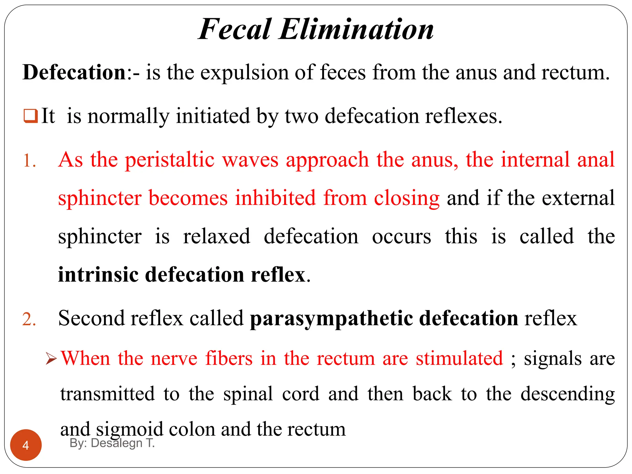
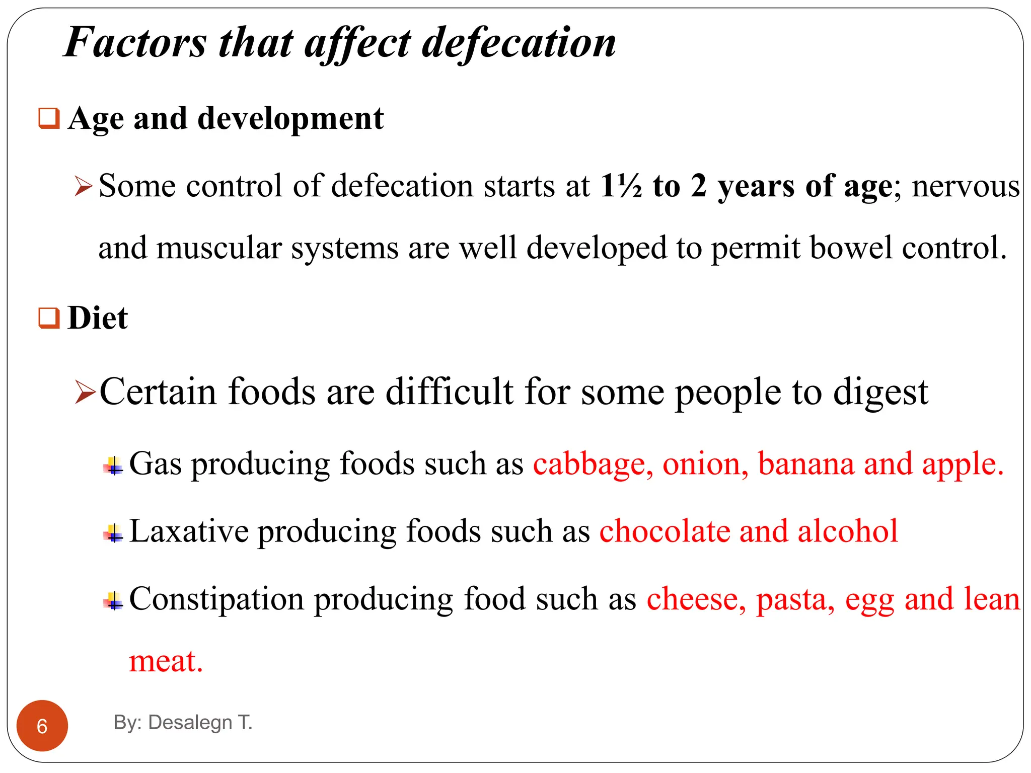
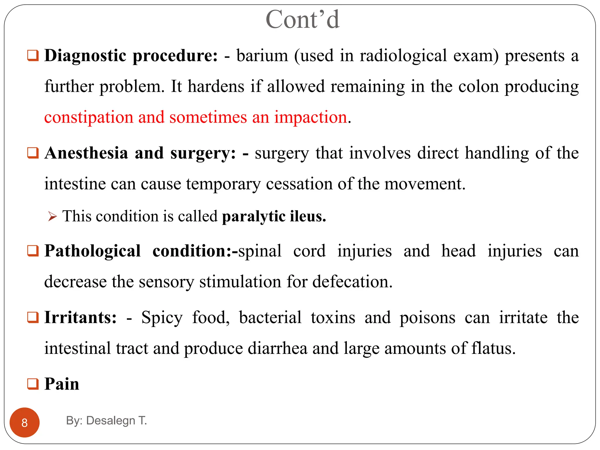
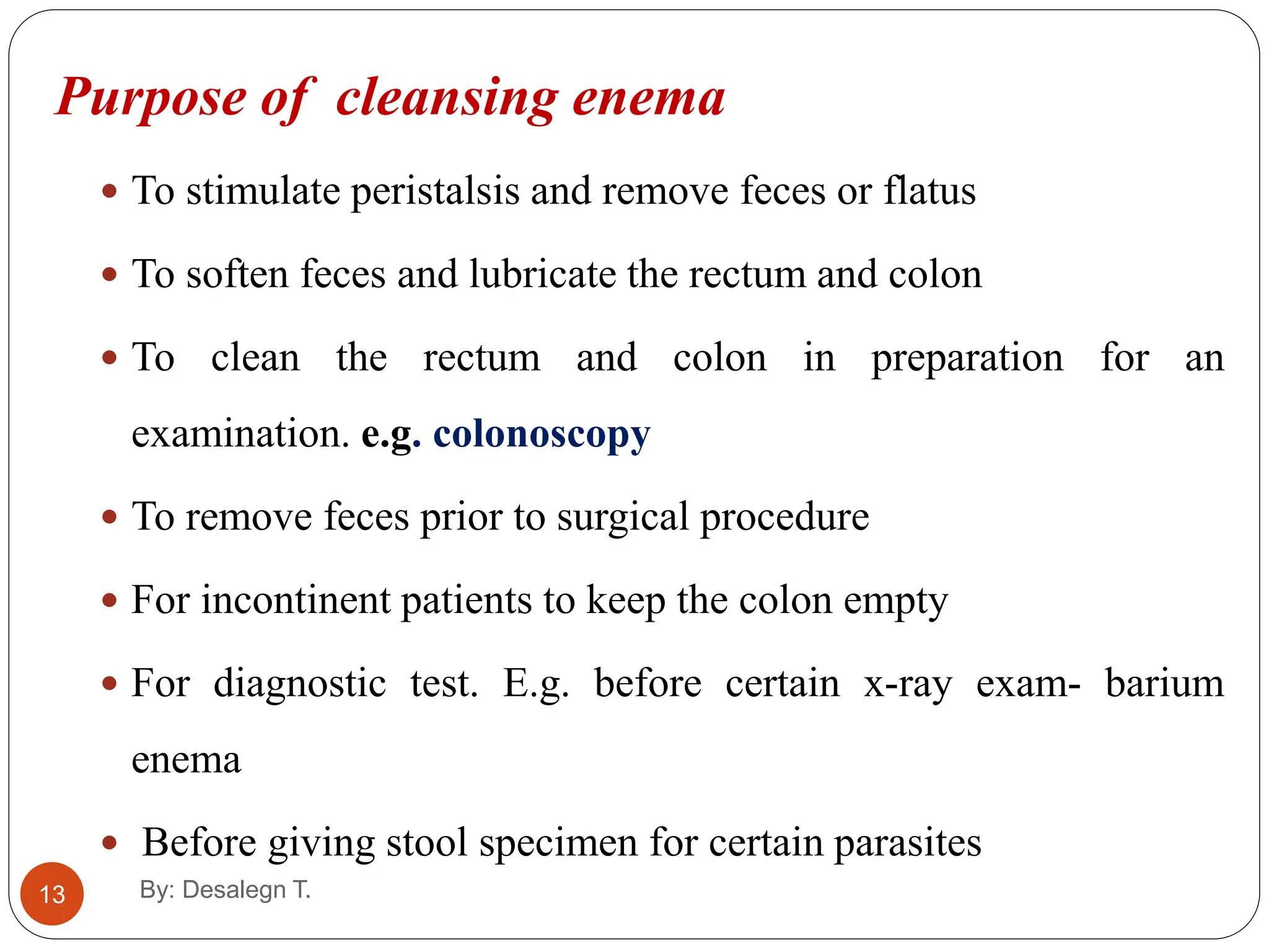
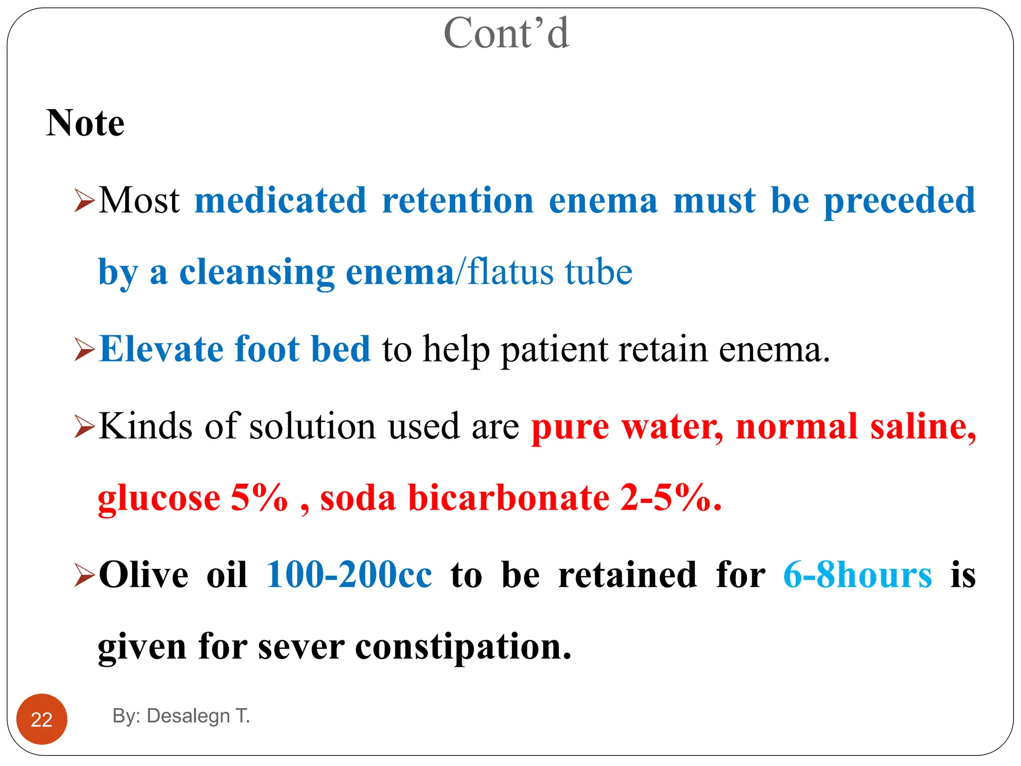
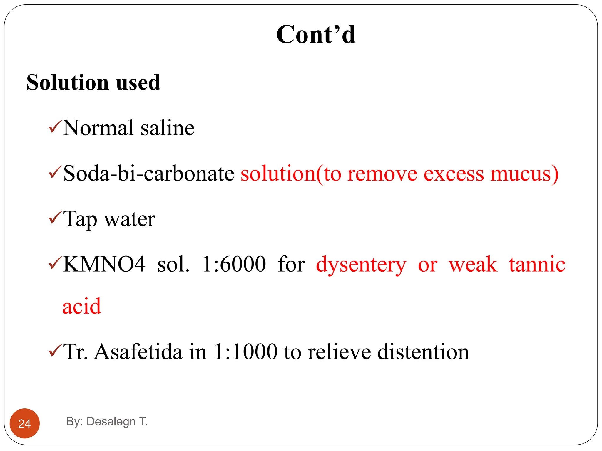
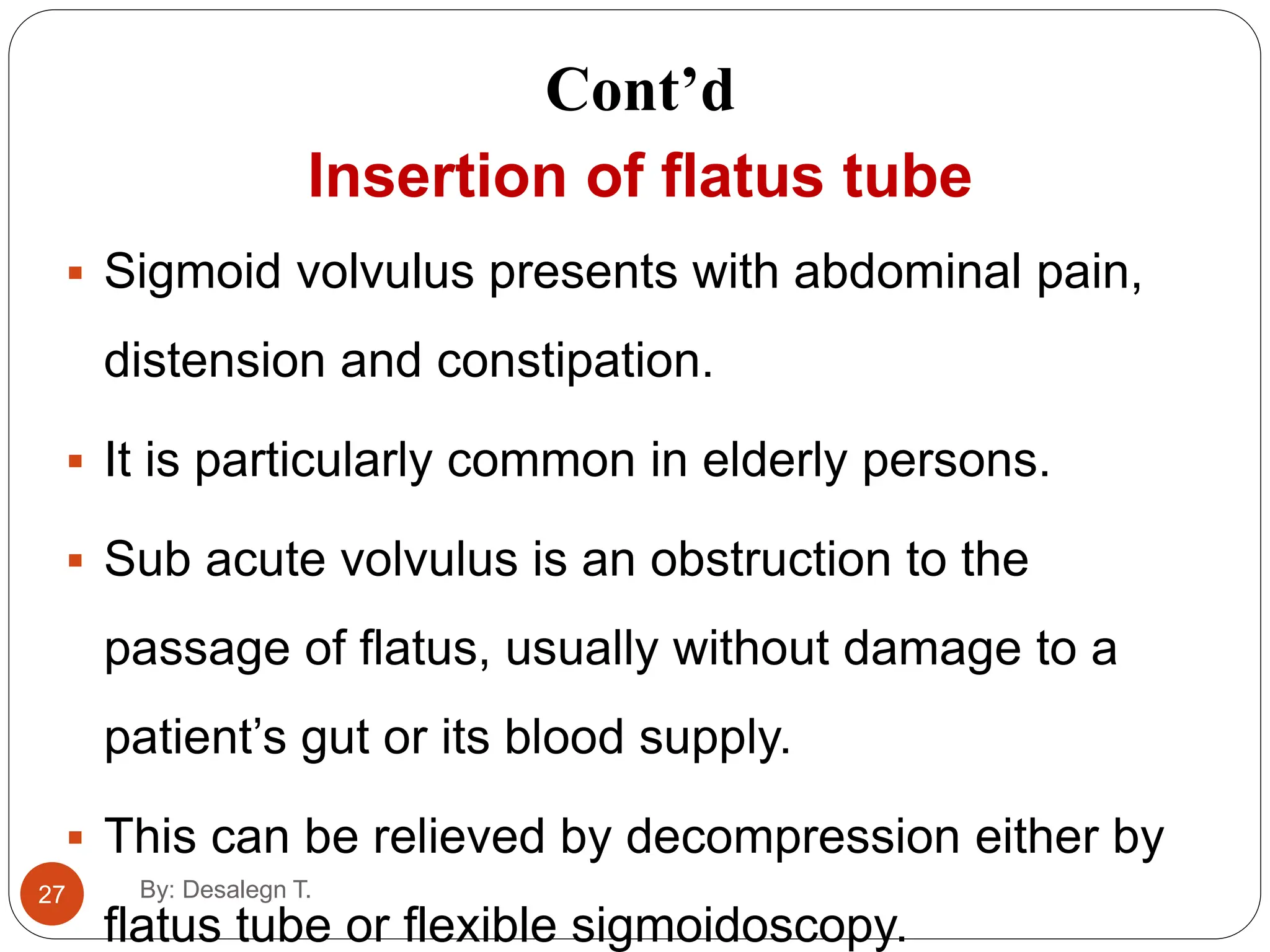
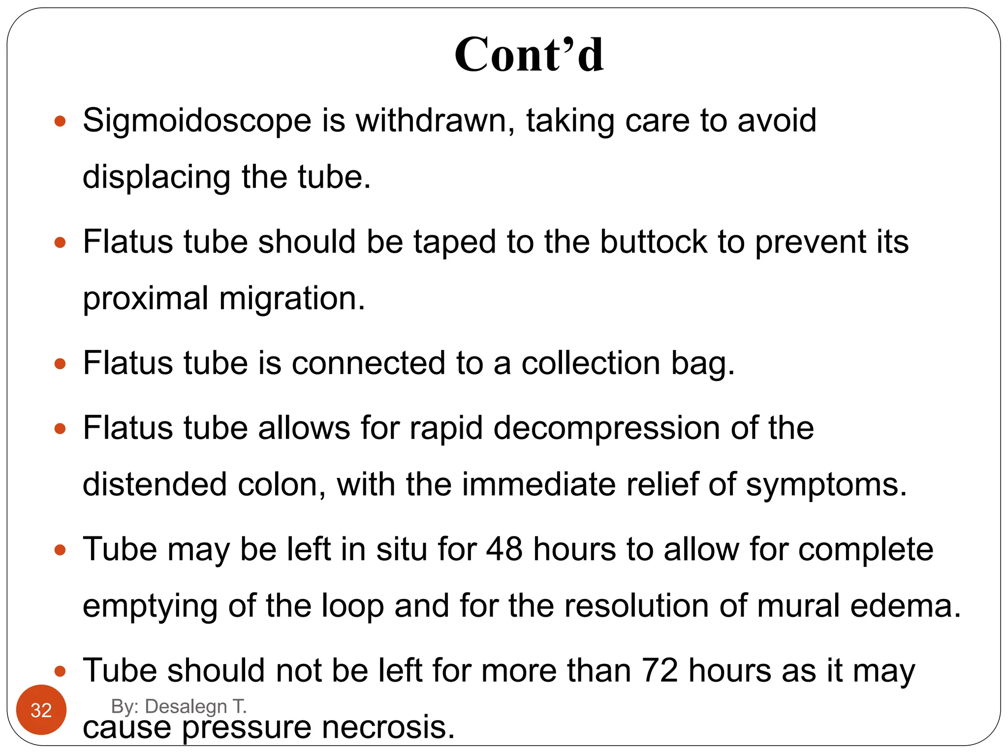
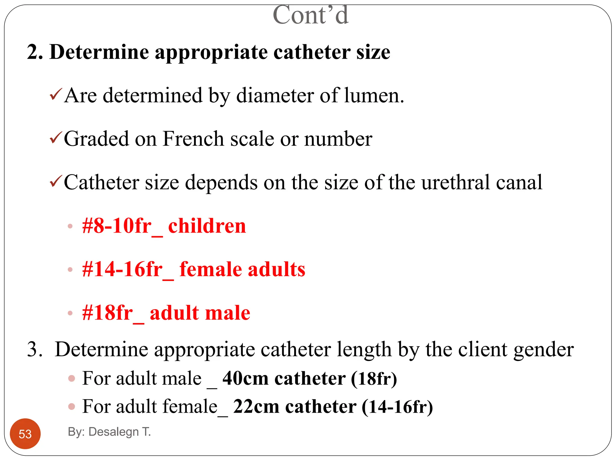
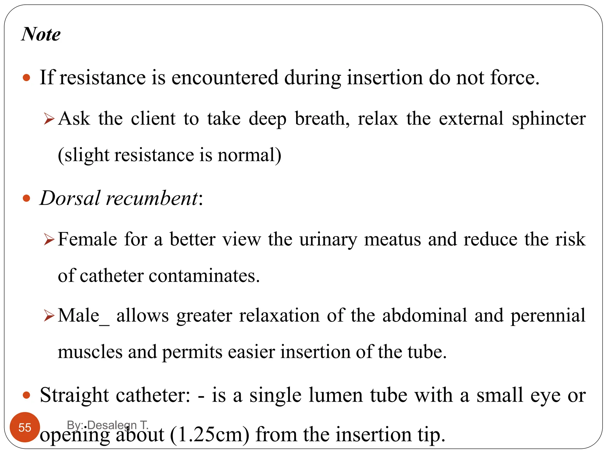
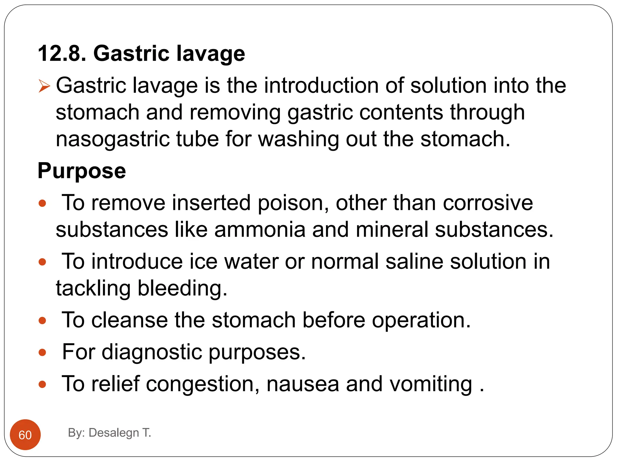
By: Desalegn T. (BscN, Msc in AH)
Urinary and bowel elimination
for second year nursing students 2022
1 By: Desalegn T.
Objectives
2
At the end of this session you will be
able to:-
Define elimination
Define and classify enema care
Demonstrate properly enema care
Perform proper colostomy care
Demonstrate proper catheterization
By: Desalegn T.
Elimination
3
 Urinary and GI systems together provide for the elimination of
body wastes.
 Urinary system filters and excretes urine from the body, there by
maintaining fluid, electrolyte, and acid-base balance.
 Normal bowel function provides for the regular elimination of
solid wastes.
 During periods of stress and illness, clients experience alterations
in elimination patterns.
 Assess for changes, identify problems, and intervene to assist
clients with maintaining proper elimination patterns
By: Desalegn T.
Fecal Elimination
4
Defecation:- is the expulsion of feces from the anus and rectum.
It is normally initiated by two defecation reflexes.
1. As the peristaltic waves approach the anus, the internal anal
sphincter becomes inhibited from closing and if the external
sphincter is relaxed defecation occurs this is called the
intrinsic defecation reflex.
2. Second reflex called parasympathetic defecation reflex
When the nerve fibers in the rectum are stimulated ; signals are
transmitted to the spinal cord and then back to the descending
and sigmoid colon and the rectum
By: Desalegn T.
Cont’d
5
Normal defecation is facilitated by
 Thigh flexion which increases the pressure within the abdomen.
 Sitting position which increases the downward pressure on the rectum.
 An adult usually forms 7 to 10 liters of flatus (gas) in the large
intestine every 24hours.
 Gases include carbon dioxide, methane, oxygen and nitrogen
some are swallowed with foods and fluids taken by mouth; others
are formed through the action of bacteria on the chime in the large
intestine and other gas diffuses from the blood into the GIT.
By: Desalegn T.
Factors that affect defecation
6
 Age and development
Some control of defecation starts at 1½ to 2 years of age; nervous
and muscular systems are well developed to permit bowel control.
 Diet
Certain foods are difficult for some people to digest
Gas producing foods such as cabbage, onion, banana and apple.
Laxative producing foods such as chocolate and alcohol
Constipation producing food such as cheese, pasta, egg and lean
meat.
By: Desalegn T.
Cont’d
7
 Fluid: - healthy fecal elimination requires a daily fluid intake of
200 to 300ml.
 Activity:- stimulate peristaltic thus facilitating the movement of
chime along the colon.
 Psychological factors:- some people who are anxious
experiences increased peristaltic activity and subsequent diarrhea.
 Life style: - early bowl training may establish the habit of
defecating at regular time.
 Medication:- Repeated administration of morphine and codeine
cause constipation.
By: Desalegn T.
Cont’d
8
 Diagnostic procedure: - barium (used in radiological exam) presents a
further problem. It hardens if allowed remaining in the colon producing
constipation and sometimes an impaction.
 Anesthesia and surgery: - surgery that involves direct handling of the
intestine can cause temporary cessation of the movement.
 This condition is called paralytic ileus.
 Pathological condition:-spinal cord injuries and head injuries can
decrease the sensory stimulation for defecation.
 Irritants: - Spicy food, bacterial toxins and poisons can irritate the
intestinal tract and produce diarrhea and large amounts of flatus.
 Pain
By: Desalegn T.
10
Enema
Introduction of fluid into rectum and sigmoid colon for
cleansing, therapeutic or diagnostic purposes.
Purpose:
 For emptying-Soap solution enema
 For diagnostic purpose- (Barium enema)
 For introducing drug/substance - (retention enema)
Solution used
 Normal saline
 Soap solution - soap 1gm in 20ml of H2O
 Epsom salt 15gm-120gm in 1000ml of H2O
By: Desalegn T.
Cont’d
11
Mechanisms of some solutions used in enema
Tap water:
Increase peristalsis by causing mechanical distension of the
colon
Soap solution:
Increases peristalsis due to irritating effect of soap to the
luminal mucosa of colon.
Epsom salt:
Causes flow of ECF(extra cellular fluid) to the lumen causing
mechanical distension resulting peristalsis
By: Desalegn T.
Cont’d
12
Classified into
- Cleansing(evacuation) - Return flow enema
- Carminative/relieving discomfort of gas/ - Retention
Cleansing enema
Kinds:
1. High enema
Is given to clean as much of the colon as possible
2. Low enema
Is administered to clean the rectum and sigmoid colon only
By: Desalegn T.
13
Purpose of cleansing enema
 To stimulate peristalsis and remove feces or flatus
 To soften feces and lubricate the rectum and colon
 To clean the rectum and colon in preparation for an
examination. e.g. colonoscopy
 To remove feces prior to surgical procedure
 For incontinent patients to keep the colon empty
 For diagnostic test. E.g. before certain x-ray exam- barium
enema
 Before giving stool specimen for certain parasites
By: Desalegn T.
Cont’d
14
Guidelines
Enema for adults are usually given at 40-43OC and
children at 37 OC
Hot-cause injury to the bowel mucous
Cold- may trigger a spasm of the sphincter muscles
The amount of solution to be administered depends on:
Kind of enema
The age of the person and
The personal ability to retain the solution
By: Desalegn T.
Cont’d
15
Age Amount
 18months 50-200ml
 18mont-5yrs 200-300ml
 5-12 yrs 300-500ml
 12yrs and older 500-1000ml
 The rectal tube should be appropriate: is measured in French scale
Age Size
 Infants/small child------------10 -12fr
 Toddler --------------14 -16fr
 Scholl age child----------------16 -18fr
 Adult’s -------------------22-30fr
By: Desalegn T.
16
Procedure
 Inform the patient about the procedure.
 Put bed side screen for privacy.
 Attach rubber tube with enema cane with nozzle and stop cock or
clamp.
 Place the patient in the lateral position with the right leg flexed
for adequate exposure of the anus (facilitate the flow of solution
by gravity in to the sigmoid and descending colon which are on
the side).
 Fill the enema can which 1000cc of solution for adults.
By: Desalegn T.
Cont’d
17
 Lubricate about 5cm of the rectal tube facilitate insertion
through the sphincter and minimize trauma.
 Hung the can at least 45cm from bed or 30cm from patient
on the stand.
 Place a piece of mackintosh under the bed
 Make the tube air free by releasing the clamp and allowing
the fluid to run down little to the bed pan and clamp open
prevents unnecessary distention.
 Lift the upper buttock to visualize the anus.
By: Desalegn T.
Cont’d
18
 Insert the tube
7-10cm in adult smoothly and slowly.
5-7.5 cm in the child.
2.5-3.75cm in an infant.
 Raise the solution container and open the clamp to allow
fluid to flow
 Administer the fluid solely, if client complains of fullness
or pain stop the flow for 30 second and restart the flow at
a slower rate
By: Desalegn T.
Cont’d
19
 Do not allow all the fluid to go as there is a possibility
of air entering the rectum or when the client cannot
hold any more and wants to defecate
 Remove bed pan clean the rectal tube.
 Document the procedure.
Note
If resistance is encountered at internal sphincter, ask the
clients to take a deep breath then run a small amount of
solution (relaxes the internal anus sphincter)
By: Desalegn T.
20
Retention enema
 Administration of solution to be retained in rectum for short
or long period
 Fluid usually medicine is retained in rectum for short or long
period- for local or general effects
E.g. oil retention enema, antispasmodic enema
Principles:
Is given slowly by means of a rectal tube
Amount of fluid is usually 150-200cc
Cleansing enema is given after the retention time is over
Temperature of enema fluid is 37.4 c or body temp
By: Desalegn T.
Cont’d
21
Purpose
To supply the body with fluid
To give medication E.g. stimulants(paraldehyde) or
antispasmodic
To soften impacted fecal matter.
Other equipment is similar except the tube for retention
enema is smaller in width.
Procedure
 Similar with the cleansing enema but the enema should be
administered very solely and always be preceded by passing a
flatus tube
By: Desalegn T.
Cont’d
22
Note
Most medicated retention enema must be preceded
by a cleansing enema/flatus tube
Elevate foot bed to help patient retain enema.
Kinds of solution used are pure water, normal saline,
glucose 5% , soda bicarbonate 2-5%.
Olive oil 100-200cc to be retained for 6-8hours is
given for sever constipation.
By: Desalegn T.
Rectal washout (siphoning enema)
23
 Colon irrigation or colonic flush
 Also called enterolysis
 Is the process of introducing large amount of fluid in to large
bowel for flush and allow return or wash out fluid
Purpose
To prepare the patient for x-ray exam and sigmoidoscopy
To prepare the patient for rectum surgery and allow return or
wash out fluid
By: Desalegn T.
Cont’d
24
Solution used
Normal saline
Soda-bi-carbonate solution(to remove excess mucus)
Tap water
KMNO4 sol. 1:6000 for dysentery or weak tannic
acid
Tr. Asafetida in 1:1000 to relieve distention
By: Desalegn T.
25
Procedure
Insert the tube like the cleansing enema.
Client lies on the bed with hips close to the side of the
bed (client assumes a right side lying position for
siphoning).
Open the clamp and allow running about 1000cc of
fluid in the bowel then siphon back into the bucket.
Carry on the procedure until the fluid return is clear.
By: Desalegn T.
Cont’d
26
Note
The procedure should not take>2hours.
Should be finished 1hour before exam or x-ray to give time
for the large intestine to absorb the rest of the fluid.
Give cleansing enema ½ hour before the rectal wash out
Allow the fluid to pass slowly
Amount of solution 5-6 liters until the wash out rectum
fluid becomes clear.
By: Desalegn T.
Cont’d
27
Insertion of flatus tube
 Sigmoid volvulus presents with abdominal pain,
distension and constipation.
 It is particularly common in elderly persons.
 Sub acute volvulus is an obstruction to the
passage of flatus, usually without damage to a
patient’s gut or its blood supply.
 This can be relieved by decompression either by
flatus tube or flexible sigmoidoscopy.
By: Desalegn T.
Cont’d
28
 Decompression can be successful in 50-90% of
cases.
 If strangulation is suspected (severe pain, toxic
or peritonitis signs, or discoloured mucosa at
sigmoidoscopy or flatus tube yields blood stained
fluid) – immediate laparotomy is needed.
 These patients should be carefully observed for
bowel ischemia (persistent abdominal pain and
blood-stained stools), as it indicates the need for
By: Desalegn T.
Cont’d
29
Role of surgery:
Surgery is indicated, if decompression fails to
alleviate the symptoms or if signs of ischemia
develop.
However, volvulus tends to recur in about 60%
of patients after conservative management with
decompression.
Surgery will be needed in recurrent cases.
By: Desalegn T.
Cont’d
By: Desalegn T.
30
PURPOSE
1. To remove flatulence from the lower bowel.
2. To relieve abdominal distension.
3. Used before giving a retention enema.
Cont’d
31
Use of flatus tube and procedure of insertion
 Long, soft flatus tube is inserted, patient in the left lateral
position.
 Obstruction is usually at 15 cms.
 Flatus tube is inserted with the help of a rigid/flexible
sigmoidoscope.
 Sigmoidoscope is held at the twist, flatus tube is passed
along it.
 With a gentle rotatory movement, the tube is eased pass
By: Desalegn T.
Cont’d
By: Desalegn T.
32
 Sigmoidoscope is withdrawn, taking care to avoid
displacing the tube.
 Flatus tube should be taped to the buttock to prevent its
proximal migration.
 Flatus tube is connected to a collection bag.
 Flatus tube allows for rapid decompression of the
distended colon, with the immediate relief of symptoms.
 Tube may be left in situ for 48 hours to allow for complete
emptying of the loop and for the resolution of mural edema.
 Tube should not be left for more than 72 hours as it may
cause pressure necrosis.
33
Bowel diversion /ostomy
 An ostomy is an opening on the abdominal wall for
elimination of feces or urine.
 Purpose of ostomy is to divert and drain fecal or urinary
material.
There are many types of ostomy
Ileostomy:- is an opening into the ileum (small bowel).
Colostomy:- is an opening into the colon (large bowel).
Ureterostomy:- is an opening into the ureter.
By: Desalegn T.
Cont’d
34
Colostomy can be either temporary or permanent.
Temporary colostomy:- performed for traumatic
injuries or inflammatory condition.
Permanent colostomy:- performed to provide a
means of elimination when the rectum or anus is non
functional as result birth defect or cancer of bowel.
Colostomy produces a malodorous mushy drainage
By: Desalegn T.
Cont’d
35
Anatomic location:- The location of the ostomy influences the
character and management of the fecal drainage
 An Ileostomy produces liquid fecal drainage. it contains
some digestive enzyme which damage the skin.
 Ascending colostomy odor is a problem requiring control
(e.g. a deodorant inside the appliance)
 A transverse colostomy some has been reabsorbed.
 A descending colostomy produces increasingly solid fecal
drainage.
By: Desalegn T.
Cont’d
36
Stoma and skin care
 The fecal material from a colostomy or Ileostomy is
irritating to the periosteal skin.
 A barrier such as Karaya is applied over the skin around
the stoma to prevent contact with any execration.
 Odor control is essential to client’s self esteem
 They need to be changed whenever the effluent leaks on
to the periostomal skin
By: Desalegn T.
37
Colostomy irrigation
 Is a form of stoma management used only for clients who have a
sigmoid or descending colostomy.
 It is not done for Ileostomy because the feces are usually liquid.
 A relatively small amount of fluid 300 to 500ml stimulates
evacuation b/se a colostomy has no sphincter and the fluid tends to
return
This problem is reduced by the use of a cone on the irrigating
catheter.
The cone helps to hold the fluid within the bowel during the
irrigation.
By: Desalegn T.
Urinary Elimination
38
 Micturition, voiding and urination all refers to the process of
emptying the urinary bladder.
 Urine collects in the bladder until pressures stimulate special
sensory nerve ending in the bladder wall called stretch receptor.
 This occurs when the adult bladder contains between 250 and
450ml of urine. In children 50 to 200ml stimulates the nerves.
 Urinary retention: - when a person unable to void even though
the bladder contains an excessive amount of urine.
By: Desalegn T.
Factors affecting voiding
39
Factors affecting volume of urine formed and process of voiding
are:
Growth and development: Changes throughout the life cycle
Psychosocial factors: Helps stimulate the micturition reflexes
include privacy , normal position, sufficient time
Fluid and food intake
 Some foods and fluids change color of urine e.g. Carrot
 Certain fluids such as alcohol increases fluid out put
Medication e.g. chlorothiazide, furosemide increases urine
formation.
By: Desalegn T.
Cont’d
40
Muscle tone and activity: exercising regularly will have good muscle tone,
increased body metabolism and a good urine production.
 The presence of an indwelling catheter can lead to poor bladder muscle
tone.
Pathological conditions: - affects the formation and /or excretion of urine.
 Febrile condition can interfere with urine formation b/s the body losses
excessive fluid through perspiration.
 Hypertrophy of prostate gland can interfere with the ability to empty the
bladder
Surgical and diagnostic procedure
 Spinal anesthesia affect the passage of urine b/s they decrease the
client’s awareness of the need to void.
By: Desalegn T.
Urinary Catheterization
41
 Urinary catheterization is introduction of a tube
(catheter) through the urethra into the urinary bladder.
 Is performed only when absolutely necessary for fear of
infection and trauma.
Note:- Strictly a sterile procedure i.e. the health care
giver should always follow aseptic technique
By: Desalegn T.
42
Cont’d
Types of catheter
Based on size, materials, and types.
1. Straight (plain or Robinson)
2. Retention (Foley, indwelling)
Based on the number of lumens they have
1. Straight catheter -has single lumen
2. Double lumen catheter- has two lumens
3. Triple lumen catheter – has three lumens.
By: Desalegn T.
Cont’d
43
 In double lumen catheter, one lumen is used to inflate
balloon at the end of the catheter and the other lumen
is to drain urine.
 The triple lumen catheter provides an additional
lumen for the instillation of irrigating solutions.
By: Desalegn T.
Cont’d
44
 Indwelling catheter
 Remain in place for continuous drainage of urine and it is also called
retention or Foley catheter.
 Are used for gradual decompression of an over distended bladder,
for intermittent bladder drainage and for continuous bladder
drainage and it has a balloon
 Intermittent or straight catheter
 Is used to drain the bladder for shorter period of time (5-10min).
 Clients can be taught to insert and remove intermittent catheter
themselves.
By: Desalegn T.
Indications of urinary catheterization
45
 To relieve urinary retention or incontinence
 To obtain sterile urine specimen
 To measure the amount of post void residual urine for
monitoring
 To empty the bladder before, during and after surgery.
 In case of bladder obstruction
 For a patient with neurologic disorders (unconscious
patients)
 For bladder irrigation or decompression
By: Desalegn T.
Contraindications
46
Urethral trauma
Pelvic fracture
Scrotal hematoma
Complications of catheterization
Urethral trauma from friction during insertion
Infections(renal inflammation, pyelonephritis,
cystitis, etc) from ascending infection following the
catheter
By: Desalegn T.
Alternatives for internal urethral catheterization
47
There are two alternatives
1. Suprapubic catheterization:-it is occasionally used for
continuous drainage.
It is inserted through a small incision above the pubic area
directly into the urinary bladder.
2. Condom catheter: - when voluntary control of urination is not
possible for male clients (if there is urinary incontinence)
 Soft, pliable device made of plastic or rubber material is
applied externally to the penis.
By: Desalegn T.
48
Equipments necessary for catheterization
- Catheter with appropriate size - Syringe (10cc)
-Sterile gloves -Lubricant (ky-
jelly)
- Sterile drape -Collecting bag
& tubing
-Cleansing solution eg. savlon -Plaster
-Cotton swab -Screen
-Forceps -Sterile water
By: Desalegn T.
Procedure
49
 Gather the necessary equipment & explain the procedure to the
client
 Position the client in supine position with legs spread and feet
together
 Open catheterization kit
 Prepare sterile field, done sterile gloves
 Check balloon for patency
 Apply lubricant to 2-5cm of the distal part of the catheter
 Apply sterile drape & bed protection materials (rubber sheet).
By: Desalegn T.
Cont’d
50
 In female separate the labia & in male hold the penis
straight with non-dominant hand.
 Cleanse peri-urethral mucosa with cleansing solution
 Pick the catheter identify the urethral meatus and gently
insert until 2-5 cm beyond where the urine started
flowing
 Inflate balloon with 10cc of sterile liquid/ water.
 Gently pull the catheter until inflation balloon snug
against the bladder neck
By: Desalegn T.
Cont’d
51
 Connect the catheter to the drainage system & secure
it to thigh or abdomen
 Place drainage bag below level of the bladder
 Evaluate catheter function & amount, color, odor,
quality of urine
 Remove gloves & wash your hands.
 Document what you have done
By: Desalegn T.
52
Cont’d
1. Selecting an appropriate catheter
May be made of
Plastic_ for one week
Latex_ 2-3weeks (rubber)
Silicon_ for 2-3 month
 Select the type of material in accordance with the
estimated length of the catheterization period.
By: Desalegn T.
Cont’d
53
2. Determine appropriate catheter size
Are determined by diameter of lumen.
Graded on French scale or number
Catheter size depends on the size of the urethral canal
• #8-10fr_ children
• #14-16fr_ female adults
• #18fr_ adult male
3. Determine appropriate catheter length by the client gender
 For adult male _ 40cm catheter (18fr)
 For adult female_ 22cm catheter (14-16fr)
By: Desalegn T.
Cont’d
54
4. Select appropriate balloon size
5-10ml _for adults
3-ml_ for children
Catheterization using intermittent/ a straight catheter
Purpose
 To relieve discomfort due to bladder distention
 To assess the residual urine
 To obtain a urine specimen
 To empty the bladder prior to surgery
By: Desalegn T.
55
Note
 If resistance is encountered during insertion do not force.
Ask the client to take deep breath, relax the external sphincter
(slight resistance is normal)
 Dorsal recumbent:
Female for a better view the urinary meatus and reduce the risk
of catheter contaminates.
Male_ allows greater relaxation of the abdominal and perennial
muscles and permits easier insertion of the tube.
 Straight catheter: - is a single lumen tube with a small eye or
opening about (1.25cm) from the insertion tip.
By: Desalegn T.
56
Inserting a Retention (Indwelling) Catheter
 Contains a second smaller tube throughout its length on the
inside, this tube is connected to a balloon near the insertion tip.
Purpose
 To manage incontinence
 To provide for intermittent or continuous bladder drainage and
irrigation.
 To prevent infection.
 To measure urine output needs to be monitored hourly.
By: Desalegn T.
Procedure
57
• Explain the procedure to the patient
 Prepare the equipment
 After catheter insertion the balloon is inflated to hold the catheter in
place within the bladder.
 The outside end of the catheter is bifurcated i.e. it has two opening
one to drain the urine the other to inflate the balloon.
 The balloon are sized by the volume of fluid or air used to inflate
them 5ml-30ml(15 commonly) indicated with the catheter size 18fr
-5ml.
By: Desalegn T.
Cont’d
58
 Test the catheter balloon.
 Follow steps as insertion straight catheter.
 Insert the catheter an additional 2.5-5cm beyond the point at
which urine began to flow.
 This ensures that the balloon is inflated inside the bladder and not the
urethra (cause trauma).
 Inflate the balloon with the pre filled syringe
 Apply slight tension on the catheter until you feel resistance
that the catheter is well anchored in the bladder.
 Release the resistance.
By: Desalegn T.
Cont’d
59
 Tape the catheter with tape to the inside of female’s thigh or to the thigh
or a body of a male client.
 Restrict the movement of the catheter and irritation in the urethra when
the client moves.
 Establish effective drainage.
 The bag should be off the floor _ the emptying spout does not become
grossly contaminated.
 Document pertinent data.
Removal
 Withdraw the solution or air from the balloon using a syringe and
remove gently.
By: Desalegn T.
12.8. Gastric lavage
 Gastric lavage is the introduction of solution into the
stomach and removing gastric contents through
nasogastric tube for washing out the stomach.
Purpose
 To remove inserted poison, other than corrosive
substances like ammonia and mineral substances.
 To introduce ice water or normal saline solution in
tackling bleeding.
 To cleanse the stomach before operation.
 For diagnostic purposes.
 To relief congestion, nausea and vomiting .
60 By: Desalegn T.
Indication
 Pyloric stenosis
 Poisoning
 Preoperative care
Equipments
1. Equipments for NG tube insertion
2. Measuring jug
3. 2-3 litters prescribed solution.
4. IV stand
5. Tap water or ice if ordered
6. 50ml syringe
7. Funnel
8. Gloves
9. Rubber and cotton sheet
10. Towel
11. Litmus paper
61 By: Desalegn T.
Cont’d…
12. Suction machine (optional)
13. Labelled specimen container
14. Laboratory request form
15. Charcoal tablets ( universal antidote)
16. Emesis basin
17. Tissue paper
18. Drainage container
19. Vital sign instruments
20. Chart
62 By: Desalegn T.
Procedure
1. Explain the procedure to the patient
2. Wash your hand
3. Assemble the necessary equipments.
4. Keep patient privacy and position the patient
· Position in left lateral position for conscious patient
but if unconscious, place in prone position with
head over the edge of the bed or head lower than
the body (semi prone position)
5. Protect client and bed linen with towel and rubber
sheet
6. Done single use examination glove
63 By: Desalegn T.
Cont…
7. Select the appropriate distances mark on the tube
by measuring the distance on the tube from the
client’s bridge of the nose to ear lobe plus the
distance form ear lobe the to the bottom of the
xiphistemu(xyphoid process).
8. Lubricate the tube.
9. Gently insert and pass the tube, the tongue, the
mouth forward the posterior pharynx (If the client is
unconscious, mouth gag may be used)
 If air bubbles, cough and cyanosis are noticed
withdraw the tube and recommence the procedure.
64 By: Desalegn T.
Cont…
10. Advance the tube slowly to prevent injury until the
pre measured mark,
11. Assess the correct placement by aspirating
stomach contents, or by listening gosh of air while
the client exhales.
12. After the NG tube is in place, allow the stomach
contents to empty in to the drainage container before
instilling any irrigating solutions. This confirms proper
placement of the tube and decreases the risk of over
filling of the stomach and inducing of vomiting.
65 By: Desalegn T.
Cont’d…
13. Once you confirm proper placement of the tube,
begin gastric lavage by instilling about 250ml of
irrigating solution to assess the patient’s tolerance
and prevent vomiting.
 If you are using simple rubber tube for the
lavage
a. Fill the small jug with water/ solution, measure and
pour gently until the funnel is empty, then invent over
the pail (the funnel is connected with the funnel end
of the oesophageal tube)
b. Take specimen, if required, and continue the
process until the returned fluid becomes clear and
the prescribed solution had been used.
66 By: Desalegn T.
Cont’d…
 If you are using a tube with a bulb
a) Clamp the tube below the bulb,
b) With right hand, squeeze the bulb thus forcing the air out
through the funnel.
c) With left hand, pinch tubing above the bulb/proximal to
you/ and at the same time
with right hand, release the clamp. This creates a suction
which will draw the stomach contents into the bulb.
d) Lower funnel and allow excess gastric contents to drain
into the pail.
e) Pour 200-300 cc of solution/water into funnel. Before
funnel is empty allow solution to drain.
f) Before a solution stops running. Turn up funnel and add
another quantity of
solution
g) Repeat this procedure until returns are clean.
67 By: Desalegn T.
Cont’d…
14. Instruct the client to take deep breath and hold it to
close epiglottis
15. Monitor patient’s vital signs, urine output, and level
of consciousness every 15 minutes and notify the
physician for any changes.
16. Give mouth wash
17. If ordered, gently remove the tube, feel the client’s
tube, and watch the respiration
18. Remove glove, hand wash, Clean or discard used
equipments.
19. Comfort the patient
20. Record the procedure, including the time, date,
type of irrigating solution and the amount of gastric
contents drained.
68 By: Desalegn T.
69
THE END !!
By: Desalegn T.

elimination power point for medical surjical

  • 1.
    By: Desalegn T.(BscN, Msc in AH) Urinary and bowel elimination for second year nursing students 2022 1 By: Desalegn T.
  • 2.
    Objectives 2 At the endof this session you will be able to:- Define elimination Define and classify enema care Demonstrate properly enema care Perform proper colostomy care Demonstrate proper catheterization By: Desalegn T.
  • 3.
    Elimination 3  Urinary andGI systems together provide for the elimination of body wastes.  Urinary system filters and excretes urine from the body, there by maintaining fluid, electrolyte, and acid-base balance.  Normal bowel function provides for the regular elimination of solid wastes.  During periods of stress and illness, clients experience alterations in elimination patterns.  Assess for changes, identify problems, and intervene to assist clients with maintaining proper elimination patterns By: Desalegn T.
  • 4.
    Fecal Elimination 4 Defecation:- isthe expulsion of feces from the anus and rectum. It is normally initiated by two defecation reflexes. 1. As the peristaltic waves approach the anus, the internal anal sphincter becomes inhibited from closing and if the external sphincter is relaxed defecation occurs this is called the intrinsic defecation reflex. 2. Second reflex called parasympathetic defecation reflex When the nerve fibers in the rectum are stimulated ; signals are transmitted to the spinal cord and then back to the descending and sigmoid colon and the rectum By: Desalegn T.
  • 5.
    Cont’d 5 Normal defecation isfacilitated by  Thigh flexion which increases the pressure within the abdomen.  Sitting position which increases the downward pressure on the rectum.  An adult usually forms 7 to 10 liters of flatus (gas) in the large intestine every 24hours.  Gases include carbon dioxide, methane, oxygen and nitrogen some are swallowed with foods and fluids taken by mouth; others are formed through the action of bacteria on the chime in the large intestine and other gas diffuses from the blood into the GIT. By: Desalegn T.
  • 6.
    Factors that affectdefecation 6  Age and development Some control of defecation starts at 1½ to 2 years of age; nervous and muscular systems are well developed to permit bowel control.  Diet Certain foods are difficult for some people to digest Gas producing foods such as cabbage, onion, banana and apple. Laxative producing foods such as chocolate and alcohol Constipation producing food such as cheese, pasta, egg and lean meat. By: Desalegn T.
  • 7.
    Cont’d 7  Fluid: -healthy fecal elimination requires a daily fluid intake of 200 to 300ml.  Activity:- stimulate peristaltic thus facilitating the movement of chime along the colon.  Psychological factors:- some people who are anxious experiences increased peristaltic activity and subsequent diarrhea.  Life style: - early bowl training may establish the habit of defecating at regular time.  Medication:- Repeated administration of morphine and codeine cause constipation. By: Desalegn T.
  • 8.
    Cont’d 8  Diagnostic procedure:- barium (used in radiological exam) presents a further problem. It hardens if allowed remaining in the colon producing constipation and sometimes an impaction.  Anesthesia and surgery: - surgery that involves direct handling of the intestine can cause temporary cessation of the movement.  This condition is called paralytic ileus.  Pathological condition:-spinal cord injuries and head injuries can decrease the sensory stimulation for defecation.  Irritants: - Spicy food, bacterial toxins and poisons can irritate the intestinal tract and produce diarrhea and large amounts of flatus.  Pain By: Desalegn T.
  • 9.
    10 Enema Introduction of fluidinto rectum and sigmoid colon for cleansing, therapeutic or diagnostic purposes. Purpose:  For emptying-Soap solution enema  For diagnostic purpose- (Barium enema)  For introducing drug/substance - (retention enema) Solution used  Normal saline  Soap solution - soap 1gm in 20ml of H2O  Epsom salt 15gm-120gm in 1000ml of H2O By: Desalegn T.
  • 10.
    Cont’d 11 Mechanisms of somesolutions used in enema Tap water: Increase peristalsis by causing mechanical distension of the colon Soap solution: Increases peristalsis due to irritating effect of soap to the luminal mucosa of colon. Epsom salt: Causes flow of ECF(extra cellular fluid) to the lumen causing mechanical distension resulting peristalsis By: Desalegn T.
  • 11.
    Cont’d 12 Classified into - Cleansing(evacuation)- Return flow enema - Carminative/relieving discomfort of gas/ - Retention Cleansing enema Kinds: 1. High enema Is given to clean as much of the colon as possible 2. Low enema Is administered to clean the rectum and sigmoid colon only By: Desalegn T.
  • 12.
    13 Purpose of cleansingenema  To stimulate peristalsis and remove feces or flatus  To soften feces and lubricate the rectum and colon  To clean the rectum and colon in preparation for an examination. e.g. colonoscopy  To remove feces prior to surgical procedure  For incontinent patients to keep the colon empty  For diagnostic test. E.g. before certain x-ray exam- barium enema  Before giving stool specimen for certain parasites By: Desalegn T.
  • 13.
    Cont’d 14 Guidelines Enema for adultsare usually given at 40-43OC and children at 37 OC Hot-cause injury to the bowel mucous Cold- may trigger a spasm of the sphincter muscles The amount of solution to be administered depends on: Kind of enema The age of the person and The personal ability to retain the solution By: Desalegn T.
  • 14.
    Cont’d 15 Age Amount  18months50-200ml  18mont-5yrs 200-300ml  5-12 yrs 300-500ml  12yrs and older 500-1000ml  The rectal tube should be appropriate: is measured in French scale Age Size  Infants/small child------------10 -12fr  Toddler --------------14 -16fr  Scholl age child----------------16 -18fr  Adult’s -------------------22-30fr By: Desalegn T.
  • 15.
    16 Procedure  Inform thepatient about the procedure.  Put bed side screen for privacy.  Attach rubber tube with enema cane with nozzle and stop cock or clamp.  Place the patient in the lateral position with the right leg flexed for adequate exposure of the anus (facilitate the flow of solution by gravity in to the sigmoid and descending colon which are on the side).  Fill the enema can which 1000cc of solution for adults. By: Desalegn T.
  • 16.
    Cont’d 17  Lubricate about5cm of the rectal tube facilitate insertion through the sphincter and minimize trauma.  Hung the can at least 45cm from bed or 30cm from patient on the stand.  Place a piece of mackintosh under the bed  Make the tube air free by releasing the clamp and allowing the fluid to run down little to the bed pan and clamp open prevents unnecessary distention.  Lift the upper buttock to visualize the anus. By: Desalegn T.
  • 17.
    Cont’d 18  Insert thetube 7-10cm in adult smoothly and slowly. 5-7.5 cm in the child. 2.5-3.75cm in an infant.  Raise the solution container and open the clamp to allow fluid to flow  Administer the fluid solely, if client complains of fullness or pain stop the flow for 30 second and restart the flow at a slower rate By: Desalegn T.
  • 18.
    Cont’d 19  Do notallow all the fluid to go as there is a possibility of air entering the rectum or when the client cannot hold any more and wants to defecate  Remove bed pan clean the rectal tube.  Document the procedure. Note If resistance is encountered at internal sphincter, ask the clients to take a deep breath then run a small amount of solution (relaxes the internal anus sphincter) By: Desalegn T.
  • 19.
    20 Retention enema  Administrationof solution to be retained in rectum for short or long period  Fluid usually medicine is retained in rectum for short or long period- for local or general effects E.g. oil retention enema, antispasmodic enema Principles: Is given slowly by means of a rectal tube Amount of fluid is usually 150-200cc Cleansing enema is given after the retention time is over Temperature of enema fluid is 37.4 c or body temp By: Desalegn T.
  • 20.
    Cont’d 21 Purpose To supply thebody with fluid To give medication E.g. stimulants(paraldehyde) or antispasmodic To soften impacted fecal matter. Other equipment is similar except the tube for retention enema is smaller in width. Procedure  Similar with the cleansing enema but the enema should be administered very solely and always be preceded by passing a flatus tube By: Desalegn T.
  • 21.
    Cont’d 22 Note Most medicated retentionenema must be preceded by a cleansing enema/flatus tube Elevate foot bed to help patient retain enema. Kinds of solution used are pure water, normal saline, glucose 5% , soda bicarbonate 2-5%. Olive oil 100-200cc to be retained for 6-8hours is given for sever constipation. By: Desalegn T.
  • 22.
    Rectal washout (siphoningenema) 23  Colon irrigation or colonic flush  Also called enterolysis  Is the process of introducing large amount of fluid in to large bowel for flush and allow return or wash out fluid Purpose To prepare the patient for x-ray exam and sigmoidoscopy To prepare the patient for rectum surgery and allow return or wash out fluid By: Desalegn T.
  • 23.
    Cont’d 24 Solution used Normal saline Soda-bi-carbonatesolution(to remove excess mucus) Tap water KMNO4 sol. 1:6000 for dysentery or weak tannic acid Tr. Asafetida in 1:1000 to relieve distention By: Desalegn T.
  • 24.
    25 Procedure Insert the tubelike the cleansing enema. Client lies on the bed with hips close to the side of the bed (client assumes a right side lying position for siphoning). Open the clamp and allow running about 1000cc of fluid in the bowel then siphon back into the bucket. Carry on the procedure until the fluid return is clear. By: Desalegn T.
  • 25.
    Cont’d 26 Note The procedure shouldnot take>2hours. Should be finished 1hour before exam or x-ray to give time for the large intestine to absorb the rest of the fluid. Give cleansing enema ½ hour before the rectal wash out Allow the fluid to pass slowly Amount of solution 5-6 liters until the wash out rectum fluid becomes clear. By: Desalegn T.
  • 26.
    Cont’d 27 Insertion of flatustube  Sigmoid volvulus presents with abdominal pain, distension and constipation.  It is particularly common in elderly persons.  Sub acute volvulus is an obstruction to the passage of flatus, usually without damage to a patient’s gut or its blood supply.  This can be relieved by decompression either by flatus tube or flexible sigmoidoscopy. By: Desalegn T.
  • 27.
    Cont’d 28  Decompression canbe successful in 50-90% of cases.  If strangulation is suspected (severe pain, toxic or peritonitis signs, or discoloured mucosa at sigmoidoscopy or flatus tube yields blood stained fluid) – immediate laparotomy is needed.  These patients should be carefully observed for bowel ischemia (persistent abdominal pain and blood-stained stools), as it indicates the need for By: Desalegn T.
  • 28.
    Cont’d 29 Role of surgery: Surgeryis indicated, if decompression fails to alleviate the symptoms or if signs of ischemia develop. However, volvulus tends to recur in about 60% of patients after conservative management with decompression. Surgery will be needed in recurrent cases. By: Desalegn T.
  • 29.
    Cont’d By: Desalegn T. 30 PURPOSE 1.To remove flatulence from the lower bowel. 2. To relieve abdominal distension. 3. Used before giving a retention enema.
  • 30.
    Cont’d 31 Use of flatustube and procedure of insertion  Long, soft flatus tube is inserted, patient in the left lateral position.  Obstruction is usually at 15 cms.  Flatus tube is inserted with the help of a rigid/flexible sigmoidoscope.  Sigmoidoscope is held at the twist, flatus tube is passed along it.  With a gentle rotatory movement, the tube is eased pass By: Desalegn T.
  • 31.
    Cont’d By: Desalegn T. 32 Sigmoidoscope is withdrawn, taking care to avoid displacing the tube.  Flatus tube should be taped to the buttock to prevent its proximal migration.  Flatus tube is connected to a collection bag.  Flatus tube allows for rapid decompression of the distended colon, with the immediate relief of symptoms.  Tube may be left in situ for 48 hours to allow for complete emptying of the loop and for the resolution of mural edema.  Tube should not be left for more than 72 hours as it may cause pressure necrosis.
  • 32.
    33 Bowel diversion /ostomy An ostomy is an opening on the abdominal wall for elimination of feces or urine.  Purpose of ostomy is to divert and drain fecal or urinary material. There are many types of ostomy Ileostomy:- is an opening into the ileum (small bowel). Colostomy:- is an opening into the colon (large bowel). Ureterostomy:- is an opening into the ureter. By: Desalegn T.
  • 33.
    Cont’d 34 Colostomy can beeither temporary or permanent. Temporary colostomy:- performed for traumatic injuries or inflammatory condition. Permanent colostomy:- performed to provide a means of elimination when the rectum or anus is non functional as result birth defect or cancer of bowel. Colostomy produces a malodorous mushy drainage By: Desalegn T.
  • 34.
    Cont’d 35 Anatomic location:- Thelocation of the ostomy influences the character and management of the fecal drainage  An Ileostomy produces liquid fecal drainage. it contains some digestive enzyme which damage the skin.  Ascending colostomy odor is a problem requiring control (e.g. a deodorant inside the appliance)  A transverse colostomy some has been reabsorbed.  A descending colostomy produces increasingly solid fecal drainage. By: Desalegn T.
  • 35.
    Cont’d 36 Stoma and skincare  The fecal material from a colostomy or Ileostomy is irritating to the periosteal skin.  A barrier such as Karaya is applied over the skin around the stoma to prevent contact with any execration.  Odor control is essential to client’s self esteem  They need to be changed whenever the effluent leaks on to the periostomal skin By: Desalegn T.
  • 36.
    37 Colostomy irrigation  Isa form of stoma management used only for clients who have a sigmoid or descending colostomy.  It is not done for Ileostomy because the feces are usually liquid.  A relatively small amount of fluid 300 to 500ml stimulates evacuation b/se a colostomy has no sphincter and the fluid tends to return This problem is reduced by the use of a cone on the irrigating catheter. The cone helps to hold the fluid within the bowel during the irrigation. By: Desalegn T.
  • 37.
    Urinary Elimination 38  Micturition,voiding and urination all refers to the process of emptying the urinary bladder.  Urine collects in the bladder until pressures stimulate special sensory nerve ending in the bladder wall called stretch receptor.  This occurs when the adult bladder contains between 250 and 450ml of urine. In children 50 to 200ml stimulates the nerves.  Urinary retention: - when a person unable to void even though the bladder contains an excessive amount of urine. By: Desalegn T.
  • 38.
    Factors affecting voiding 39 Factorsaffecting volume of urine formed and process of voiding are: Growth and development: Changes throughout the life cycle Psychosocial factors: Helps stimulate the micturition reflexes include privacy , normal position, sufficient time Fluid and food intake  Some foods and fluids change color of urine e.g. Carrot  Certain fluids such as alcohol increases fluid out put Medication e.g. chlorothiazide, furosemide increases urine formation. By: Desalegn T.
  • 39.
    Cont’d 40 Muscle tone andactivity: exercising regularly will have good muscle tone, increased body metabolism and a good urine production.  The presence of an indwelling catheter can lead to poor bladder muscle tone. Pathological conditions: - affects the formation and /or excretion of urine.  Febrile condition can interfere with urine formation b/s the body losses excessive fluid through perspiration.  Hypertrophy of prostate gland can interfere with the ability to empty the bladder Surgical and diagnostic procedure  Spinal anesthesia affect the passage of urine b/s they decrease the client’s awareness of the need to void. By: Desalegn T.
  • 40.
    Urinary Catheterization 41  Urinarycatheterization is introduction of a tube (catheter) through the urethra into the urinary bladder.  Is performed only when absolutely necessary for fear of infection and trauma. Note:- Strictly a sterile procedure i.e. the health care giver should always follow aseptic technique By: Desalegn T.
  • 41.
    42 Cont’d Types of catheter Basedon size, materials, and types. 1. Straight (plain or Robinson) 2. Retention (Foley, indwelling) Based on the number of lumens they have 1. Straight catheter -has single lumen 2. Double lumen catheter- has two lumens 3. Triple lumen catheter – has three lumens. By: Desalegn T.
  • 42.
    Cont’d 43  In doublelumen catheter, one lumen is used to inflate balloon at the end of the catheter and the other lumen is to drain urine.  The triple lumen catheter provides an additional lumen for the instillation of irrigating solutions. By: Desalegn T.
  • 43.
    Cont’d 44  Indwelling catheter Remain in place for continuous drainage of urine and it is also called retention or Foley catheter.  Are used for gradual decompression of an over distended bladder, for intermittent bladder drainage and for continuous bladder drainage and it has a balloon  Intermittent or straight catheter  Is used to drain the bladder for shorter period of time (5-10min).  Clients can be taught to insert and remove intermittent catheter themselves. By: Desalegn T.
  • 44.
    Indications of urinarycatheterization 45  To relieve urinary retention or incontinence  To obtain sterile urine specimen  To measure the amount of post void residual urine for monitoring  To empty the bladder before, during and after surgery.  In case of bladder obstruction  For a patient with neurologic disorders (unconscious patients)  For bladder irrigation or decompression By: Desalegn T.
  • 45.
    Contraindications 46 Urethral trauma Pelvic fracture Scrotalhematoma Complications of catheterization Urethral trauma from friction during insertion Infections(renal inflammation, pyelonephritis, cystitis, etc) from ascending infection following the catheter By: Desalegn T.
  • 46.
    Alternatives for internalurethral catheterization 47 There are two alternatives 1. Suprapubic catheterization:-it is occasionally used for continuous drainage. It is inserted through a small incision above the pubic area directly into the urinary bladder. 2. Condom catheter: - when voluntary control of urination is not possible for male clients (if there is urinary incontinence)  Soft, pliable device made of plastic or rubber material is applied externally to the penis. By: Desalegn T.
  • 47.
    48 Equipments necessary forcatheterization - Catheter with appropriate size - Syringe (10cc) -Sterile gloves -Lubricant (ky- jelly) - Sterile drape -Collecting bag & tubing -Cleansing solution eg. savlon -Plaster -Cotton swab -Screen -Forceps -Sterile water By: Desalegn T.
  • 48.
    Procedure 49  Gather thenecessary equipment & explain the procedure to the client  Position the client in supine position with legs spread and feet together  Open catheterization kit  Prepare sterile field, done sterile gloves  Check balloon for patency  Apply lubricant to 2-5cm of the distal part of the catheter  Apply sterile drape & bed protection materials (rubber sheet). By: Desalegn T.
  • 49.
    Cont’d 50  In femaleseparate the labia & in male hold the penis straight with non-dominant hand.  Cleanse peri-urethral mucosa with cleansing solution  Pick the catheter identify the urethral meatus and gently insert until 2-5 cm beyond where the urine started flowing  Inflate balloon with 10cc of sterile liquid/ water.  Gently pull the catheter until inflation balloon snug against the bladder neck By: Desalegn T.
  • 50.
    Cont’d 51  Connect thecatheter to the drainage system & secure it to thigh or abdomen  Place drainage bag below level of the bladder  Evaluate catheter function & amount, color, odor, quality of urine  Remove gloves & wash your hands.  Document what you have done By: Desalegn T.
  • 51.
    52 Cont’d 1. Selecting anappropriate catheter May be made of Plastic_ for one week Latex_ 2-3weeks (rubber) Silicon_ for 2-3 month  Select the type of material in accordance with the estimated length of the catheterization period. By: Desalegn T.
  • 52.
    Cont’d 53 2. Determine appropriatecatheter size Are determined by diameter of lumen. Graded on French scale or number Catheter size depends on the size of the urethral canal • #8-10fr_ children • #14-16fr_ female adults • #18fr_ adult male 3. Determine appropriate catheter length by the client gender  For adult male _ 40cm catheter (18fr)  For adult female_ 22cm catheter (14-16fr) By: Desalegn T.
  • 53.
    Cont’d 54 4. Select appropriateballoon size 5-10ml _for adults 3-ml_ for children Catheterization using intermittent/ a straight catheter Purpose  To relieve discomfort due to bladder distention  To assess the residual urine  To obtain a urine specimen  To empty the bladder prior to surgery By: Desalegn T.
  • 54.
    55 Note  If resistanceis encountered during insertion do not force. Ask the client to take deep breath, relax the external sphincter (slight resistance is normal)  Dorsal recumbent: Female for a better view the urinary meatus and reduce the risk of catheter contaminates. Male_ allows greater relaxation of the abdominal and perennial muscles and permits easier insertion of the tube.  Straight catheter: - is a single lumen tube with a small eye or opening about (1.25cm) from the insertion tip. By: Desalegn T.
  • 55.
    56 Inserting a Retention(Indwelling) Catheter  Contains a second smaller tube throughout its length on the inside, this tube is connected to a balloon near the insertion tip. Purpose  To manage incontinence  To provide for intermittent or continuous bladder drainage and irrigation.  To prevent infection.  To measure urine output needs to be monitored hourly. By: Desalegn T.
  • 56.
    Procedure 57 • Explain theprocedure to the patient  Prepare the equipment  After catheter insertion the balloon is inflated to hold the catheter in place within the bladder.  The outside end of the catheter is bifurcated i.e. it has two opening one to drain the urine the other to inflate the balloon.  The balloon are sized by the volume of fluid or air used to inflate them 5ml-30ml(15 commonly) indicated with the catheter size 18fr -5ml. By: Desalegn T.
  • 57.
    Cont’d 58  Test thecatheter balloon.  Follow steps as insertion straight catheter.  Insert the catheter an additional 2.5-5cm beyond the point at which urine began to flow.  This ensures that the balloon is inflated inside the bladder and not the urethra (cause trauma).  Inflate the balloon with the pre filled syringe  Apply slight tension on the catheter until you feel resistance that the catheter is well anchored in the bladder.  Release the resistance. By: Desalegn T.
  • 58.
    Cont’d 59  Tape thecatheter with tape to the inside of female’s thigh or to the thigh or a body of a male client.  Restrict the movement of the catheter and irritation in the urethra when the client moves.  Establish effective drainage.  The bag should be off the floor _ the emptying spout does not become grossly contaminated.  Document pertinent data. Removal  Withdraw the solution or air from the balloon using a syringe and remove gently. By: Desalegn T.
  • 59.
    12.8. Gastric lavage Gastric lavage is the introduction of solution into the stomach and removing gastric contents through nasogastric tube for washing out the stomach. Purpose  To remove inserted poison, other than corrosive substances like ammonia and mineral substances.  To introduce ice water or normal saline solution in tackling bleeding.  To cleanse the stomach before operation.  For diagnostic purposes.  To relief congestion, nausea and vomiting . 60 By: Desalegn T.
  • 60.
    Indication  Pyloric stenosis Poisoning  Preoperative care Equipments 1. Equipments for NG tube insertion 2. Measuring jug 3. 2-3 litters prescribed solution. 4. IV stand 5. Tap water or ice if ordered 6. 50ml syringe 7. Funnel 8. Gloves 9. Rubber and cotton sheet 10. Towel 11. Litmus paper 61 By: Desalegn T.
  • 61.
    Cont’d… 12. Suction machine(optional) 13. Labelled specimen container 14. Laboratory request form 15. Charcoal tablets ( universal antidote) 16. Emesis basin 17. Tissue paper 18. Drainage container 19. Vital sign instruments 20. Chart 62 By: Desalegn T.
  • 62.
    Procedure 1. Explain theprocedure to the patient 2. Wash your hand 3. Assemble the necessary equipments. 4. Keep patient privacy and position the patient · Position in left lateral position for conscious patient but if unconscious, place in prone position with head over the edge of the bed or head lower than the body (semi prone position) 5. Protect client and bed linen with towel and rubber sheet 6. Done single use examination glove 63 By: Desalegn T.
  • 63.
    Cont… 7. Select theappropriate distances mark on the tube by measuring the distance on the tube from the client’s bridge of the nose to ear lobe plus the distance form ear lobe the to the bottom of the xiphistemu(xyphoid process). 8. Lubricate the tube. 9. Gently insert and pass the tube, the tongue, the mouth forward the posterior pharynx (If the client is unconscious, mouth gag may be used)  If air bubbles, cough and cyanosis are noticed withdraw the tube and recommence the procedure. 64 By: Desalegn T.
  • 64.
    Cont… 10. Advance thetube slowly to prevent injury until the pre measured mark, 11. Assess the correct placement by aspirating stomach contents, or by listening gosh of air while the client exhales. 12. After the NG tube is in place, allow the stomach contents to empty in to the drainage container before instilling any irrigating solutions. This confirms proper placement of the tube and decreases the risk of over filling of the stomach and inducing of vomiting. 65 By: Desalegn T.
  • 65.
    Cont’d… 13. Once youconfirm proper placement of the tube, begin gastric lavage by instilling about 250ml of irrigating solution to assess the patient’s tolerance and prevent vomiting.  If you are using simple rubber tube for the lavage a. Fill the small jug with water/ solution, measure and pour gently until the funnel is empty, then invent over the pail (the funnel is connected with the funnel end of the oesophageal tube) b. Take specimen, if required, and continue the process until the returned fluid becomes clear and the prescribed solution had been used. 66 By: Desalegn T.
  • 66.
    Cont’d…  If youare using a tube with a bulb a) Clamp the tube below the bulb, b) With right hand, squeeze the bulb thus forcing the air out through the funnel. c) With left hand, pinch tubing above the bulb/proximal to you/ and at the same time with right hand, release the clamp. This creates a suction which will draw the stomach contents into the bulb. d) Lower funnel and allow excess gastric contents to drain into the pail. e) Pour 200-300 cc of solution/water into funnel. Before funnel is empty allow solution to drain. f) Before a solution stops running. Turn up funnel and add another quantity of solution g) Repeat this procedure until returns are clean. 67 By: Desalegn T.
  • 67.
    Cont’d… 14. Instruct theclient to take deep breath and hold it to close epiglottis 15. Monitor patient’s vital signs, urine output, and level of consciousness every 15 minutes and notify the physician for any changes. 16. Give mouth wash 17. If ordered, gently remove the tube, feel the client’s tube, and watch the respiration 18. Remove glove, hand wash, Clean or discard used equipments. 19. Comfort the patient 20. Record the procedure, including the time, date, type of irrigating solution and the amount of gastric contents drained. 68 By: Desalegn T.
  • 68.
    69 THE END !! By:Desalegn T.