The paper presents an algorithm utilizing wavelet transform for noise elimination in electrocardiogram (ECG) signal processing on a resource-limited microcontroller unit (MCU). The study found db4 to be the optimal mother wavelet for maximizing signal quality, and the algorithm shows promise for applications in portable ECG monitoring, particularly in remote healthcare settings. This work aims to bridge the gap in adapting wavelet transform techniques for embedded systems, thus enhancing ECG diagnostics.
![International Journal of Electrical and Computer Engineering (IJECE)
Vol. 14, No. 2, April 2024, pp. 1530~1543
ISSN: 2088-8708, DOI: 10.11591/ijece.v14i2.pp1530-1543 1530
Journal homepage: http://ijece.iaescore.com
Electrocardiogram signal processing algorithm on
microcontroller using wavelet transform method
Akkachai Phuphanin, Metha Tasakorn, Jeerapong Srivichai
Department of Electrical Engineering, Faculty of Industry and Technology, Rajamangala University of Technology Isan,
Sakon Nakhon, Thailand
Article Info ABSTRACT
Article history:
Received Sep 7, 2023
Revised Oct 26, 2023
Accepted Nov 17, 2023
The electrocardiogram (ECG) is an important parameter for analyzing the
cardiac system. It serves as the primary diagnostic tool for patients with
suspected heart disease, guiding appropriate cardiac investigations according
to the disease or condition suspected. However, ECG measurements may
generate noise, leading to false diagnoses. The wavelet transform is an
effective and widely-used technique for eliminating noise. Typically,
analysis and generation algorithms are developed on computer and using
software built in. This paper presents a noise elimination algorithm based on
the wavelet transform method, designed to operate on resource-limited Node
microcontroller unit (MCU). An efficiency study was conducted to
determine the optimum mother wavelet implementation of the algorithm,
and the results showed that, when considering synthetic ECG signals, db4
was the most suitable for eliminating interference by achieving the highest
signal to noise ratio (SNR) and correlation coefficient. In addition, this
algorithm prototype can analyze ECG signals using the wavelet transform
method processed in a microcontroller and is accurate compared to reliable
programs. It has the potential to be further developed into a low-cost
portable ECG signal measurement tool for use in remote medicine,
healthcare facilities in resource-limited areas, education and training, as well
as home monitoring for chronic patients.
Keywords:
De-noise signal
Electrocardiogram
Microcontroller
Mother wavelet
Signal processing
Wavelet transform
This is an open access article under the CC BY-SA license.
Corresponding Author:
Akkachai Phuphanin
Department of Electrical Engineering, Faculty of Industry and Technology, Rajamangala University of
Technology Isan
Sakon Nakhon Campus, 47160, Thailand
Email: akkachai.ph@rmuti.ac.th
1. INTRODUCTION
Medical professionals and patients may be shielded from coronavirus disease starting in 2019
(COVID-19) exposure by telemedicine and eHealth platforms, which use high-speed communications
networks and application software for the delivery, management, and monitoring of medical services [1]. It is
evident that eHealth and telemedicine systems are crucial for addressing COVID-19 on a worldwide scale
[2]. As the disease spreads around the globe, medical facilities utilize telemedicine and eHealth platforms in a
variety of ways to improve patient care [3]. These include decreased medical expenses, diminished worker
tiredness, more worker sustainability, diminished physician exposure, and diminished need of personal
protective equipment (PPE) suits, such as dust filter masks and respirators [4].
In addition to the remote medical software platform, there are algorithms embedded in the remote
medical system. An algorithm that makes it easier to administer patient summaries that have been analyzed
by remote analytics [5], [6]. In order to deliver the most recent information, artificial intelligence (AI)](https://image.slidesharecdn.com/v3134168ijecedbd-240405053407-3f8a792f/85/Electrocardiogram-signal-processing-algorithm-on-microcontroller-using-wavelet-transform-method-1-320.jpg)
![Int J Elec & Comp Eng ISSN: 2088-8708
Electrocardiogram signal processing algorithm on microcontroller using wavelet … (Akkachai Phuphanin)
1531
chatbot is used. Regarding COVID-19, it offers prevention recommendations as well as potential societal
remedies. It also provides real-time situation reports to medical practitioners [5]. A COVID-19 screening tool
is now being developed using AI to offer preliminary testing for patients with symptoms. and, if required,
suggest additional medical care. However, when there is sufficient and reliable data, both application
software platforms and AI chatbots will become more valuable and significant. As a result, a lot of research
has been done on developing mobile medical devices that include internet of things (IoT) technologies.
Patients can independently measure the fundamental variables required for diagnosis and transmission to
medical staff.
One of the most crucial physiological measures is pulse. The term "pulse" describes the pressure
produced as blood vessels expand and same in time with the heart's rhythmic contraction and expansion.
Therefore, the blood flow wave in the aorta that causes an aneurysm is caused by pulse rate [6]. Thus, heart
rate can be determined using pulse physiological parameters [7]. The study [6] presented an autonomous bed
shifting system for long-term bedridden patients using the patient's pulse rate as a measure of discomfort.
Disease monitoring is necessary because some medical problems necessitate long-term patient care,
especially in the case of chronic diseases [8]. Therefore, it is crucial to continuously check for such
occurrences. The monitoring of blood sugar levels, for instance, will vary depending on the individual. This
aids in scheduling activities, medications, and meals [9]. The architecture system was created using sensing
technology. The technology attempts to deliver real-time information on glucose levels and body
temperature. This also contains other data such as environmental temperature. This information is presented
graphically and is easily legible by humans for end users such as patients and healthcare professionals. A
remote monitoring system for individuals with persistent metabolic problems is described in the paper [8].
These include blood pressure and glucose monitors, smart scales, pulse oximeters, activity trackers, and
blood pressure monitors. The device is also capable of performing analyses that will help with medical
diagnostics.
Tele-ECG is a new IoT-based electrocardiogram (ECG) monitoring device that enables remote
medical monitoring of the condition of the heart [10]. The systems are still inferior to instruments used by
experts. However, by monitoring the patient's ECG over time, the procedure offers basic surveillance. The
ECG, which displays the heart's recorded electrical activity via electrodes on the body's surface, is thought to
be one of the most recognizable representations of heart function [10]. Heart electrical activity and heart rate
fluctuations are measured by physiological parameters ECG [11].
Applications for IoT and health surveillance are presented in the research [12]. Through sensors and
portable devices, the system is intended to capture ECG data together with other healthcare data. The author
also discusses on safely uploading these data to the cloud. This enables immediate access to medical
specialists. Given that earlier systems were expensive and had huge mounting locations, some researchers
[11] have suggested a 12-lead ECG system. Because of this, the suggested solution is portable and has
produced successful outcomes for home isolate systems. Additionally, ECG data can be sent from a distant
area to any point in the world to a medical specialist. Neyja et al. [13] proposed low-cost ECG health
monitoring system using internet of things with automated analytics and alarms includes energy-efficient
wearable sensor devices. The systems are linked to a smart gateway that can be accessed by cardiovascular
caregivers.
To monitor patients' ECG signals using a mobile tele-ECG application and do health assessments
without a doctor's involvement, was developed by Choudhari et al. [10]. The hidden Markov model (HMM)
and ECG sensors-based healthcare system implementation proposal was proposed by Nurdin et al. [14] with
the intention of enhancing patient monitoring and immediate patient assistance in case patients with diseases
related to the cardiovascular system. The construction of an ECG monitoring system was then described by
Rizqyawan et al. [15], in which the user can use the system while ECG signals are being transmitted. Finally,
D’Aloia et al. [16] suggested that the design, execution, and development of ECG follow-up research.
According to research articles [10]–[16], it is a remote medical system that transmits ECG signals
obtained from sensors and transmitted via the Internet to medical personnel via the internet of things. These
studies, however, only send the raw sensor data collected from them without filtering the ECG signal, which
can result in inaccuracies in the ECG. The medical professionals will diagnose patients incorrectly if they
receive muddled signals because of noise. Only study [12], [14] suggested a technique to measure R-R peaks
to calculate heart rate, and the algorithm may be prone to errors if the ECG data contains noise.
The measured ECG readings are contaminated by noise. ECG noise can take many different forms,
including: Random noise called as gaussian noise occurs during transmission, Muscular artifacts or noise
caused by muscle contractions (MA). Electrode motion interference caused by the movement of the body,
which moves the electrode and amplifies the signal. The chest region moves equally as a result of breathing,
creating baseline wander (BW) noise [17]. The ECG activity data is obscured by all four noises, which can
also lead to incorrect heart rate estimations and difficult R peak detection.](https://image.slidesharecdn.com/v3134168ijecedbd-240405053407-3f8a792f/85/Electrocardiogram-signal-processing-algorithm-on-microcontroller-using-wavelet-transform-method-2-320.jpg)
![ ISSN: 2088-8708
Int J Elec & Comp Eng, Vol. 14, No. 2, April 2024: 1530-1543
1532
In paper [18] discusses the results of using different types of wavelet filters with various thresholds
and levels to de-noise ECG signals. The study concludes that the wavelet filter is a good choice for de-
noising ECG signals due to its ability to suit many signals and applications. However, it is crucial to choose
the appropriate wavelet filter type that is similar to the signal in shape or close to its shape. The study finds
that the four-level Daubechies (db4) type is optimal for the Massachusetts institute of technology-Beth Israel
arrhythmia (MIT-BIH) database, while the level two of Symlets 4 (sym4) is suitable for ECG signals from
the monitoring system. The discrete wavelet transform (DWT) based wavelet denoising technique is used
with three different wavelet functions and four different thresholding methods [19]. The study involved the
analysis of ECG signals collected from a cohort of ten female participants aged between 20 and 25 years.
These signals were obtained while employing the Stroop color-word test as a means of inducing mental
stress. The result shows that the Coiflets 5 (coif5) wavelet function and the and rigrsure thresholding rule are
the most effective for removing noise from ECG signals, as demonstrated.
Within the scope of study [20], an innovative approach is introduced for the purpose of reducing
noise in ECG signals through the utilization of wavelets. The methodology centers around the application of
a genetic algorithm, which systematically explores an extensive array of quadrature filter banks. Its objective
is to identify the optimal filter bank that results in the minimal signal-to-noise ratio (SNR). Consequently, the
wavelet and scaling functions associated with the selected filters are acknowledged as the most effective
choice for achieving the task of de-noising. Simulation results show that using the proposed method and the
obtained wavelet improves the SNR of the noisy ECG signal by about 2.5 dB. While Majumdar [21]
describes a proposed method for de-noising ECG signals using wavelet energy and a sub-band smoothing
filter. In contrast to the conventional approach of wavelet threshold de-noising, this novel method exclusively
targets wavelet coefficients necessitating threshold de-noising, determined through wavelet energy analysis.
Meanwhile, it retains the original values of other coefficients. Furthermore, the technique incorporates a
sub-band smoothing filter to amplify the ECG signal's quality through advanced noise reduction. The
experimental results show that the proposed method effectively removes noise from the noisy ECG signals
compared to existing methods.
In recent years, advancements in medical research have led to significant breakthroughs in the field
of cardiac health and diagnostics. One notable area of progress has been the development of innovative
techniques for analyzing ECG signals, aiming to detect critical features like the QRS complex and R-peaks
with remarkable accuracy and efficiency. Research such as study [22] highlights the integration of chaos
analysis, short-time Fourier transform (STFT), and principal component analysis (PCA) to automate QRS
complex detection, offering impressive sensitivity and accuracy rates. In a similar vein, Gupta et al. [23]
introduces a novel application of PCA for R-peak detection, avoiding the need for pre-processing in noisy
ECG signals. Meanwhile, Gupta et al. [24] presents a computer-aided diagnosis system utilizing chaos
analysis to extract non-linear patterns in ECG signals, enhancing arrhythmia detection. Beyond ECG
analysis, other studies such as studies [25] and [26] apply machine learning techniques to retinal blood vessel
segmentation and the identification of high-risk carotid artery plaques, demonstrating the potential for
automated disease detection and prevention. Additionally, research like [27] explores the importance of
collateral circulation in preserving myocardium during coronary artery occlusion, emphasizing the need for
accurate collateral flow assessment in surgical planning. Furthermore, Li et al. [28] introduces a motion
compensation method for 3D coronary artery reconstruction, which proves effective in reducing artifacts and
improving image quality. Finally, Velut et al. [29] analyzes coronary trees using magnetic resonance
angiography (MRA) to estimate the capability of MRA in providing insights into the vascular network. This
collective body of research represents a significant leap forward in the diagnosis and treatment of cardiovascular
and arterial conditions, promising enhanced accuracy, automation, and patient care in the field of cardiac health.
The utilization of wavelet transform, an advanced and effective technique for noise reduction, has
gained prominence in enhancing the performance of ECG signal processing algorithms, as demonstrated in
previous studies [18]-[29]. However, it is noteworthy that, up to this point, wavelet transform-based signal
processing has primarily been confined to traditional computer-based processing platforms and software
applications like MATLAB and LabVIEW. One notable gap in the existing research landscape is the limited
exploration of implementing wavelet transform algorithms for ECG signal processing within embedded
systems, specifically microcontrollers. To date, there has been a dearth of investigations into the adaptation
of wavelet transform techniques to microcontroller-based ECG signal processing. This represents an
uncharted territory in which the potential benefits of wavelet transform, known for its efficacy in noise
reduction, have yet to be fully realized. Therefore, this research endeavors to bridge this gap by presenting a
novel approach: the application of wavelet transforms principles for ECG signal processing on a
microcontroller, specifically the node MCU. This pioneering effort seeks to unlock the advantages of wavelet
transform in noise elimination within the context of resource-constrained embedded systems, potentially
extending its utility to a broader range of real-world applications.](https://image.slidesharecdn.com/v3134168ijecedbd-240405053407-3f8a792f/85/Electrocardiogram-signal-processing-algorithm-on-microcontroller-using-wavelet-transform-method-3-320.jpg)
![Int J Elec & Comp Eng ISSN: 2088-8708
Electrocardiogram signal processing algorithm on microcontroller using wavelet … (Akkachai Phuphanin)
1533
2. ECG SIGNAL PROCESSING PROCESS
There is noise in the measured ECG data. A data processing step is required to get rid of the noise
and get a clear signal. Signal processing entails two steps: Wideband noise removal and baseline wandering
noise removal both employ the wavelet transform approach. Wavelet transform: wavelet transform is a
commonly used method for signal estimation [30], [31] and signal compression [32]. Since the Fourier
transform simply analyzes the signal in terms of frequency, the wavelet transform transforms the signal to be
considered in terms of position and frequency [30]. So, the wavelet transforms signal exhibits less distortion
as a result. The ECG signal is also a type of non-stationary signal since it represents the electrical activity of
the heart during each heartbeat, which changes over time [33]. A Fourier transform that is appropriate for the
analysis of discrete signals is therefore inappropriate. The wavelet transform's foundation is the extraction of
the signal's edge components so that each sub-characteristics components can be considered [31]. Definition
of wavelet transformation in mathematics constant is
𝑊(𝑎, 𝑏) = ∫ 𝑓(𝑡)𝜓𝑎,𝑏(𝑡)𝑑𝑡
∞
−∞
(1)
𝜓𝑎,𝑏(𝑡) =
1
√𝑎
𝜓∗
(
𝑡−𝑏
𝑎
) (2)
When 𝑓(𝑡) is signal, 𝜓𝑎,𝑏(𝑡) is an analytical function with a time shift, scalability feature. In
practice, we can use the discreate wavelet transform to calculate the coefficients for each order of
subcomponents (decomposition). The decomposition can use a set of finite impulse response (FIR) filters that
act as low pass filters and high pass filters. Then, reduce the sample frequency in half (down sampling). It is
possible to employ the discrete wavelet transform algebraically; the fundamental convolution procedures are
given
𝑎𝑛
(𝑖)
= ∑ 𝑔𝑘𝑎2𝑛−𝑘
(𝑖−1)
𝑖 = 1, 2, … , 𝑗
𝑁−1
𝑘=0 (3)
𝑑𝑛
(𝑖)
= ∑ ℎ𝑘𝑎2𝑛−𝑘
(𝑖−1)
𝑎𝑛
(0)
= 𝑥𝑛
𝑁−1
𝑘=0 (4)
When 𝑎𝑛
(𝑖)
is approximation coefficients in component 𝑖 of low pass filters and 𝑑𝑛
(𝑖)
is detail coefficients in
component 𝑖 of high pass filters. For input signal N and the coefficient of the low-pass filter (𝑔𝑘) and high
pass filter (ℎ𝑘) defined by the mother wave function [34]. The Daubechies 6 function (db06), which has a
signal structure resembling the ECG signal, was used for this study [35].
3. ECG SIGNAL PROCESSING ALGORITHM ON NODE MCU
An algorithm for ECG signal analysis using wavelet transform techniques on a node MCU
microcontroller consists of five blocks: ECG signal acquisition, moving average, wavelet transform, peak
detection, and heart rate calculation, as shown in Figure 1. The signal acquisition function stores the ECG
signal (block 1), which has a frequency of 125 Hz, in a data array. The data array requires two R-peaks for
heart rate calculations. To accurately capture both peaks of the heart rate data, a minimum of 200 data points
need to be collected in the array. Nonetheless, if the number of arrays is fewer than 200, it is possible that
two R-peaks may not be present in the louder array, potentially leading to erroneous heart rate calculations by
the algorithm. The captured ECG signal is affected by two types of noise: baseline noise, caused by breathing
or body movements, which has a low frequency, and wideband noise, caused by muscle artifacts and
instrumentation noise. Baseline noise and wideband noise are two types of noise that can affect the quality of
an electrocardiogram (ECG) signal. ECG is a vital medical test used to record the electrical activity of the
heart over time. Noise in the ECG signal can obscure important information and make it difficult to interpret
the results accurately. Therefore, both signals are incorporated into the original data for processing, allowing
us to assess the effectiveness of our proposed algorithm. Hence, within the program designed for embedded
into the microcontroller, it utilizes a total of 54,886 bytes of global memory (equivalent to 67% of the total
memory). In our simulation, all 10-second ECG signals are stored on an external memory card. The Node
MCU reads the data from the memory card and stores it in a data buffer as if it were reading from an ECG
sensor. When executing a single cycle of the program to process ECG signals, the average total execution
time is 249 milliseconds. This duration is deemed suitable for real-time display should the algorithm be
employed in real-life scenarios in the future.
The moving average function (block 2) is an alternative form of averaging that differs from the
conventional averaging method in which all data are averaged. Moving averages employ a fixed number of
windows to average and shift the windows according to the position of the data. In signal processing, moving](https://image.slidesharecdn.com/v3134168ijecedbd-240405053407-3f8a792f/85/Electrocardiogram-signal-processing-algorithm-on-microcontroller-using-wavelet-transform-method-4-320.jpg)

![ ISSN: 2088-8708
Int J Elec & Comp Eng, Vol. 14, No. 2, April 2024: 1530-1543
1536
To validate the ECG signal analysis algorithm employing the wavelet transform method
implemented on the node MCU board, we performed a comparative analysis of the results with a widely
recognized function in the reliable computer programs, known for its established credibility in ECG signal
analysis. Figure 5 illustrates an example of the comparative results, showing that the ECG signal analysis
algorithm utilizing the wavelet transform technique, executed on the node MCU board, exhibits similarities
to the use of pre-existing functions in reliable computer programs, with a correlation coefficient of 0.99. The
correlation coefficient indicates that the ECG signals processed by the algorithm running on the node MCU
exhibit similarity to those processed using the function within the robust program. This observation serves as
empirical evidence supporting the correctness and suitability of the wavelet transform algorithm implemented
on the node MCU for ECG signal analysis.
Figure 5. Comparison of the results of wavelet (Micro) with the function of computer programs
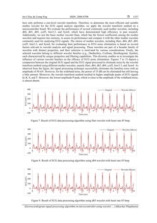
The second part of our study aims to analyze the selection of an appropriate mother wavelet for
processing ECG signals without interference to validate the true capabilities of the ECG signal processing
algorithms. To simulate pure ECG signals without any interference, we generated a simulated ECG signal
with varying heart rates of 65, 70, 75, 80, and 85 beats per minute at a sampling frequency of 125 Hz.
Investigating algorithm efficiency often involves introducing noise to the original signal. In a previous study
[36], noise was incorporated within a range of -12 to 12 dB to assess the performance of beat detection
algorithms. In our current research, we have introduced two specific types of noise: baseline noise with an
amplitude of 50 mV and wideband noise with a power level of 15 dB. Figure 6 provides an example of a
simulated ECG signal with a heart rate of 85 beats per minute. Figure 5 has been to visually represent the
ECG signal illustrating the efficacy of a window size of 49 in effectively eliminating the baseline noise in
Figure 6.
Figure 6. Synthetic ECG and Noisy synthetic ECG with heart rate 85 bmp
As mentioned earlier, the wavelet transform method relies on wavelet coefficients that function as
weights for each frequency component level. Therefore, it is essential to select a coefficient function for the
mother wavelet that is suitable for the signal being processed. However, previous studies have shown that the
use of different mother wavelets for ECG signal analysis yields varying performance results. Since ECG
signal analysis and noise elimination are typically performed on computers with ample computational
resources, wavelet transform method can be applied at multiple levels to obtain the best possible signal.
However, due to resource constraints on microcontroller boards, the ECG signal analysis algorithm presented
-100
0
100
200
300
400
0 1 2 3 4 5 6 7 8 9 10
ECG
(mV)
Time (sec)
Wavelet (computer programs) Wavelet (Micro)
-100
0
100
200
300
400
0 1 2 3 4 5 6 7 8 9 10
ECG
(mV)
Time (sec)
Original Noisy](https://image.slidesharecdn.com/v3134168ijecedbd-240405053407-3f8a792f/85/Electrocardiogram-signal-processing-algorithm-on-microcontroller-using-wavelet-transform-method-7-320.jpg)
![Int J Elec & Comp Eng ISSN: 2088-8708
Electrocardiogram signal processing algorithm on microcontroller using wavelet … (Akkachai Phuphanin)
1539
𝑆𝑁𝑅(𝑑𝑏) = 10𝑙𝑜𝑔10 (
∑ [𝑠𝑜(𝑛)]2
𝑁−1
𝑛=0
∑ [𝑠𝑟𝑒(𝑛)−𝑠𝑜(𝑛)]2
𝑁−1
𝑛=0
) (5)
𝑟 =
∑ (𝑠𝑜−𝑠𝑜
̅̅̅)∙(𝑠𝑟𝑒−𝑠𝑟𝑒
̅̅̅̅̅)
𝑁−1
𝑛=0
√∑ (𝑠𝑜−𝑠𝑜
̅̅̅)2
𝑁−1
𝑛=0 ∑ (𝑠𝑟𝑒−𝑠𝑟𝑒
̅̅̅̅̅)2
𝑁−1
𝑛=0
(6)
𝑅𝑀𝑆𝐸 = √
1
𝑁
∑ (𝑠𝑟𝑒(𝑛) − 𝑠𝑜(𝑛))2
𝑁−1
𝑛=0 (7)
when 𝑠𝑜 is the original ECG signal and 𝑠𝑟𝑒 is the processed ECG signal.
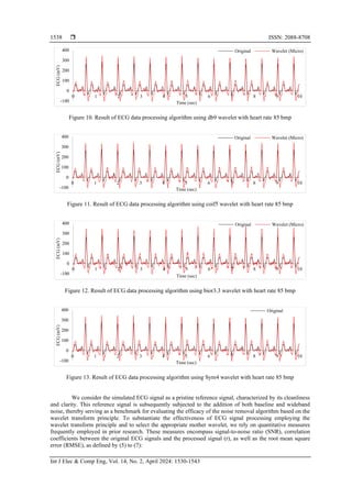
The SNR is a measure of the quality of a signal, indicating how much the signal power in a given
signal compares to the noise power. Figure 14 show the SNR of each mother wavelet used with different
heart rates, ranging from 65 to 85 bpm in increments of 5 bpm. The results indicate that the SNR of every
mother wavelet increases with increasing heart rate. Among the mother wavelets, the db4 wavelet exhibited
the highest SNR values for heart rates of 65, 70, 80, and 85 bpm, with values of 6.44, 6.67, 6.93, and 7.06,
respectively. In contrast, the db9 wavelet yielded the highest SNR at 75 bpm with a value of 6.90, while the
bior3.3 wavelet had the lowest SNR values overall. Figure 15 presents the average SNR values of all heart
rates to determine the mother wavelet that yielded the highest SNR. The db4 wavelet yielded the highest
mean SNR of 6.77, while the bior3.3 wavelet had the lowest mean SNR of 5.83. Heart rate, denoting the
frequency of heart beats per minute, serves as a fundamental physiological parameter. A higher heart rate
signifies an increased rate of cardiac contractions, resulting in a greater number of cardiac cycles occurring
within a specified temporal interval. In the context of ECG data, each cardiac cycle manifests as a distinctive
pattern characterized by key waveforms, including the P-wave, QRS complex, and T-wave. These
waveforms, collectively termed fiducial points, hold invaluable diagnostic information. Elevations in heart
rate correspond to a heightened occurrence of cardiac cycles per unit of time. Consequently, the fiducial
points (P, QRS, T) materialize more frequently within the ECG signal. This heightened frequency enhances
their distinctiveness amidst the backdrop of potential noise, thereby augmenting the SNR within the ECG
signal.
Figure 14. SNR of mother wavelets with different heart rates
Figure 15. Comparison between the mother wavelet in terms of the mean SNR
6.27
6.27
6.54
6.69
6.77
6.44
6.67
6.73
6.93
7.06
6.28
6.54
6.69
6.71
6.92
6.43
6.67
6.90
6.79
6.87
6.39
6.54
6.75
6.68
6.85
5.54
5.74
5.95
5.83
6.09
6.39
6.65
6.71
6.92
6.92
5.0
5.2
5.4
5.6
5.8
6.0
6.2
6.4
6.6
6.8
7.0
7.2
HR = 65 HR = 70 HR = 75 HR = 80 HR = 85
SNR
Heart rate (beats per minute)
Haar db4 db5 db9 coif5 bior3.3 Sym4
6.51
6.77
6.63
6.73
6.64
5.83
6.72
5.00
5.50
6.00
6.50
7.00
Haar db4 db5 db9 coif5 bior3.3 Sym4
SNR
Mother wavelet](https://image.slidesharecdn.com/v3134168ijecedbd-240405053407-3f8a792f/85/Electrocardiogram-signal-processing-algorithm-on-microcontroller-using-wavelet-transform-method-10-320.jpg)

![Int J Elec & Comp Eng ISSN: 2088-8708
Electrocardiogram signal processing algorithm on microcontroller using wavelet … (Akkachai Phuphanin)
1541
Figure 18. RMSE of mother wavelets with different heart rates
Figure 19. Comparison between the mother wavelet in terms of the mean RMSE
In the context of R-wave identification, our algorithm stands out as a straightforward and efficient
solution suitable for implementation in microcontrollers. It exhibits a commendable ability to precisely
pinpoint the R-peaks within the array data, enabling us to subsequently compute the temporal separation
between these R-peaks in alignment with the data sampling frequency. However, it is worth noting that more
sophisticated algorithms, as referenced in [22], [23] are available for the accurate localization of QRS peaks.
When such algorithms are applied to determine the positions of these peaks with precision, the timing of
these peak occurrences can prove valuable in the diagnosis of heart diseases.
5. CONCLUSION
The article describes a study on the use of wavelet transform principles for analyzing ECG signals to
eliminate interference that may occur during measurement. The article presents an algorithm that can
eliminate interference using wavelet transform principles processed in a node MCU and a peak detection
algorithm for calculating heart rate. The algorithm was tested on both simulated and real ECG signals using
parameters such as SNR, correlation coefficient, and RMSE to evaluate performance. In the study of the
performance of the ECG signal processing algorithm, we conducted two tests. The first test aimed to validate
the accuracy of the wavelet transform algorithm used in the node MCU by comparing it with the computer
program’s built-in function. The results showed that the algorithm was accurate with a correlation coefficient
of 0.99. The second test aimed to determine the most suitable mother wavelet for selecting heart rate in the
range of 65-85 beats per minute, which is the normal human heart rate. The db4 mother wavelet was found to
be the most suitable, based on the best values of average SNR, correlation coefficient, and RMSE, which
were 6.77, 0.954, and 29.74, respectively. The results showed that the most appropriate mother wavelet for
signal processing in the simulation model was the db4 wavelet, which provided the highest average SNR and
correlation coefficient values. The db9 wavelet also provided a low average RMSE value and SNR and
correlation coefficient values that were similar to those of the db4 wavelet. In this paper, we demonstrate the
feasibility of implementing complex algorithms, such as the wavelet transform, for ECG noise elimination on
a small microcontroller. Furthermore, the test results demonstrate the accurate performance of the algorithm
for detecting the two R-peaks within the ECG array data, facilitating precise heart rate calculations.
Furthermore, we present the results obtained from employing various mother waveforms to analyze and
assess their efficiency. However, due to memory constraints, the frequency sampling rate was limited to
125 Hz, and the filter bank output frequency was reduced by half. This resulted in the wavelet transform
29.50
30.07
30.47
31.08
31.76
28.95
28.99
29.81
30.23
30.69
29.49
29.14
29.97
31.00
31.18
28.96
28.71
29.24
30.70
31.37
29.10
28.71
29.76
31.09
31.46
32.10
31.96
32.62
34.31
34.32
29.12
28.78
29.88
30.25
31.21
25
26
27
28
29
30
31
32
33
34
35
HR = 65 HR = 70 HR = 75 HR = 80 HR = 85
RMSE
Heart rate (beats per minute)
Haar db4 db5 db9 coif5 bior3.3 Sym4
30.57
29.74
30.16
29.79
30.02
33.06
29.85
29.0
29.5
30.0
30.5
31.0
31.5
32.0
32.5
33.0
33.5
34.0
Haar db4 db5 db9 coif5 bior3.3 Sym4
RMSE
Mother wavelet](https://image.slidesharecdn.com/v3134168ijecedbd-240405053407-3f8a792f/85/Electrocardiogram-signal-processing-algorithm-on-microcontroller-using-wavelet-transform-method-12-320.jpg)
![ ISSN: 2088-8708
Int J Elec & Comp Eng, Vol. 14, No. 2, April 2024: 1530-1543
1542
being performed on only one level, which allowed some noise to remain in the signal. Increasing the
sampling rate or using more complex multiresolution algorithms to process the signals would be a challenge
in future research. Finally, the proposed algorithm prototype exhibits significant potential for a diverse range
of applications that extend beyond the realm of remote medicine. Notable among these applications are the
utilization in wearable health devices, remote monitoring, health and wellness Apps, healthcare facilities in
resource-limited areas, education and training, as well as home monitoring for chronic patients. These
identified applications underscore the algorithm's adaptability and its capacity to thrive within a spectrum of
healthcare and medical technology contexts, rendering it an asset for advancing cardiac care and diagnosis.
Within the constraints of price accessibility to the general public and the requisite quality standards.
REFERENCES
[1] K. Okereafor, O. Adebola, R. Djehaiche, M. El, and B. El, “Exploring the potentials of telemedicine and other non-contact
electronic health technologies in controlling the spread of the novel coronavirus disease (Covid-19),” SSRN Electronic Journal,
vol. 8, pp. 1–14, Apr. 2020.
[2] J. Kaminski, “Informatics in the time of COVID-19,” Canadian Journal of Nursing Informatics, vol. 15, no. 1, 2020.
[3] A. Doshi, Y. Platt, J. R. Dressen, B. K. Mathews, and J. C. Siy, “Keep calm and log on: Telemedicine for COVID-19 pandemic
response,” Journal of Hospital Medicine, vol. 15, no. 5, pp. 302–304, Apr. 2020, doi: 10.12788/jhm.3419.
[4] J. E. Hollander and B. G. Carr, “Virtually perfect? telemedicine for Covid-19,” New England Journal of Medicine, vol. 382,
no. 18, pp. 1679–1681, Apr. 2020, doi: 10.1056/nejmp2003539.
[5] S. Nataraja and P. Nataraja, “IoT based application for e-health an improvisation for lateral rotation,” in RTEICT 2017 - 2nd IEEE
International Conference on Recent Trends in Electronics, Information and Communication Technology, Proceedings, May 2017,
vol. 2018-Janua, pp. 1018–1021. doi: 10.1109/RTEICT.2017.8256753.
[6] M. F. Domingues et al., “Insole optical fiber sensor architecture for remote gait analysis - an e-health solution,” IEEE Internet of
Things Journal, vol. 6, no. 1, pp. 207–214, Feb. 2019, doi: 10.1109/JIOT.2017.2723263.
[7] E. Patti, M. Donatelli, E. Macii, and A. Acquaviva, “IoT software infrastructure for remote monitoring of patients with chronic
metabolic disorders,” in Proceedings - 2018 IEEE 6th International Conference on Future Internet of Things and Cloud, FiCloud
2018, Aug. 2018, pp. 311–317. doi: 10.1109/FiCloud.2018.00052.
[8] T. N. Gia et al., “IoT-based continuous glucose monitoring system: A feasibility study,” Procedia Computer Science, vol. 109,
pp. 327–334, 2017, doi: 10.1016/j.procs.2017.05.359.
[9] L. Ungurean and A. Brezulianu, “An internet of things framework for remote monitoring of the healthcare parameters,” Advances
in Electrical and Computer Engineering, vol. 17, no. 2, pp. 11–16, 2017, doi: 10.4316/AECE.2017.02002.
[10] V. Choudhari, V. Dandge, N. Choudhary, and R. G. Sutar, “A portable and low-cost 12-lead ECG device for sustainable remote
healthcare,” in Proceedings - 2018 International Conference on Communication, Information and Computing Technology,
ICCICT 2018, Feb. 2018, vol. 2018-Janua, pp. 1–6. doi: 10.1109/ICCICT.2018.8325879.
[11] M. S. Hossain and G. Muhammad, “Cloud-assisted industrial internet of things (IIoT) - enabled framework for health
monitoring,” Computer Networks, vol. 101, pp. 192–202, Jun. 2016, doi: 10.1016/j.comnet.2016.01.009.
[12] T. Nguyen Gia et al., “Low-cost fog-assisted health-care IoT system with energy-efficient sensor nodes,” in 2017 13th
International Wireless Communications and Mobile Computing Conference, IWCMC 2017, Jun. 2017, pp. 1765–1770. doi:
10.1109/IWCMC.2017.7986551.
[13] M. Neyja, S. Mumtaz, K. M. S. Huq, S. A. Busari, J. Rodriguez, and Z. Zhou, “An IoT-based e-health monitoring system using
ECG signal,” in 2017 IEEE Global Communications Conference, GLOBECOM 2017 - Proceedings, Dec. 2017, vol. 2018-Janua,
pp. 1–6. doi: 10.1109/GLOCOM.2017.8255023.
[14] M. R. F. Nurdin, S. Hadiyoso, and A. Rizal, “A low-cost Internet of Things (IoT) system for multi-patient ECG’s monitoring,” in
ICCEREC 2016 - International Conference on Control, Electronics, Renewable Energy, and Communications 2016, Conference
Proceedings, Sep. 2017, pp. 7–11. doi: 10.1109/ICCEREC.2016.7814958.
[15] M. I. Rizqyawan, M. F. Amri, R. P. Pratama, and A. Turnip, “Design and development of Android-based cloud ECG monitoring
system,” in Proceedings - 2016 3rd International Conference on Information Technology, Computer, and Electrical Engineering,
ICITACEE 2016, 2017, pp. 1–5. doi: 10.1109/ICITACEE.2016.7892444.
[16] M. D’Aloia, A. Longo, and M. Rizzi, “Noisy ECG signal analysis for automatic peak detection,” Information (Switzerland),
vol. 10, no. 2, p. 35, Jan. 2019, doi: 10.3390/info10020035.
[17] A. S. Ahmed, K. S. Rijab, and S. A. Alagha, “A study of chosen an optimum type of wavelet filter for de-noising an ECG
signal,” International Journal of Current Engineering and Technology, vol. 10, no. 05, pp. 749–756, Oct. 2020, doi:
10.14741/ijcet/v.10.5.9.
[18] P. Karthikeyan, M. Murugappan, and S. Yaacob, “ECG signal denoising using wavelet thresholding techniques in human stress
assessment,” International Journal on Electrical Engineering and Informatics, vol. 4, no. 2, pp. 306–319, Jun. 2012, doi:
10.15676/ijeei.2012.4.2.9.
[19] V. M. Kaviri, M. Sabaghi, and S. Marjani, “De-noising of ECG signals by design of an optimized wavelet,” Circuits and Systems,
vol. 07, no. 11, pp. 3746–3755, 2016, doi: 10.4236/cs.2016.711314.
[20] D. Zhang et al., “An ECG signal de-noising approach based on wavelet energy and sub-band smoothing filter,” Applied Sciences
(Switzerland), vol. 9, no. 22, p. 4968, Nov. 2019, doi: 10.3390/APP9224968.
[21] A. D. S. Majumdar, “Comparative analysis of Coiflet and Daubechies wavelets using global threshold for image de-noising,”
International Journal of Advances in Engineering and Technology, vol. 6, pp. 2247–2252, 2013.
[22] V. Gupta and M. Mittal, “QRS complex detection using STFT, chaos analysis, and PCA in standard and real-time ECG
databases,” Journal of The Institution of Engineers (India): Series B, vol. 100, no. 5, pp. 489–497, Mar. 2019, doi:
10.1007/s40031-019-00398-9.
[23] V. Gupta, N. K. Saxena, A. Kanungo, P. Kumar, and S. Diwania, “PCA as an effective tool for the detection of R-peaks in an
ECG signal processing,” International Journal of System Assurance Engineering and Management, vol. 13, no. 5, pp. 2391–2403,
Oct. 2022, doi: 10.1007/s13198-022-01650-0.
[24] V. Gupta, M. Mittal, and V. Mittal, “Chaos Theory: An emerging tool for arrhythmia detection,” Sensing and Imaging, vol. 21,
no. 1, Feb. 2020, doi: 10.1007/s11220-020-0272-9.](https://image.slidesharecdn.com/v3134168ijecedbd-240405053407-3f8a792f/85/Electrocardiogram-signal-processing-algorithm-on-microcontroller-using-wavelet-transform-method-13-320.jpg)
![Int J Elec & Comp Eng ISSN: 2088-8708
Electrocardiogram signal processing algorithm on microcontroller using wavelet … (Akkachai Phuphanin)
1543
[25] K. Balasubramanian and N. P. Ananthamoorthy, “Robust retinal blood vessel segmentation using convolutional neural network
and support vector machine,” Journal of Ambient Intelligence and Humanized Computing, vol. 12, no. 3, pp. 3559–3569, Oct.
2021, doi: 10.1007/s12652-019-01559-w.
[26] X. Xu et al., “Multi-feature fusion method for identifying carotid artery vulnerable plaque,” Irbm, vol. 43, no. 4, pp. 272–278,
Aug. 2022, doi: 10.1016/j.irbm.2021.07.004.
[27] M. Harmouche, M. Maasrani, J. P. Verhoye, H. Corbineau, and A. Drochon, “Coronary three-vessel disease with occlusion of the
right coronary artery: What are the most important factors that determine the right territory perfusion?,” Irbm, vol. 35, no. 3,
pp. 149–157, Jun. 2014, doi: 10.1016/j.irbm.2013.11.002.
[28] S. Li, J. C. Nunes, C. Toumoulin, and L. Luo, “3D coronary artery reconstruction by 2D motion compensation based on mutual
information,” Irbm, vol. 39, no. 1, pp. 69–82, Feb. 2018, doi: 10.1016/j.irbm.2017.11.005.
[29] J. Velut, P. A. Lentz, D. Boulmier, J. L. Coatrieux, and C. Toumoulin, “Assessment of qualitative and quantitative features in
coronary artery MRA,” Irbm, vol. 32, no. 4, pp. 229–242, Sep. 2011, doi: 10.1016/j.irbm.2011.05.002.
[30] D. Popov, A. Gapochkin, and A. Nekrasov, “An algorithm of daubechieswavelet transform in the final field when processing
speech signals,” Electronics (Switzerland), vol. 7, no. 7, p. 120, Jul. 2018, doi: 10.3390/electronics7070120.
[31] B. A. Rajoub, “An efficient coding algorithm for the compression of ECG signals using the wavelet transform,” IEEE
Transactions on Biomedical Engineering, vol. 49, no. 4, pp. 355–362, Apr. 2002, doi: 10.1109/10.991163.
[32] I. Haider, M. Shahbaz, M. Abdullah, and M. Nazim, “Feature extraction for identification of extension and flexion movement of
wrist using EMG signals,” in Canadian Conference on Electrical and Computer Engineering, May 2015, vol. 2015-June,
no. June, pp. 792–795. doi: 10.1109/CCECE.2015.7129375.
[33] B. R. de Oliveira, M. A. Q. Duarte, C. C. E. de Abreu, and J. V. Filho, “A wavelet-based method for power-line interference
removal in ECG signals,” Research on Biomedical Engineering, vol. 34, no. 1, pp. 73–86, 2018, doi: 10.1590/2446-4740.01817.
[34] M. K. Islam, A. N. M. M. Haque, G. Tangim, T. Ahammad, and M. R. H. Khondokar, “Study and analysis of ECG signal using
MATLAB & LABVIEW as effective tools,” International Journal of Computer and Electrical Engineering, pp. 404–408, 2012,
doi: 10.7763/ijcee.2012.v4.522.
[35] R. Haddadi, E. Abdelmounim, M. El Hanine, and A. Belaguid, “Discrete wavelet transform based algorithm for recognition of
QRS complexes,” in International Conference on Multimedia Computing and Systems -Proceedings, Apr. 2014, pp. 375–379, doi:
10.1109/ICMCS.2014.6911261.
[36] Z. F. Apandi, R. Ikeura, S. Hayakawa, and S. Tsutsumi, “An analysis of the effects of noisy electrocardiogram signal on heartbeat
detection performance,” Bioengineering, vol. 7, no. 2, pp. 1–15, Jun. 2020, doi: 10.3390/bioengineering7020053.
BIOGRAPHIES OF AUTHORS
Akkachai Phuphanin received the B.Eng., M.Eng. and Ph.D degree in
Telecommunication engineering from Suranaree University of Technology, Thailand, in 2019,
2011 and 2017, respectively. Currently, he is lecturer at the Department of Electrical
Engineering, Rajamangala University of Technology Isan Sakonnakhon Campus. His research
interests include wireless sensor networks and applications, reinforcement learning, learning
(artificial intelligence), smart farm and home technology, coverage control, internet of thing,
and signal processing. He can be contacted at email: akkachai.ph@rmuti.ac.th.
Metha Tasakorn received the B.Eng. degree in industrial electrical technology
from King Mongkut’s Institute of Technology North Bangkok (KMITNB), Bangkok,
Thailand, in 1993, M.Sc. degree in electronics electrotechnics and automatics from Université
Paris-Est Créteil Val-de-Marne, Créteil, France, in 2002. He is a lecturer with the Department
of Electrical Engineering, Faculty of Industry and Technology, Rajamangala University of
Technology Isan Sakon Nakhon Campus, Sakon Nakhon, 47160 Thailand. His research
interests include biosensor, optical tweezer, robotics, power electronics, learning (artificial
intelligence), smart farm and home technology and internet of thing. He can be contacted at
email: metha.ta@rmuti.ac.th.
Jeerapong Srivichai received the B.Eng. degree in electrical engineering from
Pathumwan Institute of Technology, Bangkok, Thailand, in 2003 and the M.S. degree in
electrical engineering from Kasetsart University, Bangkok, Thailand, in 2008 and Ph.D.
degrees in electric engineering from Suranaree University of Technology, Nakhon Ratchasima,
Thailand, in 2020. Currently, he is an assistant professor at the Department of Electrical
Engineering, Rajamangala University of Technology Isan Sakonnakhon Campus. His research
interests include railway electrification, electric vehicle, power electronics, induction heating.
He can be contacted at email: geerapong.sr@rmuti.ac.th.](https://image.slidesharecdn.com/v3134168ijecedbd-240405053407-3f8a792f/85/Electrocardiogram-signal-processing-algorithm-on-microcontroller-using-wavelet-transform-method-14-320.jpg)