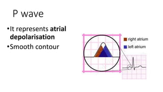
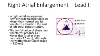
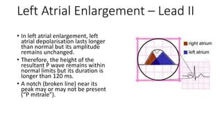
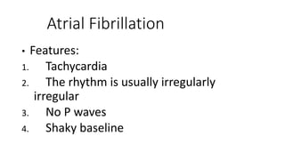
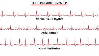
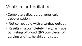
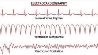
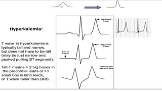
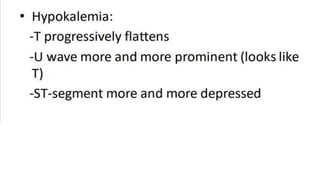
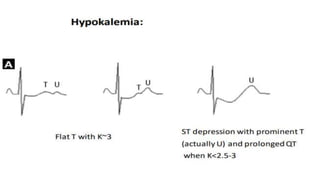
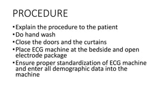
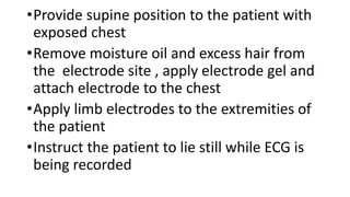
ECG
P wave
•It represents atrial
depolarisation
•Smooth contour
Right Atrial Enlargement – Lead II
• In right atrial enlargement,
right atrial depolarisation lasts
longer than normal and its
waveform extends to the end
of left atrial depolarisation.
• The combination of these two
waveforms produces a P
waves that is taller than
normal (> 2.5 mm), although
the width remains unchanged
(< 120 ms).
Left Atrial Enlargement – Lead II
• In left atrial enlargement, left
atrial depolarisation lasts longer
than normal but its amplitude
remains unchanged.
• Therefore, the height of the
resultant P wave remains within
normal limits but its duration is
longer than 120 ms.
• A notch (broken line) near its
peak may or may not be present
(“P mitrale”).
Acute STEMI may produce ST elevation with either
concave, convex or obliquely straight morphology.
Atrial Fibrillation
• Features:
1. Tachycardia
2. The rhythm is usually irregularly
irregular
3. No P waves
4. Shaky baseline
Ventricular fibrillation
•Completely disordered ventricular
depolarisation
• Not compatible with a cardiac output
• Results in a completely irregular trace
consisting of broad QRS complexes of
varying widths, heights and rates
PROCEDURE
•Explain the procedure to the patient
•Do hand wash
•Close the doors and the curtains
•Place ECG machine at the bedside and open
electrode package
•Ensure proper standardization of ECG machine
and enter all demographic data into the
machine
•Provide supine position to the patient with
exposed chest
•Remove moisture oil and excess hair from
the electrode site , apply electrode gel and
attach electrode to the chest
•Apply limb electrodes to the extremities of
the patient
•Instruct the patient to lie still while ECG is
being recorded
•After the procedure remove leads of
electrodes wipe the gel from the skin and
wash hands
•Interpret ECG and inform physician
•Replace the machine and document the
procedure
Thank you

ECG basics simplified for students .pptx

  • 1.
  • 2.
    P wave •It representsatrial depolarisation •Smooth contour
  • 3.
    Right Atrial Enlargement– Lead II • In right atrial enlargement, right atrial depolarisation lasts longer than normal and its waveform extends to the end of left atrial depolarisation. • The combination of these two waveforms produces a P waves that is taller than normal (> 2.5 mm), although the width remains unchanged (< 120 ms).
  • 4.
    Left Atrial Enlargement– Lead II • In left atrial enlargement, left atrial depolarisation lasts longer than normal but its amplitude remains unchanged. • Therefore, the height of the resultant P wave remains within normal limits but its duration is longer than 120 ms. • A notch (broken line) near its peak may or may not be present (“P mitrale”).
  • 9.
    Acute STEMI mayproduce ST elevation with either concave, convex or obliquely straight morphology.
  • 10.
    Atrial Fibrillation • Features: 1.Tachycardia 2. The rhythm is usually irregularly irregular 3. No P waves 4. Shaky baseline
  • 12.
    Ventricular fibrillation •Completely disorderedventricular depolarisation • Not compatible with a cardiac output • Results in a completely irregular trace consisting of broad QRS complexes of varying widths, heights and rates
  • 17.
    PROCEDURE •Explain the procedureto the patient •Do hand wash •Close the doors and the curtains •Place ECG machine at the bedside and open electrode package •Ensure proper standardization of ECG machine and enter all demographic data into the machine
  • 18.
    •Provide supine positionto the patient with exposed chest •Remove moisture oil and excess hair from the electrode site , apply electrode gel and attach electrode to the chest •Apply limb electrodes to the extremities of the patient •Instruct the patient to lie still while ECG is being recorded
  • 19.
    •After the procedureremove leads of electrodes wipe the gel from the skin and wash hands •Interpret ECG and inform physician •Replace the machine and document the procedure
  • 20.