Down Syndrome Chromosome Structure and Properties
•Download as PPT, PDF•
3 likes•490 views

Down syndrome is caused by trisomy 21, where a person has three copies of chromosome 21 instead of the normal two copies. About 94% of Down syndrome cases are caused by trisomy 21 due to errors in meiosis where the chromosomes do not separate properly. This can occur during the formation of either sperm or egg cells, leading to eggs or sperm with an extra copy of chromosome 21. When one of these eggs or sperm goes on to fertilization, it results in a zygote with three copies of chromosome 21, causing Down syndrome. People with Down syndrome have characteristic facial features and developmental delays. The risk of trisomy 21 increases with advanced maternal age due to a higher chance of errors in meiotic chromosome
![Let Us Pray ,[object Object],[object Object],[object Object],[object Object],[object Object],[object Object],[object Object],[object Object],[object Object],[object Object],[object Object],[object Object],Amen .](data:image/gif;base64,R0lGODlhAQABAIAAAAAAAP///yH5BAEAAAAALAAAAAABAAEAAAIBRAA7)
Recommended
More Related Content
What's hot
What's hot (20)
Similar to Down Syndrome Chromosome Structure and Properties
Similar to Down Syndrome Chromosome Structure and Properties (20)
Recently uploaded
Recently uploaded (20)
Down Syndrome Chromosome Structure and Properties
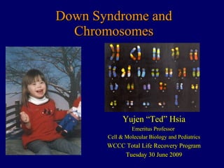
- 1. Down Syndrome and Chromosomes Yujen “Ted” Hsia Emeritus Professor Cell & Molecular Biology and Pediatrics WCCC Total Life Recovery Program Tuesday 30 June 2009
- 23. Chromatin Packaging http://www.accessexcellence.org/AB/GG/chroma_packg.html DNA double helix “ Beads on a string” Packed nucleosomes Extended chromatin Condensed chromatin Chromatid arms DNA can be packed x 50,000. nm 2 11 30 300 1400 700 It becomes condensed for cell division. The genes in DNA can only be active when not condensed. X -shaped chromosomes are formed before cell division.
- 33. Meiotic Non-Disjunction http://www.erin.utoronto.ca/~w3bio380/Lectsked/Lect04/Egg2.htm Meiosis I II 24 22 Non-disjunction Normal Non-disjunction Normal Normal Normal Maternal 21 Paternal 21 23 23 24 24 22 22
- 34. Chromosomes in Spontaneous Abortion At least 15% of first trimester pregnancies are lost. 50% Normal 50% Abnormal 46, N + 16 45, X triploidy other other autosomal trisomies
- 36. Trisomy 21 in Down Syndrome 24,X,+21 23,Y 47,XY,+21 Egg, extra 21 Sperm, normal Zygote Extra 21 Trisomy 21
- 45. Robertsonian Translocations Two acrocentrics Breaks occur at the centromere of both acrocentrics. The q arms fuse (Acrocentric p satellites have similar repetitive genes, loss of some satellites has no clinical importance). The p arms fuse 21 21 14 14 rbt ( 14q / 21q ) rbt ( 14p / 21p ) The fused p arms are lost. In effect, a 21 has fused with a 14 .
- 58. 48,XXYY 47,XYY
- 63. Maternal Serum -Fetoprotein http://www.obgyntoday.org/previous%20conf%20understanding%20AFP.htm AFP Concentration Pregnant women Normal Down syndrome
Editor's Notes
- Diffuse plaques of Ab42 begin to form by age 14. In the late 20’s, these form specific plaques with accrual of Ab40. Neurofibrillary tangles appear by the 30’s
- Photograph from:http://www.nas.com/downsyn/donations.html
- A Nucleus has DNA, RNA, Nucleous, Protein, Euchromatin, Heterochromatin
- CHROMOSOMAL RAINBOWS: NOVEL MULTIBAND PATH FILTER SETS FOR FLUOESCENCE MICROSCOPY ALLOW RAPID MAPPING OF CHROMOSOMAL REARRANGEMENTS BY IMAGING A PANEL OF 16 INDIVIDUALLY LABELED LOCUS-SPECIFIC DNA PROBES WHICH BIND EVERY 8-10 MEGABASEPAIRS ALING HUMAN CHROMOSOME 10. Benjamin O'Brien and H.-Ulli Weier, University of California, E.O. Lawrence Berkley National Laboratory, Berkeley, CA
- Non-disjunction leads to abnormal chromosome numbers If one daughter gamete has extra chromosome (i.e., 24 rather than the normal 23) the syndrome is called trisomy (3 copies of the same chromosome) at fertilization e.g., Down Syndrome--trisomy 21: this is due to an extra copy of chromosome 21 If the daughter gamete has 1 less chromosome (i.e., a total of 22 rather than the normal 23) it is referred to as monosomy (e.g., Turner Syndrome--lacks sex chromosome) Most cases of monosomy are not viable .
- Richard Hall BSc SRCS. Cytogenetics Department, Guy's & St. Thomas' NHS Foundation Trust. TRISOMY FOR ALL C’SOMES SEEN (EXCEPT 1) – ACCOUNTS FOR 60% OF ALL ABNORMALITIES MOST COMMON T16 AND 45,X (APPROX 20%) POLYPLOIDY (MOSTLY TRIPLOIDY) 15% REMAINING ABNORMALITIES (5%) ARE MOSTLY STRUCTURAL REARRANGEMENTS
- Berman: Nelson’s Pediatrics Ed 16, 2000. Judith G. Hall: Genetic Diseases. P. 329 Figure 78-4 Partial karyotypes from patients with Down syndrome. A, Patient with trisomy 21. B, Chromosome 21 from two patients and their parents. Left: Two of a patient's chromosomes with brightly fluorescent satellites were transmitted by the mother. Right: Another patient's two chromosomes with bright satellites resulted from paternal nondisjunction at second meiotic division. C, 21q21q translocation. D, 14q21q translocation in a mother (above) and her affected child (below).
- Increases to ~7 million by 6 months in utero Then # decreases due to apoptosis Only ~2 x 10 6 at birth ~0.5 x 10 6 at puberty Continual decline until menopause: ovulation and death.
- http://www.uptodate.com/online/content/image.do?imageKey=hema_pix/robertso.htm&title=Robertsonian%20translocation
- Understanding Maternal Serum Screening Lecturer: Alberto de la Vega MD, FACOG, RDMS Associate Professor of Obstetrics and Gynecology, University of Puerto Rico School of Medicine The purpose of this conference is to provide you with an understanding of the concepts behind risk assessment for both open neural tube defects (ONTD’s) and Down’s syndrome (DS) based on maternal serum screening. Alfa feto protein: · Evaluation of alfa feto protein (AFP) started the era of prenatal screening in the 1970’s. · 1972: Brock & Sutcliffe reported that measurement of an abnormally elevated value of AFP in Amniotic fluid (AF) was useful for diagnosis of ONTD’s · 1974: Wald et al and Brock et al in 2 articles published in the Lancet reported that an elevated value of AFP in maternal serum was capable of identifying patients at risk of ONTD but, due to the large overlap with the normal population, was not diagnostic. · 1977: large scale collaborative study in the United Kingdom showed that AFP was useful as a screening test for ONTD. · 1979: first large scale collaborative study in the United States showed that screening with AFP between 15 and 21 weeks was capable of identifying up to 80% of cases of ONTD’s. What is AFP? · 1956: Bergstrand and Czar reported a protein detected in fetal serum as an extra band in the Alpha-1 region. Since it was not seen in normal adults it was called AFP · Related evolutionarily to albumin: · genes for both proteins are located in chromosome 4 · both have similar molecular weight (69,000 Daltons) · both have the same amount of negative charges. · Circulating time for AFP is 4 days · Anti-sera for AFP does not cross react with albumin therefore it can be used to detect it in maternal serum. AFP metabolism: · Synthesized in yolk sac and fetal liver · Peak fetal serum concentration is 300 mg reached during the end of the first trimester · The fetal liver produces AFP until 30 weeks of gestation and then stops rather abruptly. · A specific function for AFP has not been found. Fetuses with a genetic deficiency for production of AFP have been reported without adverse effects. Amniotic fluid AFP: · Appears mostly through fetal urine due to immaturity of fetal kidneys. Thus as kidneys mature, AF concentration gradually decreases. · It reaches a peak at 12 weeks & decreases 10% per week in the second trimester (see figure 1) · In cases of ONTD and any structural defect that affects skin permeability, it appears in the AF through transudation. · In cases of fetal congenital nephrosis, the large amount of proteinuria maintains AF concentrations high.