This document provides information on various medical imaging modalities including computed tomography (CT), cone beam CT, magnetic resonance imaging (MRI), nuclear medicine, and ultrasonography. It describes the basic principles, advantages, disadvantages and uses of each technology. CT and MRI are discussed in more detail, including their history, components, image reconstruction techniques, and applications in diagnosing various oral and maxillofacial conditions.
This document discusses regenerative endodontics and its potential to regenerate dental tissues. It defines key concepts like stem cells, growth factors, and scaffolds that are important components of tissue engineering approaches. Various types of dental stem cells and growth factors are described. The document outlines regenerative endodontic procedures and notes advantages in saving teeth, while also acknowledging limitations like the pulp's blood supply. It concludes that regenerative procedures are promising for dental reconstruction but require more research to increase predictability.
Nuclear scintigraphy uses radioactive isotopes injected into the horse's bloodstream to identify areas of bone with increased metabolic activity. The isotopes accumulate in actively healing bone and damaged soft tissues, emitting gamma rays detected by a camera to create images. It is a sensitive way to diagnose subtle or complex lameness issues, monitor fracture healing, and examine areas like the pelvis or back inaccessible to other modalities. The horse is hospitalized after the scan for 48 hours as the isotope decays to ensure handler safety. Scintigraphy highlights injury locations but may require other tests to characterize findings fully.
This document provides an overview of panoramic radiography. It discusses the history and development of panoramic radiography, the principles behind it including image layer and rotation center, equipment used, procedures for taking panoramic x-rays, common errors, clinical indications, advantages, and limitations. Panoramic radiography allows visualization of all teeth and supporting structures on a single film with a relatively low radiation dose.
This document contains a 45 question multiple choice professional exam on operative dentistry. The questions cover topics like restoration techniques, endodontics, pediatric dentistry, dental materials, and crown and bridge procedures. The exam tests knowledge of cavity preparation, root canal treatment methods, dental material properties, and choices for different crown and bridge scenarios. It aims to evaluate a dentist's understanding of key areas in operative dentistry.
CBCT acquires 3D volumetric image data using a cone-shaped x-ray beam and area detector that rotates around the patient. It involves x-ray generation, detection, and image reconstruction from projection data. Key factors influencing image quality and radiation dose include field of view, voxel size, number of projections, and exposure settings. Proper patient preparation and use of optimized protocols are important for clinical applications of CBCT imaging.
This document discusses dental object localization through horizontal and vertical shifts. It notes that a tooth positioned palataly, meaning towards the palate, and that objects behind the central ray move with it and are therefore located behind other objects.
This document contains a 45 question multiple choice professional exam on operative dentistry. The exam covers topics like restoration techniques, endodontics, pediatric dentistry, dental materials, and crown and bridge work. The questions test knowledge of areas like cavity preparation and cutting instruments, root canal treatment procedures, dental material properties, and types of crown designs.
CBCT provides high-resolution 3D imaging with significantly less radiation exposure compared to medical CT. It allows visualization of detailed bony anatomy and assessment of bone quality and dimensions, which is essential for accurate implant planning and placement. CBCT imaging can evaluate the maxillary sinus, mandibular canal, alveolar bone height and width, and other anatomic structures to determine optimal implant size, position, and surgical approach. When combined with computer-guided surgery, CBCT facilitates precise placement of implants according to the presurgical plan.
CBCT provides 3D images of the jaws and teeth using low-dose x-rays and computer reconstruction. It allows visualization of bone quality, morphology, and proximity to anatomical structures for applications in dental implant planning, oral surgery, endodontics, orthodontics, and more. CBCT is particularly useful when traditional 2D imaging is limited by anatomical superimposition or inability to assess 3D bone characteristics. Some key uses of CBCT include implant planning, assessment of impacted teeth, maxillofacial trauma, airway imaging, and evaluation of periodontal defects.
CBCT imaging allows dentists to visualize anatomy in 3 dimensions. It has many applications including implant planning, assessing impacted teeth, orthodontic evaluation, and examining maxillofacial trauma and lesions. CBCT provides important information such as bone quantity and quality, location of vital structures, and relationship of pathologies to surrounding tissues. It also allows for accurate pre-surgical planning through tools like radiographic tracing and implant simulation. CBCT has advantages over medical CT such as smaller size, lower radiation dose, and software tailored for dentistry.
CBCT provides 3D imaging using a cone-shaped X-ray beam. It is useful for dental and maxillofacial applications. The scan takes 5 seconds and images can be displayed in orthogonal planes, multiplanar reformats, or 3D renders. Clinical uses include implant planning, localization of anatomical structures, assessment of impacted teeth, fractures, and lesions. CBCT allows accurate evaluation of bone dimensions for safe implant placement with less radiation than conventional CT.
The document discusses the advantages of dental CT imaging over traditional dental radiographs. Dental CT provides accurate multi-planar views of the jaw anatomy, allowing for precise measurement of bone dimensions and identification of vital structures. It indicates dental CT is particularly useful for dental implant planning and assessment of complex dental conditions like tumors, cysts, fractures and trauma. The technique of dental CT involves thin slice imaging of the jaw to generate panoramic and cross-sectional views for detailed evaluation.
The document identifies common errors that can occur when taking panoramic dental x-rays. These include the teeth being positioned too far anterior or posterior to the focal trough, the patient's head being turned or tipped in various directions, issues with the placement of the lead apron, and other errors like patient movement, double exposures, or using incorrect exposure settings. Proper patient positioning and technique are necessary to avoid these errors and ensure diagnostic quality panoramic dental x-rays.
This document discusses common artifacts and positioning errors seen on panoramic radiographs. It describes ghost images, which are duplicate images caused when an object is penetrated twice by x-rays. It also discusses errors like open lips obscuring teeth, improper positioning of the chin resulting in overlapping structures, and movement during exposure causing blurring or duplication. Positioning the patient correctly in relation to the focal trough and keeping the spine straight are important to avoid errors.
This document discusses different types of splints used in dentistry. It describes removable and fixed splints that can be used for dentulous or edentulous patients. Removable splints include labiolingual, fenestrated, lingual, and occlusal splints. Fixed splints include metal cap splints. Edentulous splints include Gunning and Kingsley splints. The document outlines the indications, advantages, and construction of various splints used to immobilize and stabilize injured jaws and dental arches.
This document provides an overview of basic implant surgery procedures. It discusses preoperative planning and patient preparation, sterile surgical techniques, flap design options, bone preparation using drills and taps, implant placement, cover screw installation, post-operative care, and recent advances like computer-guided surgery. Successful implant placement requires thorough planning, training on the selected implant system, and meticulous sterile technique to ensure predictable, long-lasting results.
Anatomical considerations for placing dental implants.
all the basic anatomical landmarks and considerations which are to be taken care off before and while placing a dental implant.
any type of implant it may be...wether endossous or subperiosteal or tranosteal.
lack of knowledge of basic anatomy will never lead to success of implant.
Indian Dental Academy: will be one of the most relevant and exciting training center with best faculty and flexible training programs for dental professionals who wish to advance in their dental practice,Offers certified courses in Dental implants,Orthodontics,Endodontics,Cosmetic Dentistry, Prosthetic Dentistry, Periodontics and General Dentistry.
Dental implants are artificial roots, usually made of titanium, that are surgically placed into the jawbone to support replacement teeth. Implants can replace one or more missing teeth and provide support for dentures or bridges. The implant surgery involves drilling into the jawbone, placing the implant, and allowing time for osseointegration where the implant fuses with surrounding bone.
This article discusses the ideal elements of a resort vacation. It recommends looking for an all-inclusive resort that provides meals, drinks and activities so travelers can fully relax without worries. The best resorts offer beautiful beaches or landscapes, a variety of on-site activities and amenities like spas, and a lively social scene to meet other guests.
Travel Fizz is a premier overseas education consultants located in Chandigarh.We provide career counselling and student visa assistance for Canada, Australia, New Zealand,USA and UK. We provide step by step guidance on admission process and visa process to student looking for admission into the top universities of the world.
El GPS es un sistema creado por el Departamento de Defensa de EE.UU. que permite localizar un elemento utilizando al menos 4 de los 24 satélites del GPS que orbitan la Tierra, midiendo la distancia a cada satélite a través del tiempo que tardan las señales en llegar al receptor para calcular de manera eficaz la localización.
Mohanad Mohamed Mahmud is a Business Administration graduate from Yarmouk University seeking a position to utilize his business knowledge and organizational skills. He currently works as a sales executive for an Abu Dhabi printing company, where his duties include maintaining client relationships, developing new customers, and presenting quotes. His graduation project focused on the impact of self-awareness on leadership style. He is proficient in MS Office, Arabic, English, and has certifications in management skills.
1. 101 級牙放共筆 W11-1
Computed Tomography (CT) 黃昭慈
● 簡介
a. CT 是一種使用斷層攝影術 tomography 的醫學影像技術。
b. 沿著單一旋轉軸 single axis of rotation 拍攝多張物體內部的 2D X 光,每
一張稱為一個 slice。
c. 再使用數位幾何處理 Digital geometry processing 將多張 2D 的 slice 合併,
組合成 3D 的影像。
● 歷史
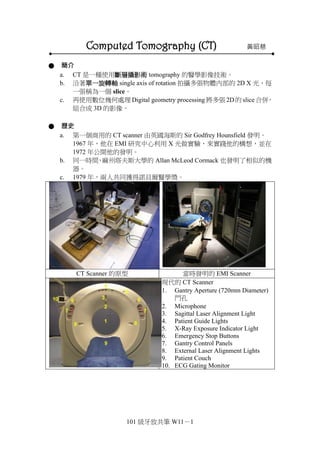
a. 第一個商用的 CT scanner 由英國海斯的 Sir Godfrey Hounsfield 發明。
1967 年,他在 EMI 研究中心利用 X 光做實驗,來實踐他的構想,並在
1972 年公開他的發明。
b. 同一時間,麻州塔夫斯大學的 Allan McLeod Cormack 也發明了相似的機
器。
c. 1979 年,兩人共同獲得諾貝爾醫學獎。
CT Scanner 的原型 當時發明的 EMI Scanner
現代的 CT Scanner
1. Gantry Aperture (720mm Diameter)
門孔
2. Microphone
3. Sagittal Laser Alignment Light
4. Patient Guide Lights
5. X-Ray Exposure Indicator Light
6. Emergency Stop Buttons
7. Gantry Control Panels
8. External Laser Alignment Lights
9. Patient Couch
10. ECG Gating Monitor
2. 101 級牙放共筆 W11-2
CT Gantry (門: 就是讓病人通過的圓孔)
–Internal Structure
1. X-Ray Tube
2. Filters, Collimator, and Reference
Detector
3. Internal Projector
4. X-Ray Tube Heat Exchanger (Oil
Cooler)
5. High Voltage Generator (0-75kv)
6. Direct Drive Gantry Motor
7. Rotation Control Unit
8. Data Acquisition System (Das)
9. Detectors
10. Slip Rings
● 基本 X 光原理 Basic Factors
1. 吸收:吸收 X 光轉換成能量
Absorption: stopping of x-rays with
transfer of energy
2. 散射:X 光折射
Scatter: deflection of x-rays
3. 入射光強度:照到物體上的 X 光粒
子數
Incident Intensity: No. of x-ray
photons falling on an object
4. 透射光強度:通過物體的 X 光粒子
數
Transmitted Intensity: No. of photons
passing through
5. 不同物體的 X 光穿透力不同,造成
透射光強度(Transmitted Intensity)不
同,這些透射光成像產生不同的灰階
● 衰減 Attenuation
1. 吸收和散射減弱了通過物體
的 X 光強度
The reduction of the beam
intensity on passing through
the material due to absorption
plus
2. 測量比較入射光和透射光強
度可以獲得 X 光的衰減程度
3. 101 級牙放共筆 W11-3
The degree of attenuation is
obtained by measuring and
comparing the incident and
transmitted intensities
3. 組織的組成會影響透射光強
度。較緻密的組織吸收掉較多
的 X 光、透過的 X 光少;相
反的,較疏鬆的組織透過的 X
光多,影像就會比較亮。
● X 光衰減和偵測的應用 Applications of X-ray attenuation & detection
a. Conventional X-ray (Radiography)
b. Conventional Tomography
c. Computed Tomography:這堂課的主角 CT
● CT Scan 原理
1. 照 CT 得到的是 Axial 向的切片
(sections/cuts/slices)
CT scan produces axial
sections/cuts/slices
2. CT 影像由掃描機記錄
The CT image is recorded through a
SCAN.
3. 繞著物體外圍一圈測量 X 光衰
減,即為一次掃描
A scan is made up of multiple
X-Ray attenuation measurements
around an objects periphery
4. X ray tube 和 Detector 分別在對角
線上,繞著物體照一圈,得到不同
面向的 slice,之後再用電腦把這
些影像拼湊起來
Principle of CT CT Image Formation
4. 101 級牙放共筆 W11-4
照 CT 的時候病人往機器裡推進(寬箭
頭),所以 X 光路徑其實是螺旋狀的(細
箭頭)。因此醫學用 CT 又稱為 Spiral CT
或 Helix CT。
那如果是物體不動、X 光繞著轉一圈
的,就是牙科比較常用的 Cone Beam
CT。
可以設定 Slice 要切多厚。每個 Slice
會再切成小 cube,以一個 cube 為單位
偵測出影像。
● X 光衰減
上圖由左至右:鉛、硫酸鋇、骨頭、肌肉、血、肝、奶油、脂肪、空氣
疏鬆的地方白,緻密的地方黑,以此判讀影像。
● CT Number
每個點具有不同的穿透值,此穿透值可以換算為 CT Number。
CT numbers are
calculated from
the measured
attenuation values
using the equation
given below.
CT 值是用右邊的
公式,與水比較,
算出 X 光衰減的
量。
公式不會考~
6. 101 級牙放共筆 W11-6
Wide Window:
相反的,如果 100-800 這段,我不想看那麼細,因為裡面結構都差不多,那
我就把它縮小,這樣叫 Wide Windowing。
中中曹曹提醒您:Narrow window→窄的放大看,Wide window→寬的放小看,考!
Gray Scale 灰色梯度表 (Hounsfield scale = Hu)
CT 值另一個名稱是 Hu,紀念發明 CT 的人。下表是組織在 CT 上看起來的顏色。
Tissue & Pathology Hu (Attenuation) Density
Metallic Material Artifact
Bone 600-1000 White
Calcification 100-500 White
Fresh Blood Clot 60-90 White
Fibrotic Tissue 50-70 Gray
Soft Tissue 35-45 Gray
Soft Tissue Edema 15-25 Gray-Dark
Thick Tissue Fluid 10-16 Dark
Pure Water 0 Dark
Fat -20~-120 Black
Air -1000 Black
以腦袋瓜來舉例,外圈白白
的就是頭骨,中間大腦有分
灰質跟白質,白質水比較
多,所以比較暗。腦室裡面
是腦脊髓液,所以顏色最
暗。
今天大腦若長一個東西,那
我們就可以跟標準組織的顏
色來比較,就能知道長的東
西是什麼質地。
由上到下:
a. 皮下脂肪
b. Sinus 裡面的空氣
c. 內層的 fat 和肌肉混
合,所以這裡的 CT 值
是一個範圍,脂肪的 CT
值會比較低,肌肉的比
較高。
d. 乳突氣室裡面的空氣
7. 101 級牙放共筆 W11-7
● 顯影劑 Contrasts
有時候為了看更清楚,會打顯影劑。顯影劑是從靜脈注射,打進去之後和血
流一起,所以有血的地方就會亮起來。大腦有血腦障壁,平常的時候顯影劑
會被擋在外面,但有發炎、感染,或已經 necrosis 了,顯影劑就會流進去。
a. 靜脈注射顯影劑可以凸顯血管,若不打,血管較難與周圍環境辨別。
b. 使用顯影劑也能得到組織的功能性資訊。
c. 顯影劑分為兩種:含碘的 Iodinated contrast 和不含碘的 Non-iodinated
contrast。過去健保給付的是含碘的,容易過敏。現在改為給付不含碘的,
比較安全。
● 使用適應症 Indications
a. 出血、腦部外傷、頭顱骨折
bleeding, brain injury and skull fractures.
b. 動脈瘤破裂造成的出血:主要症狀是突然頭部劇痛,因為顱內壓增高
bleeding due to a ruptured/leaking aneurysm in a patient with a sudden
severe headache.
c. 在病患發生中風症狀後,檢查有無血塊或顱內出血
a blood clot or bleeding within the brain shortly after a patient exhibits
symptoms of a stroke.
d. 中風
a stroke.
e. 頭頸部感染
head and neck region infection.
f. 頭頸部腫瘤
head and neck tumors.
g. 顱內積水(水腦症)患者顱腔擴大
enlarged brain cavities in patients with hydrocephalus.
h. 腦部及顱顏的畸形
diseases/malformations of the skull.
i. 評估臉部創傷的病人骨頭和軟組織的受損程度,並計畫手術重建
Evaluate the extent of bone and soft tissue damage in patients with facial
trauma, and planning surgical reconstruction.
j. 診斷 temporal bone 的疾病,其可能造成聽力的問題
diagnose diseases of the temporal bone on the side of the skull, which may
be causing hearing problems.
k. 診斷是否有副鼻竇發炎
determine whether inflammation or other changes are present in the
paranasal sinuses.
l. 放射線治療前腫瘤的定位:用 CT 定位腫瘤
plan radiation therapy for cancer.
m. 引導插針獲得組織樣本:用 CT 定位好變異細胞後,即可用針直接取切
片,不必開刀
guide the passage of a needle used to obtain a tissue sample (biopsy).
n. 評估動脈瘤或動靜脈的畸形
assess aneurysms or arteriovenous malformations.
8. 101 級牙放共筆 W11-8
● CT 的優點
a. 常有無關結構的影像疊加到我們有興趣的區域上,用 CT 可以避開此障
礙
CT completely eliminates the superimposition of images of structures
outside the area of interest.
b. 因為 CT 有高對比的特性,所以可以辨識出小於 1%的組織密度差異
Because of the inherent high-contrast resolution of CT, differences between
tissues that differ in physical density by less than 1% can be distinguished.
c. 不管是多張相鄰區域的影像,或是一張螺旋掃描影像,得到的 CT 數據
都能以三種平面來檢視:axial, coronal, or sagittal planes,稱為 multiplanar
reformatted imaging。
Data from a single CT imaging procedure consisting of either multiple
contiguous or one helical scan can be viewed as images in the axial, coronal,
or sagittal planes (multiplanar reformatted imaging).
人體的平面
Axial section Coronal section Sagittal section
照 CT 直接得到的是 Axial section,一片一片重組起來之後,改變切的方向,就
能得到不同平面的觀看角度 Coronal section、Sagittal section。
● 如何判讀 CT
四大方向:
a. Region of interest:疾病所在的地方,病人主訴的地方,你要看的地方
b. Symmetry:看對稱性,兩邊長的不一樣,一定是其中一邊有問題
c. Contrast-enhanced:打了顯影劑之後,不該有東西的地方有東西,那就
有問題
d. Continuity:看輪廓的連續性
注意,X 光上的左邊區域其實是病人的右邊!
Symmetry
病人的右邊平扁,左邊圓凸,可知左邊有問題。
此例為感染造成 Cellulites。
11. 101 級牙放共筆 W11-11
Jaw Bone Lesion
打顯影劑之前看起來是水水的,但打了顯影劑之後亮起來,所以可以知道內容物
是血液,因為顯影劑走在血管裡。
師:這是一個血管瘤,很危險,不要貿然去挖,看是要做栓塞或是找出血管打結,
但如果很多條血管就會 mosquito 一直夾,血還是一直冒。現在通常會轉給放射
師做,不要以為放射師都是宅男,打打報告啊之類的,他們也會做像這種的侵入
性治療,把他栓塞掉。
Jaw Bone Lesion
左列的圖看起來,骨頭裡面有軟組織。
右列的圖是 Bone Window,可以看到骨頭裡面有軟組織,但軟組織裡面好像又有
骨頭,白白的。
這是一個 Fibro-osseous Lesion,像 Ossifying Fibroma,骨頭發育不好的那種。
正中央的圖是 3D 重組的,可以看到該處骨頭澎起來。
結論:CT 拿來看骨頭的,或是感染的。
12. 101 級牙放共筆 W11-12
Nuclear Magnetic Resonance Imaging(MRI) 連懇
● 三大要素
a. M (magnetic):磁性。訊號的來源,人體中小磁鐵的磁化。
b. R (resonance):共振。射頻線圈(RF coil )激發與接收訊號。
c. I (imaging):影像。梯度線圈(Gradient coil)把訊號編碼。
● M (magnetic)
地球磁場=0.5 gauss,MRI 至少用 1T(=10000gauss)
磁場越強解析度越好,磁場越強,同樣變化表現就越大。
分類:
a. Permanent magnet (永久磁場)如:地球
b. Resistive magnent
c. Superconducting magnent(低溫超導)
氫原子產生磁場
a. 人體主要由水和脂肪組成,水和脂肪有很多氫原子,MRI所量測的訊號
主要由氫原子而來。
b. 每個氫原子核只含有一個質子(proton),質子帶有電荷並且不斷旋轉。電
荷旋轉就像電磁鐵運動一樣,會產生感應電流,因此可以把每一個氫原
子核都當作一個小電磁鐵。
c. 在正常情形下,人體內原子核的旋轉排列是沒有一定方向的。而因為排
列方向沒有一定,因此這些磁力就互相抵消了。
d. 將人體放入一個利用無線電波產生的外加磁場,氫原子吸收能量會改變
排列方向。當關閉外加磁場後,原子核就會恢復原來隨意排列的狀態,
同時釋放出剛剛所吸收的能量,這些放出的能量會轉換成電磁波信號,
由接收器收集、電腦分析及轉換後,計算成核磁共振影像。
R (resonance)
Radio-frequency(RF) coil(射頻線圈):
a. 以 RF pulse 激發旋進頻率相同的氫原子磁矩,使其偏轉,此即為人體
磁鐵與 RF pulse 的共振。
b. RF 去除後,慢慢回到原本的方向。