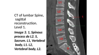
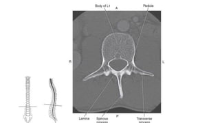
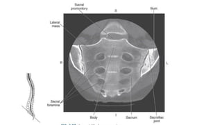
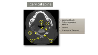
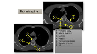
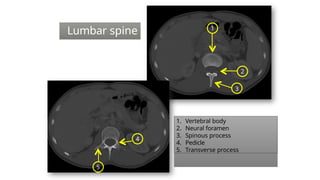
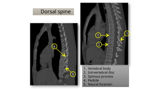
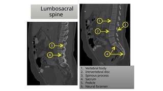
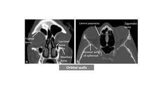
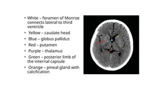
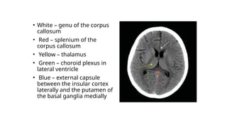
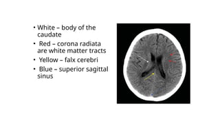
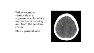
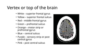
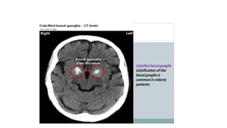
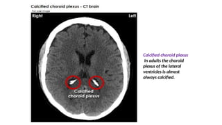
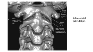
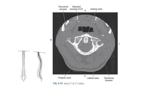
CT of lumbar Spine,
sagittal
reconstruction.
Level 1.
Image 3. 1, Spinous
process de L2. S,
Sacrum. L1, Vertebral
body, L1. L2,
Vertebral body, L2.
Cervical spine
1
3
4
5
2
1. Vertebral body
2. Spinous process
3. Pedicle
4. Lamina
5. Transverse foramen
Thoracic spine
2
1
7
5 6
1. Vertebral body
2. Neural foramen
3. Lamina
4. Pedicle
5. Transverse process
6. Spinous process
7. Rib
3
4
1
2
3
5
4
1. Vertebral body
2. Neural foramen
3. Spinous process
4. Pedicle
5. Transverse process
Lumbar spine
Dorsal spine
1
2
3
4
5
1. Vertebral body
2. Intrvertebral disc
3. Spinous process
4. Pedicle
5. Neural foramen
1
2
3
4
5
6
1. Vertebral body
2. Intrvertebral disc
3. Spinous process
4. Sacrum
5. Pedicle
6. Neural foramen
Lumbosacral
spine
• White – foramen of Monroe
connects lateral to third
ventricle
• Yellow – caudate head
• Blue – globus pallidus
• Red – putamen
• Purple – thalamus
• Green – posterior limb of
the internal capsule
• Orange – pineal gland with
calcification
• White – genu of the corpus
callosum
• Red – splenium of the
corpus callosum
• Yellow – thalamus
• Green – choroid plexus in
lateral ventricle
• Blue – external capsule
between the insular cortex
laterally and the putamen of
the basal ganglia medially
• White – body of the
caudate
• Red – corona radiata
are white matter tracts
• Yellow – falx cerebri
• Blue – superior sagittal
sinus
• Yellow – centrum
semiovale are
supraventricular white
matter tracts running to
and from the cerebral
cortex
• Blue – parietal lobe
Vertex or top of the brain
• White – superior frontal gyrus
• Yellow – superior frontal sulcus
• Red – middle frontal gyrus
• Green – prefrontal sulcus
• Orange – motor strip or
prefrontal gyrus
• Blue – central sulcus
• Purple – sensory strip or post
central gyrus
• Pink – post central sulcus
Calcified choroid plexus
In adults the choroid
plexus of the lateral
ventricles is almost
always calcified.
Atlantoaxial
articulation
Presentation 4.pptx…..kuhyyghghhhhhhhhhhhg

Presentation 4.pptx…..kuhyyghghhhhhhhhhhhg

  • 1.
    CT of lumbarSpine, sagittal reconstruction. Level 1. Image 3. 1, Spinous process de L2. S, Sacrum. L1, Vertebral body, L1. L2, Vertebral body, L2.
  • 4.
    Cervical spine 1 3 4 5 2 1. Vertebralbody 2. Spinous process 3. Pedicle 4. Lamina 5. Transverse foramen
  • 5.
    Thoracic spine 2 1 7 5 6 1.Vertebral body 2. Neural foramen 3. Lamina 4. Pedicle 5. Transverse process 6. Spinous process 7. Rib 3 4
  • 6.
    1 2 3 5 4 1. Vertebral body 2.Neural foramen 3. Spinous process 4. Pedicle 5. Transverse process Lumbar spine
  • 7.
    Dorsal spine 1 2 3 4 5 1. Vertebralbody 2. Intrvertebral disc 3. Spinous process 4. Pedicle 5. Neural foramen
  • 8.
    1 2 3 4 5 6 1. Vertebral body 2.Intrvertebral disc 3. Spinous process 4. Sacrum 5. Pedicle 6. Neural foramen Lumbosacral spine
  • 10.
    • White –foramen of Monroe connects lateral to third ventricle • Yellow – caudate head • Blue – globus pallidus • Red – putamen • Purple – thalamus • Green – posterior limb of the internal capsule • Orange – pineal gland with calcification
  • 11.
    • White –genu of the corpus callosum • Red – splenium of the corpus callosum • Yellow – thalamus • Green – choroid plexus in lateral ventricle • Blue – external capsule between the insular cortex laterally and the putamen of the basal ganglia medially
  • 12.
    • White –body of the caudate • Red – corona radiata are white matter tracts • Yellow – falx cerebri • Blue – superior sagittal sinus
  • 13.
    • Yellow –centrum semiovale are supraventricular white matter tracts running to and from the cerebral cortex • Blue – parietal lobe
  • 14.
    Vertex or topof the brain • White – superior frontal gyrus • Yellow – superior frontal sulcus • Red – middle frontal gyrus • Green – prefrontal sulcus • Orange – motor strip or prefrontal gyrus • Blue – central sulcus • Purple – sensory strip or post central gyrus • Pink – post central sulcus
  • 16.
    Calcified choroid plexus Inadults the choroid plexus of the lateral ventricles is almost always calcified.
  • 17.