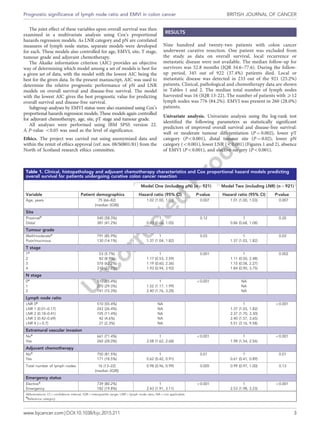
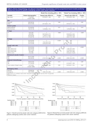
Prognostic value of lymph node ratio and
extramural vascular invasion on survival for
patients undergoing curative colon cancer
resection
C N Parnaby*,1
, N W Scott2
, G Ramsay1
, C MacKay1
, L Samuel3
, G I Murray4
and M A Loudon1
1
Department of Surgery, Aberdeen Royal Infirmary, Aberdeen, UK; 2
Medical Statistics Team, University of Aberdeen, Aberdeen,
UK; 3
Department of Oncology, Aberdeen Royal Infirmary, Aberdeen, UK and 4
Department of Pathology, Aberdeen Royal
Infirmary, Aberdeen, UK
Background: Increasing lymph node ratio (LNR) (ratio of metastatic lymph nodes to the total number of harvested lymph nodes)
and extramural vascular invasion (EMVI) have been proposed as adverse prognostic indicators in colorectal cancer, although their
use remains variable and controversial. The aim of the present study was to assess the prognostic value of LNR and EMVI in
predicting survival for patients undergoing curative colon cancer resection.
Methods: Between 2006 and 2012, 922 patients underwent curative colon cancer resection. Surgical technique and pathological
assessment did not change during the study period. Clinical and pathological data were collected from a prospectively
maintained database. The primary outcome measure was overall survival and disease-free survival. LNR was separated into five
categories based on three previously calculated cutoff values: LNR 0 (no lymph nodes involved), LNR 1 (ratio 0.01o0.17), LNR 2
(ratio 0.18–0.41), LNR 3 (ratio 0.42–0.69), and LNR 4 (ratio 40.70).
Results: Nine hundred and twenty-two patients underwent colon cancer resection. The median follow-up for survivors was 52.8
months (IQR 34.6–77.6). The median total number of lymph nodes harvested was 16 (IQR13-22). On multivariate analysis, both pN
and LNR were strongly associated with overall and disease-free survival. Using the Akaike information criterion (AIC), LNR had
greater prognostic value compared with pN. For overall survival, compared with patients in LNR category 0, hazard ratios (95% CI)
for those in categories 1, 2, 3 and 4 were 1.37 (1.03,1.82), 2.37 (1.70,3.30), 2.40 (1.57,3.65) and 5.51 (3.16,9.58), respectively. For
disease-free survival, patients had hazard ratios (95% CI) of 1.78 (1.25,2.52), 3.79 (2.56,5.61), 2.60 (1.50,4.48) and 4.76 (2.21,10.27),
respectively. The presence of EMVI was a significant predictor of decreased overall and disease-free survival (Po0.001).
Conclusions: This study demonstrated, in the presence of high surgical, oncology and pathological standards, EMVI and
increasing LNR were independent predictors of decreased overall and disease-free survival for patients undergoing curative colon
cancer resection. LNR was superior to pN stage in predicting overall and disease-free survival.
Colorectal cancer is the third most common cancer in Europe and
North America (International Agency for Research on Cancer,
2008). Pathological stagingQ1 not only provides important prognostic
information but also are used to inform discussions and decisions
on the use of adjuvant chemotherapy. The most widely used
colorectal staging tool is the TNM (tumours/nodes/metastases)
system (Sobin and Wittekind, 1997). The fifth edition of TNM
(TNM 5), introduced in 1995, is Q2still predominantly used in the
*Correspondence: Dr CN Parnaby; E-mail: craigparnaby@nhs.net
Received 21 December 2014; revised 20 April 2015; accepted 14 May 2015
& 2015 Cancer Research UK. All rights reserved 0007 – 0920/15
FULL PAPER
Keywords: lymph node ratio; extramural vascular invasion; colon cancer; prognosis
British Journal of Cancer (2015), 1–8 | doi: 10.1038/bjc.2015.211
www.bjcancer.com | DOI:10.1038/bjc.2015.211 1
United Kingdom to ensure consistency and comparability of data.
Subsequent revisions have not demonstrated an improvement on
prognosis (Ueno et al, 2012).
The presence of lymph node metastases are an important
prognostic factor for survival of patients with colonic cancer. The
current pN stage classifies patients into three categories: pN0
(no lymph node metastases), pN1 (1–3 lymph node metastases),
and pN2 (44 lymph node metastases). A number of studies have
suggested the lymph node ratio (LNR) (defined as the ratio of
metastatic lymph nodes to the total number of harvested lymph
nodes) is a superior prognostic factor compared with the pN stage
(Berger et al, 2005; Rosenberg et al, 2008; Moug et al, 2011; Lykke
et al, 2012). These studies have demonstrated the marked changes
in survival within the pN1 or pN2 categories depending on the
value of LNR. A number of concerns remain, for example: the LNR
categories were arbitrarily defined, colon and rectal cancer were
analysed together, pathological assessment changed over time, the
study period covered a large timeframe, or the use of adjuvant
chemotherapy was not reported.
Additional pathological features have been identified as adverse
prognostic indicators for survival in patients undergoing potential
curative surgery for colorectal cancer. Extramural vascular invasion
(EMVI) has been reported to be an important adverse prognostic
indicator (Courtney et al, 2009; Bhangu et al, 2013). Detection of
EMVI may help improve selection of patients who would benefit
from adjuvant chemotherapy. EMVI is one of the core parameters
to be reported in the minimum data set for colorectal cancer
histopathology reporting as described by the Royal College of
Pathologists (Williams et al, 2007). Despite this, the detection of
EMVI remains highly variable betweenQ3 pathologists (Quirke and
Morris, 2007).
The aim of the present study was to compare the ability of LNR,
EMVI and other pathological factors in predicting overall survival
and disease-free survival for patients undergoing curative colon
cancer resection.
Q7
MATERIALS AND METHODS
Inclusion criteria. All patients undergoing curative colon adeno-
carcinoma resection from 2006 to 2012 were included. This
included all patients undergoing elective or emergency surgery.
Colon cancer was defined as a cancer 415 cm from the anal verge.
Tumour site was separated into proximal and distal tumours.
Proximal colon cancers were defined proximal to the splenic
flexure. Distal colon cancers were defined as involving splenic
flexure to the rectosigmoid junction. Curative colon cancer
resection was defined as no evidence of metastases on computed
tomography (CT) of the chest, abdomen and pelvis and curative
tumour resection (R0) on pathology assessment.
Patients with metastases, concurrent malignancy and rectal
cancers (o15 cm from the anal verge) were excluded from the
study.
Data collection. Data on patient demographics and tumour
characteristics were obtained from a prospectively maintained
database. Survival data were collated by analysis of a weekly
updated local cancer registry. Adjuvant chemotherapy data were
collected by retrospective review of patient electronic records. All
patients were subject to multidisciplinary review following colonic
resection. The primary outcomes were overall survival (the time
from surgery to the date of death from any cause) and disease-free
survival (the time to death or the discovery of local recurrence or
regional metastasis on follow-up).
Data on age, gender, tumour site, tumour differentiation, TNM
classification, lymph node yield, total number of tumour involved
lymph nodes, LNR, EMVI, adjuvant chemotherapy, overall survival
and disease-free survival were collected. LNR was defined as the
ratio of number of metastatic lymph nodes/total number of
harvested lymph nodes. LNR was separated into five categories
based on three previously calculated cutoff values for LNRs that
had the highest prognostic discrimination of survival in a cohort of
3026 patients with colon and rectal cancer (Rosenberg et al, 2008):
LNR ¼ 0 (no lymph nodes involved), LNR ¼ 1 (ratio 0.01-0.17),
LNR ¼ 2 (ratio 0.18–0.41), LNR ¼ 3 (ratio 0.42–0.69), and LNR ¼ 4
(ratio 40.70). These values have been validated on a population-
based study of colorectal cancer patients (Rosenberg et al, 2010).
Patients with no lymph nodes involved (LNR ¼ 0) were included in
the study to compare survival outcomes with the other four LNR
categories both in the main analyses and sub group analyses.
Clinical management. All patients were subject to multidisciplin-
ary review following colonic resection. Clinical assessment and
staging was based on endoscopic evaluation and CT of the chest,
abdomen and pelvis. Operative technique did not change during
the study period and consisted of complete mesocolic mobilisation
and division of the supplying arteries at their origin. For right-
sided colon cancer resection, more radical mobilisation as
described by Hohenberger et al (2009), involving mesenteric root,
duodenum and pancreatic head mobilisation was not routinely
performed.
Histopathological assessment of the colonic cancer excision
specimens followed a uniform protocol throughout the study
period. The colonic cancer excision specimens were received fresh
in the diagnostic histopathological laboratory, opened along the
antimesenteric border proximal and, when appropriate distal to the
tumour, washed in cold water and then fixed in 10% neutral
buffered formalin for at least 48 h at room temperature prior to
further dissection and block selection. Representative tissue blocks
were embedded in wax, sections were then stained with
haematoxylin and eosin for histopathological diagnosis and when
required tumour sections were also stained with elastic haematox-
ylin and eosin to permit further assessment of EMVI. The tumours
were reported according to the guidelines of Royal College of
Pathologists for the histopathological reporting of colorectal cancer
resection specimens, which incorporates guidance from version
five of the TNM staging system (Williams et al, 2007).
The follow-up programme included outpatient visits every 6
months for 3 years. Blood was takn at these visits to measure liver
function and carcinoembryonic antigen. Imaging was annual CT
scanning (thorax, abdomen and pelvis), with 6 monthly interval
abdominal ultrasound scanning. A follow-up colonoscopy was
performed at 5 years unless it was incomplete at preoperative
assessment. If this occurred, a repeat colonoscopy was performed
in the postoperative period.
Adjuvant chemotherapy was considered for fit patients with
adverse prognostic factors, such as the presence of metastatic
lymph nodes, the presence of EMVI, serosal involvement or poorly
differentiated tumours. The majority of patients with colon cancer
were age 470 years, any decision on the use of adjuvant
chemotherapy considered co-morbidities and the reduced physio-
logical reserve of an older population of patients. During the study
period, suitable patients were offered participation into adjuvant
chemotherapy trials. These were the AVANT study (de Gramont
et al, 2012) and the SCOT study (SCOT-Short Course Oncology
Therapy, 2007). Outside of these trials, suitable patients would
have been considered for oxaliplatin combined with folinic acid/5-
FU or capecitabine.
Statistical analysis. The effect of sex, tumour site, tumour grade, T
stage, N stage, adjuvant chemotherapy, LNR and EMVI on overall
survival and disease-free survival was first examined individually
using Kaplan-Meier survival analysis. Differences in survival
between categories were assessed using the log-rank test.
BRITISH JOURNAL OF CANCER Prognostic significance of lymph node ratio and EMVI in colon cancer
2 www.bjcancer.com | DOI:10.1038/bjc.2015.211
The joint effect of these variables upon overall survival was then
examined in a multivariate analysis using Cox’s proportional
hazards regression models. As LNR category and pN are correlated
measures of lymph node status, separate models were developed
for each. These models also controlled for age, EMVI, site, T stage,
tumour grade and adjuvant chemotherapy.
The Akaike information criterion (AIC) provides an objective
way of determining which model among a set of models is best for
a given set of data, with the model with the lowest AIC being the
best for the given data. In the present manuscript, AIC was used to
determine the relative prognostic performance of pN and LNR
models on overall survival and disease-free survival. The model
with the lowest AIC gives the best prognostic value for predicting
overall survival and disease-free survival.
Subgroup analyses by EMVI status were also examined using Cox’s
proportional hazards regression models. These models again controlled
for adjuvant chemotherapy, age, site, pT stage and tumour grade.
All analyses were performed using IBM SPSS version 22.
A P-value o0.05 was used as the level of significance.
Ethics. The project was carried out using anonymised data and
within the remit of ethics approval (ref. nos. 08/S0801/81) from the
North of Scotland research ethics committee.
RESULTS
Nine hundred and twenty-two patients with colon cancer
underwent curative resection. One patient was excluded from
the study as data on overall survival, local recurrence or
metastatic disease were not available. The median follow-up for
survivors was 52.8 months (IQR 34.6–77.6). During the follow-
up period, 345 out of 922 (37.4%) patients died. Local or
metastatic disease was detected in 233 out of the 921 (25.2%)
patients. Clinical, pathological and chemotherapy data are shown
in Tables 1 and 2. The median total number of lymph nodes
harvested was 16 (IQR 13-22). The number of patients with X12
lymph nodes was 776 (84.2%). EMVI was present in 260 (28.0%)
patients.
Univariate analysis. Univariate analysis using the log-rank test
identified the following parameters as statistically significant
predictors of improved overall survival and disease-free survival:
well or moderate tumour differentiation (P ¼ 0.002), lower pT
category (Po0.001), distal tumour site (P ¼ 0.02), lower pN
category (o0.001), lower LNR (o0.001) (Figures 1 and 2), absence
of EMVI (Po0.001), and elective surgery (Po0.001).
Table 1. Clinical, histopathology and adjuvant chemotherapy characteristics and Cox proportional hazard models predicting
overall survival for patients undergoing curative colon cancer resection
Model One (including pN) (n ¼ 921) Model Two (including LNR) (n ¼ 921)
Variable Patient demographics Hazard ratio (95% CI) P-value Hazard ratio (95% CI) P-value
Age, years 75 (66–82)
(median (IQR))
1.02 (1.00, 1.03) 0.007 1.01 (1.00, 1.03) 0.007
Site
Proximala
540 (58.3%) 1 0.12 1 0.20
Distal 381 (41.2%) 0.83 (0.66, 1.05) 0.86 (0.68, 1.08)
Tumour grade
Well/moderatea
791 (85.9%) 1 0.03 1 0.03
Poor/mucinous 130 (14.1%) 1.37 (1.04, 1.82) 1.37 (1.03, 1.82)
T stage
1a
53 (5.7%) 1 0.001 1 0.002
2 82 (8.9%) 1.17 (0.53, 2.59) 1.11 (0.50, 2.48)
3 576 (62.2%) 1.19 (0.60, 2.36) 1.15 (0.58, 2.27)
4 210 (22.7%) 1.93 (0.94, 3.92) 1.84 (0.90, 3.75)
N stage
0a
510 (55.4%) 1 o0.001 NA
1 270 (29.3%) 1.52 (1.17, 1.99) NA
2 141 (15.3%) 2.40 (1.76, 3.28) NA
Lymph node ratio
LNR 0a
510 (55.4%) NA 1 o0.001
LNR 1 (0.01–0.17) 243 (26.4%) NA 1.37 (1.03, 1.82)
LNR 2 (0.18–0.41) 105 (11.4%) NA 2.37 (1.70, 3.30)
LNR 3 (0.42–0.69) 42 (4.6%) NA 2.40 (1.57, 3.65)
LNR 4 (40.7) 21 (2.3%) NA 5.51 (3.16, 9.58)
Extramural vascular invasion
Noa
661 (71.4%) 1 o0.001 1 o0.001
Yes 260 (28.0%) 2.08 (1.62, 2.68) 1.98 (1.54, 2.56)
Adjuvant chemotherapy
Noa
750 (81.5%) 1 0.01 1 0.01
Yes 171 (18.5%) 0.62 (0.42, 0.91) 0.61 (0.41, 0.89)
Total number of lymph nodes 16 (13–22)
(median (IQR))
0.98 (0.96, 0.99) 0.005 0.99 (0.97, 1.00) 0.13
Emergency status
Electivea
739 (80.2%) 1 o0.001 1 o0.001
Emergency 182 (19.8%) 2.43 (1.91, 3.11) 2.53 (1.98, 3.23)
Abbreviations: CI ¼ confidence interval; IQR ¼ interquartile range; LNR ¼ lymph node ratio; NA ¼ not applicable.
a
Reference category.
Prognostic significance of lymph node ratio and EMVI in colon cancer BRITISH JOURNAL OF CANCER
www.bjcancer.com | DOI:10.1038/bjc.2015.211 3
Table 2. Clinical, histopathology and adjuvant chemotherapy characteristics and Cox proportional hazard models predicting
disease-free survival for patients undergoing curative colon cancer resection
Model One (including pN) (n ¼ 921) Model Two (including LNR) (n ¼ 921)
Variable Patient demographics Hazard ratio (95% CI) P-value Hazard ratio (95% CI) P-value
Age, years 75 (66–82)
(median (IQR))
0.99 (0.97, 1.00) 0.07 0.99 (0.98, 1.00) 0.10
Site
Proximala
540 (58.3%) 1 0.48 1 0.69
Distal 381 (41.2%) 0.91 (0.69, 1.19) 0.95 (0.72, 1.25)
Tumour grade
Well/moderatea
791 (85.9%) 1 0.27 1 0.16
Poor/mucinous 130 (14.1%) 1.22 (0.86, 1.73) 1.29 (0.90, 1.83)
T stage
1a
53 (5.7%) 1 0.001 1 0.001
2 82 (8.9%) 0.87 (0.28, 2.77) 0.84 (0.26, 2.65)
3 576 (62.2%) 1.78 (0.72, 4.42) 1.69 (0.68, 4.18)
4 210 (22.7%) 2.91 (1.14, 7.44) 2.82 (1.10, 7.20)
N stage
0a
510 (55.4%) 1 o0.001 NA
1 270 (29.3%) 1.96 (1.40, 2.73) NA
2 141 (15.3%) 3.35 (2.28, 4.92) NA
Lymph node ratio
LNR 0a
510 (55.4%) NA 1 o0.001
LNR 1 (0.01–0.17) 243 (26.4%) NA 1.78 (1.25, 2.52)
LNR 2 (0.18–0.41) 105 (11.4%) NA 3.79 (2.56, 5.61)
LNR 3 (0.42–0.69) 42 (4.6%) NA 2.60 (1.50, 4.48)
LNR 4 (40.7) 21 (2.3%) NA 4.76 (2.21, 10.27)
Extramural vascular invasion
Noa
661 (71.4%) 1 1
Yes 260 (28.0%) 2.10 (1.55, 2.83) o0.001 2.08 (1.54, 2.81) o0.001
Adjuvant chemotherapy
Noa
750 (81.5%) 1 0.12 1 0.09
Yes 171 (18.5%) 0.73 (0.49, 1.09) 0.70 (0.47, 1.05)
Total number of lymph nodes 16 (13–22)
(median (IQR))
0.98 (0.96, 1.00) 0.02 0.99 (0.97, 1.01) 0.22
Emergency status
Electivea
739 (80.2%) 1 1
Emergency 182 (19.8%) 1.88 (1.39, 2.54) o0.001 1.90 (1.40, 2.57) o0.001
Abbreviations: CI ¼ confidence interval; IQR ¼ interquartile range; LNR ¼ lymph node ratio; NA ¼ not applicable.
a
Reference category.
0.00
0.25
0.50
0.75
1.00
21 6 2 1 0LNR1 = 4
42 18 8 3 2LNR1 = 3
105 59 30 21 6LNR1 = 2
243 169 91 47 21LNR1 = 1
511 410 239 118 39LNR1 = 0
Number at risk
24 48 72 96
Time from operation to death or last follow-up (months)
LNR1 = 0 LNR1 = 1
LNR1 = 2 LNR1 = 3
LNR1 = 4
Figure 1. Overall survival by lymph node ratio category (LNR 0–4) for
patients undergoing curative colon cancer resection.
0.00
0.25
0.50
0.75
1.00
21 5 2 1 0LNR1 = 4
42 17 7 2 1LNR1 = 3
105 43 29 18 5LNR1 = 2
243 152 85 45 20LNR1 = 1
510 389 221 110 37LNR1 = 0
Number at risk
24 48 72 96
Time from operation to death or last follow-up (months)
LNR1 = 0 LNR1 = 1
LNR1 = 2 LNR1 = 3
LNR1 = 4
Figure 2. Disease-free survival by lymph node ratio category (LNR 0–4)
for patients undergoing curative colon cancer resection.
BRITISH JOURNAL OF CANCER Prognostic significance of lymph node ratio and EMVI in colon cancer
4 www.bjcancer.com | DOI:10.1038/bjc.2015.211
Adjuvant chemotherapy and sex were not statistically significant
predictors of overall mortality (P ¼ 0.065 and P ¼ 0.381,
respectively).
Multivariate analysis. Table 2 shows the results of the two Cox
regression models for overall survival, one including pN and the
other LNR category. Table 3 shows the results of the corresponding
Cox regression models for disease-free survival. There was strong
evidence that, after controlling for other variables, both N stage
and LNR category were associated with overall survival and
disease-free survival. For overall survival, compared with patients
with pN 0, those with pN 1 and 2 had hazard ratios (95% CI) of
1.52 (1.17, 1.99) and 2.40 (1.76, 3.28), respectively. For disease-free
survival, patients had hazard ratios (95% CI) of 1.96 (1.40, 2.73)
and 3.55 (2.28, 4.92), respectively.
For overall survival, compared with patients in LNR category 0,
hazard ratios (95% CI) for those in categories 1, 2, 3 and 4 were
1.37 (1.03, 1.82), 2.37 (1.70, 3.30), 2.40 (1.57, 3.65) and 5.51 (3.16,
9.58), respectively. For disease-free survival, patients had hazard
ratios (95% CI) of 1.78 (1.25, 2.52), 3.79 (2.56, 5.61), 2.60 (1.50,
4.48) and 4.76 (2.21, 10.27), respectively.
The multivariate analyses identified the presence of EMVI as a
statistically significant predictor of decreased overall and disease-
free survival (Po0.001).
Other parameters associated with statistically significant
decreased overall and disease-free survival included: higher pT
category and emergency surgery. The administration of adjuvant
chemotherapy was a significant predictor of improved overall
survival but not disease-free survival in the adjusted analysis.
Using pN in the Cox regression model, the total number of
lymph nodes harvested was associated with significant decreased
overall and disease-free survival. This association was not present
when LNR was used in the model.
Prognostic value of LNR and pN on overall survival. The AIC
was used to determine the relative performance of pN and LNR.
LNR model was found to have lower AIC values for both overall
survival (13.1 points) and disease-free survival (4.4 points),
indicating that LNR may have the greater prognostic value
(Table 4).
Subgroup analysis. The relationship of LNR and EMVI on overall
survival was evaluated using subgroup analyses. Separate Cox’s
proportional hazard regression models were performed for the
LNR categories in patients with and without EMVI (Figures 3 and 4).
LNR categories 2, 3 and 4 were combined because of small
numbers in these individual categories.
Patients with EMVI had a worse overall survival. For both
subgroups (those with and without EMVI), there was a similar
statistically significant relationship (Po0.001) between LNR
categories (LNR 0, LNR 1 and LNR 2-4) and overall survival
(Figures 3 and 4).
DISCUSSION
The present study has shown LNR and EMVI were independent
predictors of overall and disease-free survival for patients under-
going curative colon cancer resection. LNR category was of greater
prognostic value for predicting overall survival compared with pN
category.
Table 3. Prognostic value of pN and LNR on overall survival
using Akaike information criterion (AIC)
AIC
Base modela
4169.5
Base model plus pN 4144.1
Base model plus LNR 4131.0
Abbreviation: LNR ¼ lymph node ratio.
a
Base model is a Cox proportional hazards model including the following variables: age,
site, grade, T stage, extramural vascular invasion, adjuvant chemotherapy, total nodes, and
emergency status.
Table 4. Prognostic value of pN and LNR on disease-free
survival using Akaike information criterion (AIC)
AIC
Base modela
2866.9
Base model plus pN 2832.7
Base model plus LNR 2828.3
Abbreviation: LNR ¼ lymph node ratio.
a
Base model is a Cox proportional hazards model including the following variables: age,
site, grade, T stage, extramural vascular invasion, adjuvant chemotherapy, total nodes, and
emergency status.
0.00
0.25
0.50
0.75
1.00
24 48 72 96
Time from operation to death or last follow-up (months)
LNR1cat = 2–4
LNR1cat = 1
100 31 12 7 2
80 42 16 7 3
80 48 14 7 1LNR1cat = 0
Number at risk
LNR1cat = 0 LNR1cat = 1
LNR1cat = 2–4
Figure 3. Overall survival of patients with extramural vascular invasion
by LNR category (LNR 0, LNR 1 and LNR 2–4 together) undergoing
curative colon cancer resection.
0.00
0.25
0.50
0.75
1.00
68 52 28 18 6LNR1cat = 2–4
163 127 75 40 18LNR1cat = 1
431 362 225 111 38LNR1cat = 0
Number at risk
24 48 72 96
Time from operation to death or last follow-up (months)
LNR1cat = 0 LNR1cat = 1
LNR1cat = 2–4
Figure 4. Overall survival of patients without extramural vascular
invasion by LNR category (LNR 0, LNR 1 and LNR 2–4 together)
undergoing curative colon cancer resection.
Prognostic significance of lymph node ratio and EMVI in colon cancer BRITISH JOURNAL OF CANCER
www.bjcancer.com | DOI:10.1038/bjc.2015.211 5
A number of studies have reported the prognostic value of LNR
in predicting survival for patients undergoing colorectal cancer
resection. Different groups have used different methods for
stratifying the LNR categories.
Schiffmann et al (2013), in a cohort of 142 stage III colon cancer
patients, compared LNR with pN category. Using a single LNR
statistical cutoff of 0.2 to separate patients into two groups, the
authors concluded LNR did not improve the prognostic value in
predicting survival. Limitations included the small number of
patients and the variable preoperative staging strategy. No details
of pathological assessment were given. Using only two groups for
LNR may reduce its prognostic value.
Berger et al (2005) stratified patients with stage II and III colon
cancer into four LNR categories based on quartiles. They
demonstrated that LNR was an important predictor of overall
survival when 410 lymph nodes were harvested. There were
marked differences in the 5-year survival between the lowest and
highest LNR categories for patients within the pN1 and N2
categories (19% and 28%, respectively).
From 1988 to 2003, Wang et al (2008) in a population-based
study of 24 477 patients with node-positive colon cancer from the
Surveillance, Epidemiology and End Results cancer (SEER) registry
demonstrated that LNR was a more accurate prognostic method
for staging node-positive colon cancers than the number of positive
lymph nodes. LNR was categorised based on four cutoff points: 1
out of 14, 25%, 50%, 100%. The results of this multi-centre study
need to be interpreted with caution as the clinical and pathological
techniques may change over time or vary between centres. In
addition, not all treatment data were available, including the
administration of adjuvant chemotherapy.
Rosenberg et al (2008), using statistical analysis, identified three
cutoff values for LNR (0.17, 0.41 and 0.69) and demonstrated
superior prognostic impact on overall survival compared with pN
category. There were some limitations to the study in that the study
period covered 25 years (raising concerns regarding changes in
pathological assessment over the time period) and colon and rectal
cancers were analysed together. A follow-up of large population-
based study of colorectal cancer patients by the same leading
author and using the same LNR cutoff values confirmed the strong
independent prognostic value of these LNR categories (Rosenberg
et al, 2010). There were marked differences in the 10-year survival
between the lowest and highest LNR categories for patients within
the pN1 and N2 categories (20% and 50%, respectively). The
authors concluded that LNR should be routinely included in the
pathological reporting.
The present study, in a different patient population, used the
same LNR cutoff values as Rosenberg et al (2008). This
demonstrated the statistically significant difference in overall and
disease-free survival using LNR categories from Rosenberg et al
(2008) for patients undergoing curative colon cancer resection. The
models including LNR category were found to have lower AIC
values (indicating better model fit) than the models including pN
category when predicting overall and disease-free survival. It is not
possible to include both within the same scoring system due to the
relationship between the categories; our results lend support to the
view that LNR should be used instead of pN status. This has several
advantages over other studies: surgical and pathological technique
did not change, adjuvant chemotherapy was controlled for in the
statistical model, and colon cancer was analysed separately.
The optimal total number of lymph nodes harvested for colon
cancer resection is controversial. Despite the lack of evidence, The
Royal College of Pathologists UK recommends a minimum of
12 lymph nodes need to be harvested (Williams et al, 2007).
A number of observational studies have suggested different cutoffs
for total number of lymph nodes to be harvested (Cianchi et al,
2002; BuiQ4 et al, 2006; Chen and Bilchik, 2006; Choi et al, 2010).
O’Shea et al (2014) compared total lymph node yield of 3216
patients undergoing surgery for colorectal cancer. The authors
demonstrated an increasing lymph node yield over time (14.91 in
2005 rising to 21.38 in 2012), but this was not associated with
increased number of lymph node-positive cancers. Baxter et al
(2010), in a population study of 11 044 patients with pT3 colon
cancers (SEER registry), reported that the proportion of patients
found to be node positive increases with increasing total lymph
node counts up to 5 or 6 but, at higher lymph node count (X7),
has marginal effects on staging.
Sjo et al (2012), in an observational study, looked at the
prognostic impact of total lymph node harvest and LNR in 950
patients undergoing curative colon cancer resection. The authors
found that, on multivariate analysis, the total number of lymph
nodes was not a prognostic factor when analysed with LNR for
patients with lymph node metastasis. Also, Wang et al (2008) in
the previously mentioned population-based study assessing
patients with node-positive colon cancer concluded that the
prognostic capability of LNR was not dependent on the total
number of lymph nodes.
In the present study, 84% of patients had 412 lymph nodes
harvested. On multivariate analysis, using the LNR model, the total
number of lymph nodes harvested was not associated with overall
survival or disease-free survival.
The presence of EMVI has been shown to be an adverse
prognostic indicator of survival for patients with colorectal cancer.
The majority of studies have focussed on node-negative colorectal
cancers. Courtney et al (2009), in 378 patients undergoing
potentially curative colorectal cancer resection, demonstrated that
the presence of EMVI was associated with a significant decrease in
survival. The authors did not perform any further analysis for
patients with metastatic lymph nodes. Morris et al (2006), in a
population-based study of 1306 patients with node-negative
colonic carcinoma treated with surgery alone demonstrated a
worse prognosis in the presence of EMVI. It is possible that these
studies have concentrated on patients with node-negative colonic
cancers as nodal disease is an indicator for adjuvant chemotherapy
and the presence of EMVI is therefore unlikely to alter manage-
ment in these patients. Sternberg et al (1999) assessed 171 patients
with node-positive colorectal cancer and found that those patients
without venous invasion had a better cancer-related survival
compared with those with venous invasion. Stratifying patients to
pN1 and pN2 lymph node categories did not show any significant
cancer-related survival benefit. The authors concluded that venous
invasion was a better indicator of prognosis than pN status. The
authors assessed colon and rectal cancers together and the study
did not control for administration of adjuvant chemotherapy. The
differentiation between intramural and extramural venous invasion
was not routinely made. Betge et al (2012) randomly selected 381
patients with colorectal cancer from 1992 to 2000. Differentiation
between intramural and extramural venous invasion was made.
EMVI was detected 14% of patients and a significant predictor of
both disease progression and cancer-specific survival.
Quirke found in routine UK practice that the detection of EMVI
was poor at 10% (Quirke and Morris, 2007). He felt that a
pathologist should be able to detect EMVI in approximately 30% of
cases. The Royal College of Pathologists have recommended that
EMVI should be detected in at least 25% of cases (Williams et al,
2007).
In the present study, EMVI status was examined in all patients
using the technique recommended by the Royal College of
Pathologists. The incidence of EMVI in this study was 28%. In
this cohort of patients undergoing curative colon cancer resection,
we have demonstrated that the presence of EMVI was a significant
independent predictor of decreased overall survival and disease-
free survival even when adjusted for lymph node status.
A number of studies have found that patients with a low LNR
have a similar long-term survival as node-negative patients. Moug
BRITISH JOURNAL OF CANCER Prognostic significance of lymph node ratio and EMVI in colon cancer
6 www.bjcancer.com | DOI:10.1038/bjc.2015.211
et al (2011), in a study of 206 patients undergoing curative
colorectal cancer patients, demonstrated that there was no
statistically significant difference in overall survival between the
two lowest LNR groups (pLNRo0.05 and pLNR 0.05–0.19).
Patients in the pLNR category o0.05 were mainly node negative.
The study did not control for adjuvant chemotherapy and did not
assess for EMVI status. In a prospective nationwide Danish study
of 8901 patients with Stage I-III colon cancer, the 5-year survival
rate in LNR o0.08 was the same as those with node-negative
disease (Lykke et al, 2012). It was not possible to differentiate
between those patients receiving adjuvant chemotherapy.
In this study, which controlled for adjuvant chemotherapy, the
presence of EMVI was associated with a worse prognosis regardless
of lymph node status. For patients with or without EMVI, there
was a similar statistically significant relationship between LNR
categories as the overall sample. However, it was necessary to
combine patients in LNR categories 2-4 due to smaller numbers.
For patients without EMVI, those with low LNR (LNR1) have
improved overall survival compared with those in the LNR 2-4
group. It may be that, on an individual basis for patients with a low
LNR (0.01–0.17), adjuvant chemotherapy can be omitted in those
deemed at risk of developing significant side effects as they have a
relatively good prognosis.
This study has limitations. This was a retrospective review of
routinely prospectively collected data. Although the overall number
of patients in the study was reasonable, the number of patients in
each lymph node category on sub-group analysis of EMVI status
was small. The results of these subgroup analyses should only be
interpreted as exploratory analyses.
CONCLUSION
This study has demonstrated, in the presence of consistently
high surgical, pathological, oncological standards and long
follow-up, the presence of EMVI and increasing LNR were
independent predictors of adverse overall and disease-free
survival in patients undergoing potentially curative colon cancer
resection. LNR was superior to pN stage in predicting overall
and disease-free survival. We believe that LNR and EMVI should
be routinely included in the minimum data set for colon cancer
pathological reporting. This would allow improved prognostic
stratification (compared with TNM stage) and may help
influence decisions on adjuvant chemotherapy for more frail
patients.
ACKNOWLEDGEMENTS
There was no study funding. We are grateful to Tony Rafferty
(Tailored Information for the People of Scotland, TIPs) for
providing survival data.
CONFLICT OF INTEREST
The authors declare no conflict of interest.
REFERENCES
Baxter NN, Ricciardi R, Simunovic M, Urbach DR, Virnig BA (2010)
An evaluation of the relationship between lymph node number and
staging in pT3 colon cancer using population-based data. Dis Colon
Rectum 53: 65–70.
Berger AC, Sigurdson ER, LeVoyer T, Hanlon A, Mayer RJ, Macdonald JS,
Catalano PJ, Haller DG (2005) Colon cancer survival is associated with
decreasing ratio of metastatic to examined lymph nodes. J Clin Oncol 23:
8706–8712.
Betge J, Polheimer MJ, Lindtner RA, Komprat P, Schlemmer A, Rehak P,
Vieth M, Hoefler G, Langner C (2012) Intramural and extramural vascular
invasion in colorectal cancer. Cancer 1: 628–638.
Bhangu A, Fitzgerald JEF, Slesser A, Northover JM, Faiz O, Tekkis P (2013)
Prognostic significance of extramural vascular invasion in T4 rectal
cancer. Colorectal Dis 15(11): e665–e671.
Bui L, Rempel E, Reeson D, Simunovic M (2006) Lymph node counts, rates of
positive lymph nodes, and patient survival for colon cancer surgery
in Ontario, Canada: a population-based study. J Surg Oncol 93(6):
439–445.
Chen SL, Bilchik AJ (2006) More extensive nodal dissection improves survival
for stages I to III of colon cancer: a population-based study. Ann Surg
244(4): 602–610.
Choi HK, Law WL, Poon JT (2010) The optimal number of lymph nodes
examined in stage II colorectal cancer and its impact of on outcomes.
BMC Cancer 10: 267.
Cianchi F, Palomba A, Boddi V, Messerini L, Pucciani F, Perigli G, Bechi P,
Cortesini C (2002) Lymph node recovery from colorectal tumor
specimens: recommendation for a minimum number of lymph nodes to
be examined. World J Surg 26(3): 384–389.
Courtney ED, West NJ, Kaur C, Ho J, Kalber B, Hagger R, Finlayson C,
Leicester RJ (2009) Extramural vascular invasion is an adverse prognostic
indicator of survival in patients with colorectal cancer. Colorectal Dis 11:
150–156.
de Gramont A, Van Cutsem E, Schmoll HJ, Tabernero J, Clarke S, Moore MJ,
Cunningham D, Cartwright TH, Hecht JR, Rivera F, Im SA, Bodoky G,
Salazar R, Maindrault-Goebel F, Shacham-Shmueli E, Bajetta E,
Makrutzki M, Shang A, Andre T, Hoff PM (2012) Bevacizumab plus
oxaliplatin as adjuvant treatment for colon cancer (AVANT): a phase 3
randomised controlled trial. Lancet Oncol 13: 1225–1233.
Hohenberger W, Weber K, Matzel K, Papadopoulos T, Merkel S (2009)
Standardized surgery for colonic cancer: complete mesocolic excision
and central ligation–technical notes and outcome. Colorectal Dis 11:
354–365.
International Agency for Research on Cancer (2008) GLOBOCAN: Cancer Q5
Incidence, Mortality and Prevalence Worldwide (2008 Estimates).
Lykke J, Roikjaer O, Jess P. Danish Colorectal Cancer Group (2012) The
relation between lymph node status and survival in stage I-III colon
cancer: results from a prospective nationwide cohort study. Colorectal Dis
15: 559–565.
Morris M, Platell C, de Boer B, McCaul K, Iacopetta B (2006) Population-
based study of prognostic factors in stage II colonic cancer. Br J Surg 93:
866–871.
Moug SJ, McColl G, Lloyd SM, Wilson G, Saldanha JD, Diament RH (2011)
Comparison of positive lymph node ratio with an inflammation-based
prognostic score in colorectal cancer. Br J Surg 98: 282–286.
O’Shea A, Aly O, Parnaby CN, Loudon MA, Samuel LM, Murray GI (2014)
Increased lymph node yield in colorectal cancer is not necessarily
associated with greater number of lymph node positive cancers. PLoS One
9: e104991.
Quirke P, Morris E (2007) Reporting colorectal cancer. Histopathology 50:
103–112.
Rosenberg R, Engel J, Bruns C, Hettland W, Hermes N, Jauch KW, Kopp R,
Putterich E, Ruppert R, Schuster T, Friess H, Hulzel D (2010)
The prognostic value of lymph node ratio in a population-based collective
of colorectal cancer patients. Ann Surg 251: 1070–1078.
Rosenberg R, Friederichs J, Schuster T, Gertler R, Maak M, Becker K,
Grebner A, Ulm K, Hofler H, Nekarda H, Siewert JR (2008) Prognosis of
patients with colorectal cancer is associated with lymph node ratio.
Ann Surg 248: 1067–1073.
Schiffmann L, Eiken AK, Gock M, Klar E (2013) Is the lymph node ratio
superior to the Union for International Cancer Control (UICC) TNM in
prognosis of colon cancer. World J Surg Oncol 11: 79.
SCOT-Short Course Oncology Therapy (2007) SCOT—Short Q6Course
Oncology Therapy: a study of adjuvant chemotherapy in colorectal cancer
by the CACTUS and QUASAR 3 groups.
Sjo OH, Merok MA, Svindland A, Nesbakken A (2012) Prognostic impact of
lymph node harvest and lymph node ratio in patients with colon cancer.
Dis Colon Rectum 55: 307–315.
Sobin LH, Wittekind Ch (eds): (1997) TNM Classification of Malignant
Tumours, 5th edn. Wiley-Liss: New York, NY, USA.
Prognostic significance of lymph node ratio and EMVI in colon cancer BRITISH JOURNAL OF CANCER
www.bjcancer.com | DOI:10.1038/bjc.2015.211 7
Sternberg A, Sibirsky O, Cohen D, Blemenson LE, Rodriguez-Bigas MA,
Petrelli NJ (1999) New approach to the substaging of node-positive
colorectal adenocarcinoma. Ann Surg Oncol 6: 161–165.
Ueno H, Mochizuki H, Akagi Y, Kusumi T, Yamada K, Ikegami M, Kawachi H,
Kameoka S, Ohkura Y, Masaki T, Kushima R, Takahashi K, Ajioka Y,
Hase K, Ochiai A, Wada R, Iwaya K, Shimazaki H, Nakamura T, Sugihara K
(2012) Optimal colorectal cancer staging criteria in TNM classification.
J Clin Oncol 30: 1519–1526.
Wang J, Hassett JM, Dayton MT, Kulaylat MN (2008) Lymph node ratio: role
in the staging of node-positive colon cancer. Ann Surg Oncol 15: 1600–1608.
Williams GT, Quirke P, Shepherd NA (2007) Standards and Datasets for
Reporting Cancers. Dataset for Colorectal Cancer, 2nd edn. Royal College
of Pathologists: London, UK.
This work is published under the standard license to publish agree-
ment. After 12 months the work will become freely available and
the license terms will switch to a Creative Commons Attribution-
NonCommercial-Share Alike 4.0 Unported License
BRITISH JOURNAL OF CANCER Prognostic significance of lymph node ratio and EMVI in colon cancer
8 www.bjcancer.com | DOI:10.1038/bjc.2015.211

CRC_PNR & EMVI_prognosis_BJCpaper

  • 1.
    Prognostic value oflymph node ratio and extramural vascular invasion on survival for patients undergoing curative colon cancer resection C N Parnaby*,1 , N W Scott2 , G Ramsay1 , C MacKay1 , L Samuel3 , G I Murray4 and M A Loudon1 1 Department of Surgery, Aberdeen Royal Infirmary, Aberdeen, UK; 2 Medical Statistics Team, University of Aberdeen, Aberdeen, UK; 3 Department of Oncology, Aberdeen Royal Infirmary, Aberdeen, UK and 4 Department of Pathology, Aberdeen Royal Infirmary, Aberdeen, UK Background: Increasing lymph node ratio (LNR) (ratio of metastatic lymph nodes to the total number of harvested lymph nodes) and extramural vascular invasion (EMVI) have been proposed as adverse prognostic indicators in colorectal cancer, although their use remains variable and controversial. The aim of the present study was to assess the prognostic value of LNR and EMVI in predicting survival for patients undergoing curative colon cancer resection. Methods: Between 2006 and 2012, 922 patients underwent curative colon cancer resection. Surgical technique and pathological assessment did not change during the study period. Clinical and pathological data were collected from a prospectively maintained database. The primary outcome measure was overall survival and disease-free survival. LNR was separated into five categories based on three previously calculated cutoff values: LNR 0 (no lymph nodes involved), LNR 1 (ratio 0.01o0.17), LNR 2 (ratio 0.18–0.41), LNR 3 (ratio 0.42–0.69), and LNR 4 (ratio 40.70). Results: Nine hundred and twenty-two patients underwent colon cancer resection. The median follow-up for survivors was 52.8 months (IQR 34.6–77.6). The median total number of lymph nodes harvested was 16 (IQR13-22). On multivariate analysis, both pN and LNR were strongly associated with overall and disease-free survival. Using the Akaike information criterion (AIC), LNR had greater prognostic value compared with pN. For overall survival, compared with patients in LNR category 0, hazard ratios (95% CI) for those in categories 1, 2, 3 and 4 were 1.37 (1.03,1.82), 2.37 (1.70,3.30), 2.40 (1.57,3.65) and 5.51 (3.16,9.58), respectively. For disease-free survival, patients had hazard ratios (95% CI) of 1.78 (1.25,2.52), 3.79 (2.56,5.61), 2.60 (1.50,4.48) and 4.76 (2.21,10.27), respectively. The presence of EMVI was a significant predictor of decreased overall and disease-free survival (Po0.001). Conclusions: This study demonstrated, in the presence of high surgical, oncology and pathological standards, EMVI and increasing LNR were independent predictors of decreased overall and disease-free survival for patients undergoing curative colon cancer resection. LNR was superior to pN stage in predicting overall and disease-free survival. Colorectal cancer is the third most common cancer in Europe and North America (International Agency for Research on Cancer, 2008). Pathological stagingQ1 not only provides important prognostic information but also are used to inform discussions and decisions on the use of adjuvant chemotherapy. The most widely used colorectal staging tool is the TNM (tumours/nodes/metastases) system (Sobin and Wittekind, 1997). The fifth edition of TNM (TNM 5), introduced in 1995, is Q2still predominantly used in the *Correspondence: Dr CN Parnaby; E-mail: craigparnaby@nhs.net Received 21 December 2014; revised 20 April 2015; accepted 14 May 2015 & 2015 Cancer Research UK. All rights reserved 0007 – 0920/15 FULL PAPER Keywords: lymph node ratio; extramural vascular invasion; colon cancer; prognosis British Journal of Cancer (2015), 1–8 | doi: 10.1038/bjc.2015.211 www.bjcancer.com | DOI:10.1038/bjc.2015.211 1
  • 2.
    United Kingdom toensure consistency and comparability of data. Subsequent revisions have not demonstrated an improvement on prognosis (Ueno et al, 2012). The presence of lymph node metastases are an important prognostic factor for survival of patients with colonic cancer. The current pN stage classifies patients into three categories: pN0 (no lymph node metastases), pN1 (1–3 lymph node metastases), and pN2 (44 lymph node metastases). A number of studies have suggested the lymph node ratio (LNR) (defined as the ratio of metastatic lymph nodes to the total number of harvested lymph nodes) is a superior prognostic factor compared with the pN stage (Berger et al, 2005; Rosenberg et al, 2008; Moug et al, 2011; Lykke et al, 2012). These studies have demonstrated the marked changes in survival within the pN1 or pN2 categories depending on the value of LNR. A number of concerns remain, for example: the LNR categories were arbitrarily defined, colon and rectal cancer were analysed together, pathological assessment changed over time, the study period covered a large timeframe, or the use of adjuvant chemotherapy was not reported. Additional pathological features have been identified as adverse prognostic indicators for survival in patients undergoing potential curative surgery for colorectal cancer. Extramural vascular invasion (EMVI) has been reported to be an important adverse prognostic indicator (Courtney et al, 2009; Bhangu et al, 2013). Detection of EMVI may help improve selection of patients who would benefit from adjuvant chemotherapy. EMVI is one of the core parameters to be reported in the minimum data set for colorectal cancer histopathology reporting as described by the Royal College of Pathologists (Williams et al, 2007). Despite this, the detection of EMVI remains highly variable betweenQ3 pathologists (Quirke and Morris, 2007). The aim of the present study was to compare the ability of LNR, EMVI and other pathological factors in predicting overall survival and disease-free survival for patients undergoing curative colon cancer resection. Q7 MATERIALS AND METHODS Inclusion criteria. All patients undergoing curative colon adeno- carcinoma resection from 2006 to 2012 were included. This included all patients undergoing elective or emergency surgery. Colon cancer was defined as a cancer 415 cm from the anal verge. Tumour site was separated into proximal and distal tumours. Proximal colon cancers were defined proximal to the splenic flexure. Distal colon cancers were defined as involving splenic flexure to the rectosigmoid junction. Curative colon cancer resection was defined as no evidence of metastases on computed tomography (CT) of the chest, abdomen and pelvis and curative tumour resection (R0) on pathology assessment. Patients with metastases, concurrent malignancy and rectal cancers (o15 cm from the anal verge) were excluded from the study. Data collection. Data on patient demographics and tumour characteristics were obtained from a prospectively maintained database. Survival data were collated by analysis of a weekly updated local cancer registry. Adjuvant chemotherapy data were collected by retrospective review of patient electronic records. All patients were subject to multidisciplinary review following colonic resection. The primary outcomes were overall survival (the time from surgery to the date of death from any cause) and disease-free survival (the time to death or the discovery of local recurrence or regional metastasis on follow-up). Data on age, gender, tumour site, tumour differentiation, TNM classification, lymph node yield, total number of tumour involved lymph nodes, LNR, EMVI, adjuvant chemotherapy, overall survival and disease-free survival were collected. LNR was defined as the ratio of number of metastatic lymph nodes/total number of harvested lymph nodes. LNR was separated into five categories based on three previously calculated cutoff values for LNRs that had the highest prognostic discrimination of survival in a cohort of 3026 patients with colon and rectal cancer (Rosenberg et al, 2008): LNR ¼ 0 (no lymph nodes involved), LNR ¼ 1 (ratio 0.01-0.17), LNR ¼ 2 (ratio 0.18–0.41), LNR ¼ 3 (ratio 0.42–0.69), and LNR ¼ 4 (ratio 40.70). These values have been validated on a population- based study of colorectal cancer patients (Rosenberg et al, 2010). Patients with no lymph nodes involved (LNR ¼ 0) were included in the study to compare survival outcomes with the other four LNR categories both in the main analyses and sub group analyses. Clinical management. All patients were subject to multidisciplin- ary review following colonic resection. Clinical assessment and staging was based on endoscopic evaluation and CT of the chest, abdomen and pelvis. Operative technique did not change during the study period and consisted of complete mesocolic mobilisation and division of the supplying arteries at their origin. For right- sided colon cancer resection, more radical mobilisation as described by Hohenberger et al (2009), involving mesenteric root, duodenum and pancreatic head mobilisation was not routinely performed. Histopathological assessment of the colonic cancer excision specimens followed a uniform protocol throughout the study period. The colonic cancer excision specimens were received fresh in the diagnostic histopathological laboratory, opened along the antimesenteric border proximal and, when appropriate distal to the tumour, washed in cold water and then fixed in 10% neutral buffered formalin for at least 48 h at room temperature prior to further dissection and block selection. Representative tissue blocks were embedded in wax, sections were then stained with haematoxylin and eosin for histopathological diagnosis and when required tumour sections were also stained with elastic haematox- ylin and eosin to permit further assessment of EMVI. The tumours were reported according to the guidelines of Royal College of Pathologists for the histopathological reporting of colorectal cancer resection specimens, which incorporates guidance from version five of the TNM staging system (Williams et al, 2007). The follow-up programme included outpatient visits every 6 months for 3 years. Blood was takn at these visits to measure liver function and carcinoembryonic antigen. Imaging was annual CT scanning (thorax, abdomen and pelvis), with 6 monthly interval abdominal ultrasound scanning. A follow-up colonoscopy was performed at 5 years unless it was incomplete at preoperative assessment. If this occurred, a repeat colonoscopy was performed in the postoperative period. Adjuvant chemotherapy was considered for fit patients with adverse prognostic factors, such as the presence of metastatic lymph nodes, the presence of EMVI, serosal involvement or poorly differentiated tumours. The majority of patients with colon cancer were age 470 years, any decision on the use of adjuvant chemotherapy considered co-morbidities and the reduced physio- logical reserve of an older population of patients. During the study period, suitable patients were offered participation into adjuvant chemotherapy trials. These were the AVANT study (de Gramont et al, 2012) and the SCOT study (SCOT-Short Course Oncology Therapy, 2007). Outside of these trials, suitable patients would have been considered for oxaliplatin combined with folinic acid/5- FU or capecitabine. Statistical analysis. The effect of sex, tumour site, tumour grade, T stage, N stage, adjuvant chemotherapy, LNR and EMVI on overall survival and disease-free survival was first examined individually using Kaplan-Meier survival analysis. Differences in survival between categories were assessed using the log-rank test. BRITISH JOURNAL OF CANCER Prognostic significance of lymph node ratio and EMVI in colon cancer 2 www.bjcancer.com | DOI:10.1038/bjc.2015.211
  • 3.
    The joint effectof these variables upon overall survival was then examined in a multivariate analysis using Cox’s proportional hazards regression models. As LNR category and pN are correlated measures of lymph node status, separate models were developed for each. These models also controlled for age, EMVI, site, T stage, tumour grade and adjuvant chemotherapy. The Akaike information criterion (AIC) provides an objective way of determining which model among a set of models is best for a given set of data, with the model with the lowest AIC being the best for the given data. In the present manuscript, AIC was used to determine the relative prognostic performance of pN and LNR models on overall survival and disease-free survival. The model with the lowest AIC gives the best prognostic value for predicting overall survival and disease-free survival. Subgroup analyses by EMVI status were also examined using Cox’s proportional hazards regression models. These models again controlled for adjuvant chemotherapy, age, site, pT stage and tumour grade. All analyses were performed using IBM SPSS version 22. A P-value o0.05 was used as the level of significance. Ethics. The project was carried out using anonymised data and within the remit of ethics approval (ref. nos. 08/S0801/81) from the North of Scotland research ethics committee. RESULTS Nine hundred and twenty-two patients with colon cancer underwent curative resection. One patient was excluded from the study as data on overall survival, local recurrence or metastatic disease were not available. The median follow-up for survivors was 52.8 months (IQR 34.6–77.6). During the follow- up period, 345 out of 922 (37.4%) patients died. Local or metastatic disease was detected in 233 out of the 921 (25.2%) patients. Clinical, pathological and chemotherapy data are shown in Tables 1 and 2. The median total number of lymph nodes harvested was 16 (IQR 13-22). The number of patients with X12 lymph nodes was 776 (84.2%). EMVI was present in 260 (28.0%) patients. Univariate analysis. Univariate analysis using the log-rank test identified the following parameters as statistically significant predictors of improved overall survival and disease-free survival: well or moderate tumour differentiation (P ¼ 0.002), lower pT category (Po0.001), distal tumour site (P ¼ 0.02), lower pN category (o0.001), lower LNR (o0.001) (Figures 1 and 2), absence of EMVI (Po0.001), and elective surgery (Po0.001). Table 1. Clinical, histopathology and adjuvant chemotherapy characteristics and Cox proportional hazard models predicting overall survival for patients undergoing curative colon cancer resection Model One (including pN) (n ¼ 921) Model Two (including LNR) (n ¼ 921) Variable Patient demographics Hazard ratio (95% CI) P-value Hazard ratio (95% CI) P-value Age, years 75 (66–82) (median (IQR)) 1.02 (1.00, 1.03) 0.007 1.01 (1.00, 1.03) 0.007 Site Proximala 540 (58.3%) 1 0.12 1 0.20 Distal 381 (41.2%) 0.83 (0.66, 1.05) 0.86 (0.68, 1.08) Tumour grade Well/moderatea 791 (85.9%) 1 0.03 1 0.03 Poor/mucinous 130 (14.1%) 1.37 (1.04, 1.82) 1.37 (1.03, 1.82) T stage 1a 53 (5.7%) 1 0.001 1 0.002 2 82 (8.9%) 1.17 (0.53, 2.59) 1.11 (0.50, 2.48) 3 576 (62.2%) 1.19 (0.60, 2.36) 1.15 (0.58, 2.27) 4 210 (22.7%) 1.93 (0.94, 3.92) 1.84 (0.90, 3.75) N stage 0a 510 (55.4%) 1 o0.001 NA 1 270 (29.3%) 1.52 (1.17, 1.99) NA 2 141 (15.3%) 2.40 (1.76, 3.28) NA Lymph node ratio LNR 0a 510 (55.4%) NA 1 o0.001 LNR 1 (0.01–0.17) 243 (26.4%) NA 1.37 (1.03, 1.82) LNR 2 (0.18–0.41) 105 (11.4%) NA 2.37 (1.70, 3.30) LNR 3 (0.42–0.69) 42 (4.6%) NA 2.40 (1.57, 3.65) LNR 4 (40.7) 21 (2.3%) NA 5.51 (3.16, 9.58) Extramural vascular invasion Noa 661 (71.4%) 1 o0.001 1 o0.001 Yes 260 (28.0%) 2.08 (1.62, 2.68) 1.98 (1.54, 2.56) Adjuvant chemotherapy Noa 750 (81.5%) 1 0.01 1 0.01 Yes 171 (18.5%) 0.62 (0.42, 0.91) 0.61 (0.41, 0.89) Total number of lymph nodes 16 (13–22) (median (IQR)) 0.98 (0.96, 0.99) 0.005 0.99 (0.97, 1.00) 0.13 Emergency status Electivea 739 (80.2%) 1 o0.001 1 o0.001 Emergency 182 (19.8%) 2.43 (1.91, 3.11) 2.53 (1.98, 3.23) Abbreviations: CI ¼ confidence interval; IQR ¼ interquartile range; LNR ¼ lymph node ratio; NA ¼ not applicable. a Reference category. Prognostic significance of lymph node ratio and EMVI in colon cancer BRITISH JOURNAL OF CANCER www.bjcancer.com | DOI:10.1038/bjc.2015.211 3
  • 4.
    Table 2. Clinical,histopathology and adjuvant chemotherapy characteristics and Cox proportional hazard models predicting disease-free survival for patients undergoing curative colon cancer resection Model One (including pN) (n ¼ 921) Model Two (including LNR) (n ¼ 921) Variable Patient demographics Hazard ratio (95% CI) P-value Hazard ratio (95% CI) P-value Age, years 75 (66–82) (median (IQR)) 0.99 (0.97, 1.00) 0.07 0.99 (0.98, 1.00) 0.10 Site Proximala 540 (58.3%) 1 0.48 1 0.69 Distal 381 (41.2%) 0.91 (0.69, 1.19) 0.95 (0.72, 1.25) Tumour grade Well/moderatea 791 (85.9%) 1 0.27 1 0.16 Poor/mucinous 130 (14.1%) 1.22 (0.86, 1.73) 1.29 (0.90, 1.83) T stage 1a 53 (5.7%) 1 0.001 1 0.001 2 82 (8.9%) 0.87 (0.28, 2.77) 0.84 (0.26, 2.65) 3 576 (62.2%) 1.78 (0.72, 4.42) 1.69 (0.68, 4.18) 4 210 (22.7%) 2.91 (1.14, 7.44) 2.82 (1.10, 7.20) N stage 0a 510 (55.4%) 1 o0.001 NA 1 270 (29.3%) 1.96 (1.40, 2.73) NA 2 141 (15.3%) 3.35 (2.28, 4.92) NA Lymph node ratio LNR 0a 510 (55.4%) NA 1 o0.001 LNR 1 (0.01–0.17) 243 (26.4%) NA 1.78 (1.25, 2.52) LNR 2 (0.18–0.41) 105 (11.4%) NA 3.79 (2.56, 5.61) LNR 3 (0.42–0.69) 42 (4.6%) NA 2.60 (1.50, 4.48) LNR 4 (40.7) 21 (2.3%) NA 4.76 (2.21, 10.27) Extramural vascular invasion Noa 661 (71.4%) 1 1 Yes 260 (28.0%) 2.10 (1.55, 2.83) o0.001 2.08 (1.54, 2.81) o0.001 Adjuvant chemotherapy Noa 750 (81.5%) 1 0.12 1 0.09 Yes 171 (18.5%) 0.73 (0.49, 1.09) 0.70 (0.47, 1.05) Total number of lymph nodes 16 (13–22) (median (IQR)) 0.98 (0.96, 1.00) 0.02 0.99 (0.97, 1.01) 0.22 Emergency status Electivea 739 (80.2%) 1 1 Emergency 182 (19.8%) 1.88 (1.39, 2.54) o0.001 1.90 (1.40, 2.57) o0.001 Abbreviations: CI ¼ confidence interval; IQR ¼ interquartile range; LNR ¼ lymph node ratio; NA ¼ not applicable. a Reference category. 0.00 0.25 0.50 0.75 1.00 21 6 2 1 0LNR1 = 4 42 18 8 3 2LNR1 = 3 105 59 30 21 6LNR1 = 2 243 169 91 47 21LNR1 = 1 511 410 239 118 39LNR1 = 0 Number at risk 24 48 72 96 Time from operation to death or last follow-up (months) LNR1 = 0 LNR1 = 1 LNR1 = 2 LNR1 = 3 LNR1 = 4 Figure 1. Overall survival by lymph node ratio category (LNR 0–4) for patients undergoing curative colon cancer resection. 0.00 0.25 0.50 0.75 1.00 21 5 2 1 0LNR1 = 4 42 17 7 2 1LNR1 = 3 105 43 29 18 5LNR1 = 2 243 152 85 45 20LNR1 = 1 510 389 221 110 37LNR1 = 0 Number at risk 24 48 72 96 Time from operation to death or last follow-up (months) LNR1 = 0 LNR1 = 1 LNR1 = 2 LNR1 = 3 LNR1 = 4 Figure 2. Disease-free survival by lymph node ratio category (LNR 0–4) for patients undergoing curative colon cancer resection. BRITISH JOURNAL OF CANCER Prognostic significance of lymph node ratio and EMVI in colon cancer 4 www.bjcancer.com | DOI:10.1038/bjc.2015.211
  • 5.
    Adjuvant chemotherapy andsex were not statistically significant predictors of overall mortality (P ¼ 0.065 and P ¼ 0.381, respectively). Multivariate analysis. Table 2 shows the results of the two Cox regression models for overall survival, one including pN and the other LNR category. Table 3 shows the results of the corresponding Cox regression models for disease-free survival. There was strong evidence that, after controlling for other variables, both N stage and LNR category were associated with overall survival and disease-free survival. For overall survival, compared with patients with pN 0, those with pN 1 and 2 had hazard ratios (95% CI) of 1.52 (1.17, 1.99) and 2.40 (1.76, 3.28), respectively. For disease-free survival, patients had hazard ratios (95% CI) of 1.96 (1.40, 2.73) and 3.55 (2.28, 4.92), respectively. For overall survival, compared with patients in LNR category 0, hazard ratios (95% CI) for those in categories 1, 2, 3 and 4 were 1.37 (1.03, 1.82), 2.37 (1.70, 3.30), 2.40 (1.57, 3.65) and 5.51 (3.16, 9.58), respectively. For disease-free survival, patients had hazard ratios (95% CI) of 1.78 (1.25, 2.52), 3.79 (2.56, 5.61), 2.60 (1.50, 4.48) and 4.76 (2.21, 10.27), respectively. The multivariate analyses identified the presence of EMVI as a statistically significant predictor of decreased overall and disease- free survival (Po0.001). Other parameters associated with statistically significant decreased overall and disease-free survival included: higher pT category and emergency surgery. The administration of adjuvant chemotherapy was a significant predictor of improved overall survival but not disease-free survival in the adjusted analysis. Using pN in the Cox regression model, the total number of lymph nodes harvested was associated with significant decreased overall and disease-free survival. This association was not present when LNR was used in the model. Prognostic value of LNR and pN on overall survival. The AIC was used to determine the relative performance of pN and LNR. LNR model was found to have lower AIC values for both overall survival (13.1 points) and disease-free survival (4.4 points), indicating that LNR may have the greater prognostic value (Table 4). Subgroup analysis. The relationship of LNR and EMVI on overall survival was evaluated using subgroup analyses. Separate Cox’s proportional hazard regression models were performed for the LNR categories in patients with and without EMVI (Figures 3 and 4). LNR categories 2, 3 and 4 were combined because of small numbers in these individual categories. Patients with EMVI had a worse overall survival. For both subgroups (those with and without EMVI), there was a similar statistically significant relationship (Po0.001) between LNR categories (LNR 0, LNR 1 and LNR 2-4) and overall survival (Figures 3 and 4). DISCUSSION The present study has shown LNR and EMVI were independent predictors of overall and disease-free survival for patients under- going curative colon cancer resection. LNR category was of greater prognostic value for predicting overall survival compared with pN category. Table 3. Prognostic value of pN and LNR on overall survival using Akaike information criterion (AIC) AIC Base modela 4169.5 Base model plus pN 4144.1 Base model plus LNR 4131.0 Abbreviation: LNR ¼ lymph node ratio. a Base model is a Cox proportional hazards model including the following variables: age, site, grade, T stage, extramural vascular invasion, adjuvant chemotherapy, total nodes, and emergency status. Table 4. Prognostic value of pN and LNR on disease-free survival using Akaike information criterion (AIC) AIC Base modela 2866.9 Base model plus pN 2832.7 Base model plus LNR 2828.3 Abbreviation: LNR ¼ lymph node ratio. a Base model is a Cox proportional hazards model including the following variables: age, site, grade, T stage, extramural vascular invasion, adjuvant chemotherapy, total nodes, and emergency status. 0.00 0.25 0.50 0.75 1.00 24 48 72 96 Time from operation to death or last follow-up (months) LNR1cat = 2–4 LNR1cat = 1 100 31 12 7 2 80 42 16 7 3 80 48 14 7 1LNR1cat = 0 Number at risk LNR1cat = 0 LNR1cat = 1 LNR1cat = 2–4 Figure 3. Overall survival of patients with extramural vascular invasion by LNR category (LNR 0, LNR 1 and LNR 2–4 together) undergoing curative colon cancer resection. 0.00 0.25 0.50 0.75 1.00 68 52 28 18 6LNR1cat = 2–4 163 127 75 40 18LNR1cat = 1 431 362 225 111 38LNR1cat = 0 Number at risk 24 48 72 96 Time from operation to death or last follow-up (months) LNR1cat = 0 LNR1cat = 1 LNR1cat = 2–4 Figure 4. Overall survival of patients without extramural vascular invasion by LNR category (LNR 0, LNR 1 and LNR 2–4 together) undergoing curative colon cancer resection. Prognostic significance of lymph node ratio and EMVI in colon cancer BRITISH JOURNAL OF CANCER www.bjcancer.com | DOI:10.1038/bjc.2015.211 5
  • 6.
    A number ofstudies have reported the prognostic value of LNR in predicting survival for patients undergoing colorectal cancer resection. Different groups have used different methods for stratifying the LNR categories. Schiffmann et al (2013), in a cohort of 142 stage III colon cancer patients, compared LNR with pN category. Using a single LNR statistical cutoff of 0.2 to separate patients into two groups, the authors concluded LNR did not improve the prognostic value in predicting survival. Limitations included the small number of patients and the variable preoperative staging strategy. No details of pathological assessment were given. Using only two groups for LNR may reduce its prognostic value. Berger et al (2005) stratified patients with stage II and III colon cancer into four LNR categories based on quartiles. They demonstrated that LNR was an important predictor of overall survival when 410 lymph nodes were harvested. There were marked differences in the 5-year survival between the lowest and highest LNR categories for patients within the pN1 and N2 categories (19% and 28%, respectively). From 1988 to 2003, Wang et al (2008) in a population-based study of 24 477 patients with node-positive colon cancer from the Surveillance, Epidemiology and End Results cancer (SEER) registry demonstrated that LNR was a more accurate prognostic method for staging node-positive colon cancers than the number of positive lymph nodes. LNR was categorised based on four cutoff points: 1 out of 14, 25%, 50%, 100%. The results of this multi-centre study need to be interpreted with caution as the clinical and pathological techniques may change over time or vary between centres. In addition, not all treatment data were available, including the administration of adjuvant chemotherapy. Rosenberg et al (2008), using statistical analysis, identified three cutoff values for LNR (0.17, 0.41 and 0.69) and demonstrated superior prognostic impact on overall survival compared with pN category. There were some limitations to the study in that the study period covered 25 years (raising concerns regarding changes in pathological assessment over the time period) and colon and rectal cancers were analysed together. A follow-up of large population- based study of colorectal cancer patients by the same leading author and using the same LNR cutoff values confirmed the strong independent prognostic value of these LNR categories (Rosenberg et al, 2010). There were marked differences in the 10-year survival between the lowest and highest LNR categories for patients within the pN1 and N2 categories (20% and 50%, respectively). The authors concluded that LNR should be routinely included in the pathological reporting. The present study, in a different patient population, used the same LNR cutoff values as Rosenberg et al (2008). This demonstrated the statistically significant difference in overall and disease-free survival using LNR categories from Rosenberg et al (2008) for patients undergoing curative colon cancer resection. The models including LNR category were found to have lower AIC values (indicating better model fit) than the models including pN category when predicting overall and disease-free survival. It is not possible to include both within the same scoring system due to the relationship between the categories; our results lend support to the view that LNR should be used instead of pN status. This has several advantages over other studies: surgical and pathological technique did not change, adjuvant chemotherapy was controlled for in the statistical model, and colon cancer was analysed separately. The optimal total number of lymph nodes harvested for colon cancer resection is controversial. Despite the lack of evidence, The Royal College of Pathologists UK recommends a minimum of 12 lymph nodes need to be harvested (Williams et al, 2007). A number of observational studies have suggested different cutoffs for total number of lymph nodes to be harvested (Cianchi et al, 2002; BuiQ4 et al, 2006; Chen and Bilchik, 2006; Choi et al, 2010). O’Shea et al (2014) compared total lymph node yield of 3216 patients undergoing surgery for colorectal cancer. The authors demonstrated an increasing lymph node yield over time (14.91 in 2005 rising to 21.38 in 2012), but this was not associated with increased number of lymph node-positive cancers. Baxter et al (2010), in a population study of 11 044 patients with pT3 colon cancers (SEER registry), reported that the proportion of patients found to be node positive increases with increasing total lymph node counts up to 5 or 6 but, at higher lymph node count (X7), has marginal effects on staging. Sjo et al (2012), in an observational study, looked at the prognostic impact of total lymph node harvest and LNR in 950 patients undergoing curative colon cancer resection. The authors found that, on multivariate analysis, the total number of lymph nodes was not a prognostic factor when analysed with LNR for patients with lymph node metastasis. Also, Wang et al (2008) in the previously mentioned population-based study assessing patients with node-positive colon cancer concluded that the prognostic capability of LNR was not dependent on the total number of lymph nodes. In the present study, 84% of patients had 412 lymph nodes harvested. On multivariate analysis, using the LNR model, the total number of lymph nodes harvested was not associated with overall survival or disease-free survival. The presence of EMVI has been shown to be an adverse prognostic indicator of survival for patients with colorectal cancer. The majority of studies have focussed on node-negative colorectal cancers. Courtney et al (2009), in 378 patients undergoing potentially curative colorectal cancer resection, demonstrated that the presence of EMVI was associated with a significant decrease in survival. The authors did not perform any further analysis for patients with metastatic lymph nodes. Morris et al (2006), in a population-based study of 1306 patients with node-negative colonic carcinoma treated with surgery alone demonstrated a worse prognosis in the presence of EMVI. It is possible that these studies have concentrated on patients with node-negative colonic cancers as nodal disease is an indicator for adjuvant chemotherapy and the presence of EMVI is therefore unlikely to alter manage- ment in these patients. Sternberg et al (1999) assessed 171 patients with node-positive colorectal cancer and found that those patients without venous invasion had a better cancer-related survival compared with those with venous invasion. Stratifying patients to pN1 and pN2 lymph node categories did not show any significant cancer-related survival benefit. The authors concluded that venous invasion was a better indicator of prognosis than pN status. The authors assessed colon and rectal cancers together and the study did not control for administration of adjuvant chemotherapy. The differentiation between intramural and extramural venous invasion was not routinely made. Betge et al (2012) randomly selected 381 patients with colorectal cancer from 1992 to 2000. Differentiation between intramural and extramural venous invasion was made. EMVI was detected 14% of patients and a significant predictor of both disease progression and cancer-specific survival. Quirke found in routine UK practice that the detection of EMVI was poor at 10% (Quirke and Morris, 2007). He felt that a pathologist should be able to detect EMVI in approximately 30% of cases. The Royal College of Pathologists have recommended that EMVI should be detected in at least 25% of cases (Williams et al, 2007). In the present study, EMVI status was examined in all patients using the technique recommended by the Royal College of Pathologists. The incidence of EMVI in this study was 28%. In this cohort of patients undergoing curative colon cancer resection, we have demonstrated that the presence of EMVI was a significant independent predictor of decreased overall survival and disease- free survival even when adjusted for lymph node status. A number of studies have found that patients with a low LNR have a similar long-term survival as node-negative patients. Moug BRITISH JOURNAL OF CANCER Prognostic significance of lymph node ratio and EMVI in colon cancer 6 www.bjcancer.com | DOI:10.1038/bjc.2015.211
  • 7.
    et al (2011),in a study of 206 patients undergoing curative colorectal cancer patients, demonstrated that there was no statistically significant difference in overall survival between the two lowest LNR groups (pLNRo0.05 and pLNR 0.05–0.19). Patients in the pLNR category o0.05 were mainly node negative. The study did not control for adjuvant chemotherapy and did not assess for EMVI status. In a prospective nationwide Danish study of 8901 patients with Stage I-III colon cancer, the 5-year survival rate in LNR o0.08 was the same as those with node-negative disease (Lykke et al, 2012). It was not possible to differentiate between those patients receiving adjuvant chemotherapy. In this study, which controlled for adjuvant chemotherapy, the presence of EMVI was associated with a worse prognosis regardless of lymph node status. For patients with or without EMVI, there was a similar statistically significant relationship between LNR categories as the overall sample. However, it was necessary to combine patients in LNR categories 2-4 due to smaller numbers. For patients without EMVI, those with low LNR (LNR1) have improved overall survival compared with those in the LNR 2-4 group. It may be that, on an individual basis for patients with a low LNR (0.01–0.17), adjuvant chemotherapy can be omitted in those deemed at risk of developing significant side effects as they have a relatively good prognosis. This study has limitations. This was a retrospective review of routinely prospectively collected data. Although the overall number of patients in the study was reasonable, the number of patients in each lymph node category on sub-group analysis of EMVI status was small. The results of these subgroup analyses should only be interpreted as exploratory analyses. CONCLUSION This study has demonstrated, in the presence of consistently high surgical, pathological, oncological standards and long follow-up, the presence of EMVI and increasing LNR were independent predictors of adverse overall and disease-free survival in patients undergoing potentially curative colon cancer resection. LNR was superior to pN stage in predicting overall and disease-free survival. We believe that LNR and EMVI should be routinely included in the minimum data set for colon cancer pathological reporting. This would allow improved prognostic stratification (compared with TNM stage) and may help influence decisions on adjuvant chemotherapy for more frail patients. ACKNOWLEDGEMENTS There was no study funding. We are grateful to Tony Rafferty (Tailored Information for the People of Scotland, TIPs) for providing survival data. CONFLICT OF INTEREST The authors declare no conflict of interest. REFERENCES Baxter NN, Ricciardi R, Simunovic M, Urbach DR, Virnig BA (2010) An evaluation of the relationship between lymph node number and staging in pT3 colon cancer using population-based data. Dis Colon Rectum 53: 65–70. Berger AC, Sigurdson ER, LeVoyer T, Hanlon A, Mayer RJ, Macdonald JS, Catalano PJ, Haller DG (2005) Colon cancer survival is associated with decreasing ratio of metastatic to examined lymph nodes. J Clin Oncol 23: 8706–8712. Betge J, Polheimer MJ, Lindtner RA, Komprat P, Schlemmer A, Rehak P, Vieth M, Hoefler G, Langner C (2012) Intramural and extramural vascular invasion in colorectal cancer. Cancer 1: 628–638. Bhangu A, Fitzgerald JEF, Slesser A, Northover JM, Faiz O, Tekkis P (2013) Prognostic significance of extramural vascular invasion in T4 rectal cancer. Colorectal Dis 15(11): e665–e671. Bui L, Rempel E, Reeson D, Simunovic M (2006) Lymph node counts, rates of positive lymph nodes, and patient survival for colon cancer surgery in Ontario, Canada: a population-based study. J Surg Oncol 93(6): 439–445. Chen SL, Bilchik AJ (2006) More extensive nodal dissection improves survival for stages I to III of colon cancer: a population-based study. Ann Surg 244(4): 602–610. Choi HK, Law WL, Poon JT (2010) The optimal number of lymph nodes examined in stage II colorectal cancer and its impact of on outcomes. BMC Cancer 10: 267. Cianchi F, Palomba A, Boddi V, Messerini L, Pucciani F, Perigli G, Bechi P, Cortesini C (2002) Lymph node recovery from colorectal tumor specimens: recommendation for a minimum number of lymph nodes to be examined. World J Surg 26(3): 384–389. Courtney ED, West NJ, Kaur C, Ho J, Kalber B, Hagger R, Finlayson C, Leicester RJ (2009) Extramural vascular invasion is an adverse prognostic indicator of survival in patients with colorectal cancer. Colorectal Dis 11: 150–156. de Gramont A, Van Cutsem E, Schmoll HJ, Tabernero J, Clarke S, Moore MJ, Cunningham D, Cartwright TH, Hecht JR, Rivera F, Im SA, Bodoky G, Salazar R, Maindrault-Goebel F, Shacham-Shmueli E, Bajetta E, Makrutzki M, Shang A, Andre T, Hoff PM (2012) Bevacizumab plus oxaliplatin as adjuvant treatment for colon cancer (AVANT): a phase 3 randomised controlled trial. Lancet Oncol 13: 1225–1233. Hohenberger W, Weber K, Matzel K, Papadopoulos T, Merkel S (2009) Standardized surgery for colonic cancer: complete mesocolic excision and central ligation–technical notes and outcome. Colorectal Dis 11: 354–365. International Agency for Research on Cancer (2008) GLOBOCAN: Cancer Q5 Incidence, Mortality and Prevalence Worldwide (2008 Estimates). Lykke J, Roikjaer O, Jess P. Danish Colorectal Cancer Group (2012) The relation between lymph node status and survival in stage I-III colon cancer: results from a prospective nationwide cohort study. Colorectal Dis 15: 559–565. Morris M, Platell C, de Boer B, McCaul K, Iacopetta B (2006) Population- based study of prognostic factors in stage II colonic cancer. Br J Surg 93: 866–871. Moug SJ, McColl G, Lloyd SM, Wilson G, Saldanha JD, Diament RH (2011) Comparison of positive lymph node ratio with an inflammation-based prognostic score in colorectal cancer. Br J Surg 98: 282–286. O’Shea A, Aly O, Parnaby CN, Loudon MA, Samuel LM, Murray GI (2014) Increased lymph node yield in colorectal cancer is not necessarily associated with greater number of lymph node positive cancers. PLoS One 9: e104991. Quirke P, Morris E (2007) Reporting colorectal cancer. Histopathology 50: 103–112. Rosenberg R, Engel J, Bruns C, Hettland W, Hermes N, Jauch KW, Kopp R, Putterich E, Ruppert R, Schuster T, Friess H, Hulzel D (2010) The prognostic value of lymph node ratio in a population-based collective of colorectal cancer patients. Ann Surg 251: 1070–1078. Rosenberg R, Friederichs J, Schuster T, Gertler R, Maak M, Becker K, Grebner A, Ulm K, Hofler H, Nekarda H, Siewert JR (2008) Prognosis of patients with colorectal cancer is associated with lymph node ratio. Ann Surg 248: 1067–1073. Schiffmann L, Eiken AK, Gock M, Klar E (2013) Is the lymph node ratio superior to the Union for International Cancer Control (UICC) TNM in prognosis of colon cancer. World J Surg Oncol 11: 79. SCOT-Short Course Oncology Therapy (2007) SCOT—Short Q6Course Oncology Therapy: a study of adjuvant chemotherapy in colorectal cancer by the CACTUS and QUASAR 3 groups. Sjo OH, Merok MA, Svindland A, Nesbakken A (2012) Prognostic impact of lymph node harvest and lymph node ratio in patients with colon cancer. Dis Colon Rectum 55: 307–315. Sobin LH, Wittekind Ch (eds): (1997) TNM Classification of Malignant Tumours, 5th edn. Wiley-Liss: New York, NY, USA. Prognostic significance of lymph node ratio and EMVI in colon cancer BRITISH JOURNAL OF CANCER www.bjcancer.com | DOI:10.1038/bjc.2015.211 7
  • 8.
    Sternberg A, SibirskyO, Cohen D, Blemenson LE, Rodriguez-Bigas MA, Petrelli NJ (1999) New approach to the substaging of node-positive colorectal adenocarcinoma. Ann Surg Oncol 6: 161–165. Ueno H, Mochizuki H, Akagi Y, Kusumi T, Yamada K, Ikegami M, Kawachi H, Kameoka S, Ohkura Y, Masaki T, Kushima R, Takahashi K, Ajioka Y, Hase K, Ochiai A, Wada R, Iwaya K, Shimazaki H, Nakamura T, Sugihara K (2012) Optimal colorectal cancer staging criteria in TNM classification. J Clin Oncol 30: 1519–1526. Wang J, Hassett JM, Dayton MT, Kulaylat MN (2008) Lymph node ratio: role in the staging of node-positive colon cancer. Ann Surg Oncol 15: 1600–1608. Williams GT, Quirke P, Shepherd NA (2007) Standards and Datasets for Reporting Cancers. Dataset for Colorectal Cancer, 2nd edn. Royal College of Pathologists: London, UK. This work is published under the standard license to publish agree- ment. After 12 months the work will become freely available and the license terms will switch to a Creative Commons Attribution- NonCommercial-Share Alike 4.0 Unported License BRITISH JOURNAL OF CANCER Prognostic significance of lymph node ratio and EMVI in colon cancer 8 www.bjcancer.com | DOI:10.1038/bjc.2015.211