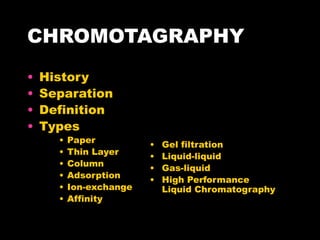
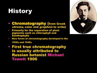
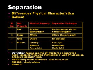
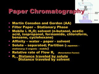
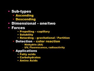
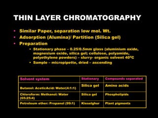
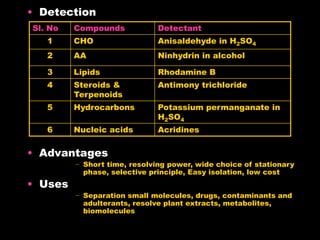
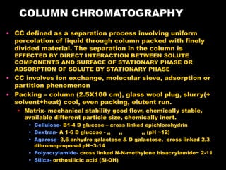
CHROMOTAGRAPHY
• History
• Separation
• Definition
• Types
• Paper
• Thin Layer
• Column
• Adsorption
• Ion-exchange
• Affinity
• Gel filtration
• Liquid-liquid
• Gas-liquid
• High Performance
Liquid Chromatography
History
• Chromatography (from Greek
chroma, color and graphein to write)
• Primarily for the separation of plant
pigments such as chlorophyll and
Xanthophylls
• New forms of chromatography developed in the
1930s and 1940s
• First true chromatography
is usually attributed to
Russian botanist Michael
Tswett 1906
Separation
• Differences Physical Characteristics
• Solvent
• Definition-Components of mixtures separated -
RATE – CARRIED THROUGH STATIONARY PHASE BY LIQUID OR
GASEOUS MOBILE PHASE.
• ‘SOME’ components held firmly – stationary phase
• ADSORB – sheet, column
• AFFINITY
Sl.
No
Mol.
Property
Physical Property Separation Technique
1 Size Diffusion
Sedimentation
Gel Permeation, Dialysis
Ultracentrifugation
2 Shape Affinity Affinity Chromatography
3 Ionic Charge Ion exchange
4 Polarity Volatility
Solubility
Adsorptivity
Gas-Liquid
Liquid-liquid
Liquid-Solid
• Martin Consden and Gordon (AA)
• Filter Paper – Stationary Phase
• Mobile i. H2O; solvent (n-butanol, acetic
acid, isopropanol, formamide, chloroform,
benzene, cyclohexane)
• Affinity – water – paper - solvent
• Solute – separated; Partition (i aqueous –
stationary; ii organic - mobile)
• Relative rate of Flow Rf (Retardation Factor)
• Rf = Distance traveled by solute
Distance traveled by solvent
Paper Chromatography
• Sub-types
• Ascending
• Descending
• Dimensional - one/two
• Forces
• Propelling – capillary
• Solubility
• Retarding – gravitational / Partition
• Detection – color reaction
− Ninhydrin (AA)
− UV, Fluorescence, radioactivity
• Applications
• Fatty acids
• Carbohydrates
• Amino Acids
THIN LAYER CHROMATOGRAPHY
• Similar Paper, separation low mol. Wt.
• Adsorption (Alumina)/ Partition (Silica gel)
• Preparation
• Stationary phase – 0.25/0.5mm glass (aluminium oxide,
magnesium oxide, silica gel; cellulose, polyamide,
polyethylene powders) – slurry- organic solvent 400C
• Sample – micropipette, dried – ascending
Solvent system Stationary Compounds separated
Butanol: AceticAcid: Water(4:1:1)
Silica gel Amino acids
Chloroform: Methanol: Water
(65:25:4)
Silica gel Phospholipids
Petroleum ether: Propanol (99:1) Kieselghur Plant pigments
• Detection
• Advantages
− Short time, resolving power, wide choice of stationary
phase, selective principle, Easy isolation, low cost
• Uses
− Separation small molecules, drugs, contaminants and
adulterants, resolve plant extracts, metabolites,
biomolecules
Sl. No Compounds Detectant
1 CHO Anisaldehyde in H2SO4
2 AA Ninhydrin in alcohol
3 Lipids Rhodamine B
4 Steroids &
Terpenoids
Antimony trichloride
5 Hydrocarbons Potassium permanganate in
H2SO4
6 Nucleic acids Acridines
COLUMN CHROMATOGRAPHY
• CC defined as a separation process involving uniform
percolation of liquid through column packed with finely
divided material. The separation in the column is
EFFECTED BY DIRECT INTERACTION BETWEEN SOLUTE
COMPONENTS AND SURFACE OF STATIONARY PHASE OR
ADSORPTION OF SOLUTE BY STATIONARY PHASE
• CC involves ion exchange, molecular sieve, adsorption or
partition phenomenon
• Packing – column (2.5X100 cm), glass wool plug, slurry(+
solvent+heat) cool, even packing, elutent run.
• Matrix- mechanical stability good flow, chemically stable,
available different particle size, chemically inert.
• Cellulose- B1-4 D glucose – cross linked epichlorohydrin
• Dextran- A 1-6 D glucose - ,, ,, ,, (pH ~12)
• Agarose- 3,6 anhydro galactose & D galactose, cross linked 2,3
dibromoproponal pH~3-14
• Polyacrylamide- cross linked N-N-methylene bisacrylamide~ 2-11
• Silica- orthosilicic acid (Si-OH)
• Sample application
• Remove mobile phase suction. Sample
allowed to run slowly –column- pipette slow
addition mobile 2-5 cm; connect reservoir –
constant flow.
• High density solution packing (1% sucrose,
sinks)
• Capillary/ syringe
• Factors
• Dimension
• Particle size
• Low viscosity
• Temperature
• Flow rate
• Packing
• Elution
• Isocratic
• Stepwise
• Gradient
• Retention Time
• Time taken each compound to emerge
• Volume mobile phase – elution volume
• Chromatogram
− Detector end ~ signals plotted – function
time or volume- peaks
ION-EXCHANGE CHROMATOGRAPHY
Process that allows the separation
of ions based on their charge. It can
be used for almost any kind of
charged molecule including
large proteins, small nucleotides
and amino acids. The solution to be
injected is usually called a sample
and the individually separated
components are called analytes. It
is often used in protein purification,
water analysis, and quality control
etc.
HISTORY
 Ion methods have been in use since 1850, when H.
Thompson and J. T. Way, researchers in England, treated
various clays with ammonium sulfate or carbonate in
solution to extract the ammonia and release calcium.
 In 1927, the first zeolite mineral column was used to
remove interfering calcium and magnesium ions from
solution to determine the sulfate content of water.
 The modern version of IEC was developed during the
wartime. A technique was required to separate and
concentrate the radioactive elements needed to make the
atom bomb. Researchers chose adsorbents that would
attach onto charged transuranium elements, which could
then be differentially eluted.
 In the early 1970s, ion exchange chromatography was
developed at Dow Chemical Company as a novel method
of IEC usable in automated analysis.
PRINCIPLE
Ion exchange chromatography retains
analyte molecules on the column based on ionic
interactions. The stationary phase surface displays
ionic functional groups that interact with analyte ions
of opposite charge. This type of chromatography is
further subdivided into cation exchange
chromatography and anion exchange chromatography.
The ionic compound consisting of the cationic species
and the anionic species are retained by the stationary
phase.
Cation exchange chromatography retains positively
charged cation because the stationary phase displays
a negatively charged functional group.
Anion exchange chromatography retains anions using
positively charged functional group.
ANION EXCHANGER
 The resin in an anion exchanger is positively charged. Anion
exchangers are named so because of their affinity to anions.
 Anion exchangers can be classified as:-
Weak:- Weak anion exchangers tend to lose their charge
as the pH changes (weak base).
Eg:-DEAE (diethylaminoethane).
Strong:- Strong anion exchangers are able to maintain
their positive charges across a variable pH range (strong
base). Eg:-Q (quaternary resin).
CATION EXCHANGERS
 Cation exchangers are named for their ability to attract
cations or positively charged particles. In this case, the
resin of the chromatography system is negatively
charged.
 Cation exchangers can be classified as:-
Weak-A weak cation exchanger is comprised of a weak
acid that gradually loses its charge as the pH decreases.
Eg.. Carboxymethal groups
Strong- A Strong cation exchanger is comprised of a
strong acid that is able to sustain its charge over a wide
pH range. Eg..Sulfopropyl groups
STAGES
EQUILIBRATION
 In this step a buffer with the desired
conditions is applied, and all of the
charged group in the stationary phase
are associated with ions of the opposite
charge. When this process in complete,
equilibrium has been reached.
APPLICATION OF SAMPLE
 In this step, the sample is applied to the
stationary phase. Only samples carrying
a charge opposite to the stationary
phase will bind to it while those with the
same charge or no charge will not bind.
These unbound particles will wash out
during this stage.
ELUTION
 This step involves changing the buffer conditions to
particles that have bound to the stationary phase. Done
by several ways:-
A. By Changing the pH of the buffer solution. When the pI of a
protein is the same as the pH of a solution, the net charge of
the protein will be zero. Therefore, when the buffer
solution's pH reaches the pI of the protein, the protein's net
charge will be zero. The protein will no longer bind to the
stationary phase and will be released and washed out.
B. By Increasing the salt concentration. As the salt
concentration of the buffer increases, salt ions replace
the bound protein. Proteins with weaker ion interactions
will be released at lower salt concentrations. Proteins
with stronger charges will have a higher affinity for the
stationary phase and will remain bound to the column
longer.
REGENERATION
 This step simply involves removing all bound protein from
the stationary phase so that it is ready for another
process
APPLICATIONS
 The treatment of water for drinking purpose (commercial,
industrial, and residential), and wastewater treatment. Ion
exchangers can soften the water, deionize it, and even be
used in desalination.
 Seperation of Protiens and Polysacharides, Nucleotides,
Amino acids, Vitamins
 The recovery of valuable metals is also possible.
 In food industry – wine making, sugar manufacture.
 In the medical world, development and preparation of key
drugs and antibiotics, such as streptomycin and quinine.
Treatments for ulcers, kidneys etc.
ADSORPTION CHROMATOGRAPHY
• Principle
Certain solids - ABILITY TO HOLD MOLECULES - SURFACE –
INVOLVES WEAK ATTRACTIVE FORCES – VAN DER WAAL AND
HYDROGEN BONDING. Phenomena exploited separation.
Solute different components – interact differently –
stationary phase – some settle, others migrate –
separation achieved
WEAK INTERMEDIATE STRONG
Sucrose
Cellulose
Starch
Calcium Carbonate
Calcium sulphate
Calcium phosphate
Charcoal
Magnesium
Aluminium
• Preparation
• Column
• Packing
– Dry
– Wet
• Factors
– Adsorbent, solvent choice, flow rate, dimension,
temperature
• Removal separated components
– Extrusion
– Elution
• Applications
• Polycyclic aromatic compounds, phenols, amines etc
• Plasma corticol
• Aliphatic hydrocarbons from aromatic hydrocarbons
• Geometrical isomers
Adsorbent Substance separated
Activated carbon
Alumina
Calcium phosphate gel
Silica gel
Peptides, AA, CHO
Small organic molecules, proteins
Proteins, polynucleotides
Sterols, AA
GEL PERMEATION/ GEL FILTERATION
CHROMATOGRAPHY
• Principle
Molecular sieve/ Gel exclusion
chromatography. SEPARATION – PARTITION-
MOLECULAR SIZE. Column packed tiny inert
substance – small pores. Larger molecules
pass – eluted
• Gels – cross linked polymers, inert
• Dextran – Polysaccride, dry beads – Sephadex
• Polyacrylamide – Acrylamide with NN methylene-
bisacrylamide. Size regulated – Biogel
• Agarose – Linear polymer D-glucose and 3,6-
anhydro galactose - Sepharose
Name Fraction range for
protein (Daltons)
Dextran (Sephadex)
G – 10
G – 15
G – 25
G – 50
G – 75
G – 100
G – 150
G – 200
0-700
0-1500
1000-5000
1500 - 30,000
3,000 - 80,000
4,000- 1,50,000
5,000- 3,00,000
5,000- 6,00,000
Poly-acrylamide (Bio-Gels)
P – 2
P – 4
P – 6
P – 100
P - 300
100-1800
800-4000
1000-6000
5000-1,00,000
60,000-4,00,000
Agarose
Bio-gel A - 0.5
Bio-gel A- 1.5
Bio-gel A- 150
10,000-5,00,000
10,000-15,00,000
1,00,000 – 50,00,000
• Applications
• Separation low mol.wt compounds from
high mol.wt compounds
• Separtion of desired protein from mixer of
proteins
• Determination of molecular wt of protein
AFFINITY CHROMATOGRAPHY
• Principle
Make use – SPECIFIC AFFINITY
BETWEEN SUBSTANCE – ISOLATE
MOLECULE – SPECIFICALLY BIND –
LIGAND. Column prepared –
covalently coupling binding molecule
– matrix. Elution – changing
conditions.
• Structure – binding nature – pre-requisite
– Sephacryl S, Sepharose, Bio-gel, Bio-beads
• Spacer arm – 1,6, diaminohexane, 6-aminohexanoic acid,
1,4-bis(2,3-epoxypropoxy)butane
Ligands used in affinity chromatography
LIGAND MACROMOLECULE
Tryptophan
Benzamidine
Heparin
Poly U
Lysine
Avidin
5’-AMP
2’,5’-ADP
Concanavalin A
Protein A & G
Soybean lectin
A-Chymotrypsin
Thrombin
Coagulation factor
Poly A messenger RNA
Ribosomal RNA
Biotin containing enzymes
NAD+ dependent dehydrogenase
NADP+ dependent dehydrogenase
Glycoprotein containing A-D-glucose & A-D-
mannose
Immunoglobins
Glycoproteins N-acetyl-galactopyranosyl
residues

COURSEPAGE-DUMP_AnalyticalInstrumentation_CHROMOTOGRAPHY.ppt

  • 1.
    CHROMOTAGRAPHY • History • Separation •Definition • Types • Paper • Thin Layer • Column • Adsorption • Ion-exchange • Affinity • Gel filtration • Liquid-liquid • Gas-liquid • High Performance Liquid Chromatography
  • 2.
    History • Chromatography (fromGreek chroma, color and graphein to write) • Primarily for the separation of plant pigments such as chlorophyll and Xanthophylls • New forms of chromatography developed in the 1930s and 1940s • First true chromatography is usually attributed to Russian botanist Michael Tswett 1906
  • 3.
    Separation • Differences PhysicalCharacteristics • Solvent • Definition-Components of mixtures separated - RATE – CARRIED THROUGH STATIONARY PHASE BY LIQUID OR GASEOUS MOBILE PHASE. • ‘SOME’ components held firmly – stationary phase • ADSORB – sheet, column • AFFINITY Sl. No Mol. Property Physical Property Separation Technique 1 Size Diffusion Sedimentation Gel Permeation, Dialysis Ultracentrifugation 2 Shape Affinity Affinity Chromatography 3 Ionic Charge Ion exchange 4 Polarity Volatility Solubility Adsorptivity Gas-Liquid Liquid-liquid Liquid-Solid
  • 4.
    • Martin Consdenand Gordon (AA) • Filter Paper – Stationary Phase • Mobile i. H2O; solvent (n-butanol, acetic acid, isopropanol, formamide, chloroform, benzene, cyclohexane) • Affinity – water – paper - solvent • Solute – separated; Partition (i aqueous – stationary; ii organic - mobile) • Relative rate of Flow Rf (Retardation Factor) • Rf = Distance traveled by solute Distance traveled by solvent Paper Chromatography
  • 5.
    • Sub-types • Ascending •Descending • Dimensional - one/two • Forces • Propelling – capillary • Solubility • Retarding – gravitational / Partition • Detection – color reaction − Ninhydrin (AA) − UV, Fluorescence, radioactivity • Applications • Fatty acids • Carbohydrates • Amino Acids
  • 6.
    THIN LAYER CHROMATOGRAPHY •Similar Paper, separation low mol. Wt. • Adsorption (Alumina)/ Partition (Silica gel) • Preparation • Stationary phase – 0.25/0.5mm glass (aluminium oxide, magnesium oxide, silica gel; cellulose, polyamide, polyethylene powders) – slurry- organic solvent 400C • Sample – micropipette, dried – ascending Solvent system Stationary Compounds separated Butanol: AceticAcid: Water(4:1:1) Silica gel Amino acids Chloroform: Methanol: Water (65:25:4) Silica gel Phospholipids Petroleum ether: Propanol (99:1) Kieselghur Plant pigments
  • 7.
    • Detection • Advantages −Short time, resolving power, wide choice of stationary phase, selective principle, Easy isolation, low cost • Uses − Separation small molecules, drugs, contaminants and adulterants, resolve plant extracts, metabolites, biomolecules Sl. No Compounds Detectant 1 CHO Anisaldehyde in H2SO4 2 AA Ninhydrin in alcohol 3 Lipids Rhodamine B 4 Steroids & Terpenoids Antimony trichloride 5 Hydrocarbons Potassium permanganate in H2SO4 6 Nucleic acids Acridines
  • 8.
    COLUMN CHROMATOGRAPHY • CCdefined as a separation process involving uniform percolation of liquid through column packed with finely divided material. The separation in the column is EFFECTED BY DIRECT INTERACTION BETWEEN SOLUTE COMPONENTS AND SURFACE OF STATIONARY PHASE OR ADSORPTION OF SOLUTE BY STATIONARY PHASE • CC involves ion exchange, molecular sieve, adsorption or partition phenomenon • Packing – column (2.5X100 cm), glass wool plug, slurry(+ solvent+heat) cool, even packing, elutent run. • Matrix- mechanical stability good flow, chemically stable, available different particle size, chemically inert. • Cellulose- B1-4 D glucose – cross linked epichlorohydrin • Dextran- A 1-6 D glucose - ,, ,, ,, (pH ~12) • Agarose- 3,6 anhydro galactose & D galactose, cross linked 2,3 dibromoproponal pH~3-14 • Polyacrylamide- cross linked N-N-methylene bisacrylamide~ 2-11 • Silica- orthosilicic acid (Si-OH)
  • 9.
    • Sample application •Remove mobile phase suction. Sample allowed to run slowly –column- pipette slow addition mobile 2-5 cm; connect reservoir – constant flow. • High density solution packing (1% sucrose, sinks) • Capillary/ syringe • Factors • Dimension • Particle size • Low viscosity • Temperature • Flow rate • Packing
  • 10.
    • Elution • Isocratic •Stepwise • Gradient • Retention Time • Time taken each compound to emerge • Volume mobile phase – elution volume • Chromatogram − Detector end ~ signals plotted – function time or volume- peaks
  • 11.
    ION-EXCHANGE CHROMATOGRAPHY Process thatallows the separation of ions based on their charge. It can be used for almost any kind of charged molecule including large proteins, small nucleotides and amino acids. The solution to be injected is usually called a sample and the individually separated components are called analytes. It is often used in protein purification, water analysis, and quality control etc.
  • 12.
    HISTORY  Ion methodshave been in use since 1850, when H. Thompson and J. T. Way, researchers in England, treated various clays with ammonium sulfate or carbonate in solution to extract the ammonia and release calcium.  In 1927, the first zeolite mineral column was used to remove interfering calcium and magnesium ions from solution to determine the sulfate content of water.  The modern version of IEC was developed during the wartime. A technique was required to separate and concentrate the radioactive elements needed to make the atom bomb. Researchers chose adsorbents that would attach onto charged transuranium elements, which could then be differentially eluted.  In the early 1970s, ion exchange chromatography was developed at Dow Chemical Company as a novel method of IEC usable in automated analysis.
  • 13.
    PRINCIPLE Ion exchange chromatographyretains analyte molecules on the column based on ionic interactions. The stationary phase surface displays ionic functional groups that interact with analyte ions of opposite charge. This type of chromatography is further subdivided into cation exchange chromatography and anion exchange chromatography. The ionic compound consisting of the cationic species and the anionic species are retained by the stationary phase. Cation exchange chromatography retains positively charged cation because the stationary phase displays a negatively charged functional group. Anion exchange chromatography retains anions using positively charged functional group.
  • 14.
    ANION EXCHANGER  Theresin in an anion exchanger is positively charged. Anion exchangers are named so because of their affinity to anions.  Anion exchangers can be classified as:- Weak:- Weak anion exchangers tend to lose their charge as the pH changes (weak base). Eg:-DEAE (diethylaminoethane). Strong:- Strong anion exchangers are able to maintain their positive charges across a variable pH range (strong base). Eg:-Q (quaternary resin). CATION EXCHANGERS  Cation exchangers are named for their ability to attract cations or positively charged particles. In this case, the resin of the chromatography system is negatively charged.  Cation exchangers can be classified as:- Weak-A weak cation exchanger is comprised of a weak acid that gradually loses its charge as the pH decreases. Eg.. Carboxymethal groups Strong- A Strong cation exchanger is comprised of a strong acid that is able to sustain its charge over a wide pH range. Eg..Sulfopropyl groups
  • 16.
    STAGES EQUILIBRATION  In thisstep a buffer with the desired conditions is applied, and all of the charged group in the stationary phase are associated with ions of the opposite charge. When this process in complete, equilibrium has been reached. APPLICATION OF SAMPLE  In this step, the sample is applied to the stationary phase. Only samples carrying a charge opposite to the stationary phase will bind to it while those with the same charge or no charge will not bind. These unbound particles will wash out during this stage.
  • 17.
    ELUTION  This stepinvolves changing the buffer conditions to particles that have bound to the stationary phase. Done by several ways:- A. By Changing the pH of the buffer solution. When the pI of a protein is the same as the pH of a solution, the net charge of the protein will be zero. Therefore, when the buffer solution's pH reaches the pI of the protein, the protein's net charge will be zero. The protein will no longer bind to the stationary phase and will be released and washed out. B. By Increasing the salt concentration. As the salt concentration of the buffer increases, salt ions replace the bound protein. Proteins with weaker ion interactions will be released at lower salt concentrations. Proteins with stronger charges will have a higher affinity for the stationary phase and will remain bound to the column longer. REGENERATION  This step simply involves removing all bound protein from the stationary phase so that it is ready for another process
  • 18.
    APPLICATIONS  The treatmentof water for drinking purpose (commercial, industrial, and residential), and wastewater treatment. Ion exchangers can soften the water, deionize it, and even be used in desalination.  Seperation of Protiens and Polysacharides, Nucleotides, Amino acids, Vitamins  The recovery of valuable metals is also possible.  In food industry – wine making, sugar manufacture.  In the medical world, development and preparation of key drugs and antibiotics, such as streptomycin and quinine. Treatments for ulcers, kidneys etc.
  • 20.
    ADSORPTION CHROMATOGRAPHY • Principle Certainsolids - ABILITY TO HOLD MOLECULES - SURFACE – INVOLVES WEAK ATTRACTIVE FORCES – VAN DER WAAL AND HYDROGEN BONDING. Phenomena exploited separation. Solute different components – interact differently – stationary phase – some settle, others migrate – separation achieved WEAK INTERMEDIATE STRONG Sucrose Cellulose Starch Calcium Carbonate Calcium sulphate Calcium phosphate Charcoal Magnesium Aluminium
  • 21.
    • Preparation • Column •Packing – Dry – Wet • Factors – Adsorbent, solvent choice, flow rate, dimension, temperature • Removal separated components – Extrusion – Elution • Applications • Polycyclic aromatic compounds, phenols, amines etc • Plasma corticol • Aliphatic hydrocarbons from aromatic hydrocarbons • Geometrical isomers Adsorbent Substance separated Activated carbon Alumina Calcium phosphate gel Silica gel Peptides, AA, CHO Small organic molecules, proteins Proteins, polynucleotides Sterols, AA
  • 23.
    GEL PERMEATION/ GELFILTERATION CHROMATOGRAPHY • Principle Molecular sieve/ Gel exclusion chromatography. SEPARATION – PARTITION- MOLECULAR SIZE. Column packed tiny inert substance – small pores. Larger molecules pass – eluted • Gels – cross linked polymers, inert • Dextran – Polysaccride, dry beads – Sephadex • Polyacrylamide – Acrylamide with NN methylene- bisacrylamide. Size regulated – Biogel • Agarose – Linear polymer D-glucose and 3,6- anhydro galactose - Sepharose
  • 25.
    Name Fraction rangefor protein (Daltons) Dextran (Sephadex) G – 10 G – 15 G – 25 G – 50 G – 75 G – 100 G – 150 G – 200 0-700 0-1500 1000-5000 1500 - 30,000 3,000 - 80,000 4,000- 1,50,000 5,000- 3,00,000 5,000- 6,00,000 Poly-acrylamide (Bio-Gels) P – 2 P – 4 P – 6 P – 100 P - 300 100-1800 800-4000 1000-6000 5000-1,00,000 60,000-4,00,000 Agarose Bio-gel A - 0.5 Bio-gel A- 1.5 Bio-gel A- 150 10,000-5,00,000 10,000-15,00,000 1,00,000 – 50,00,000
  • 26.
    • Applications • Separationlow mol.wt compounds from high mol.wt compounds • Separtion of desired protein from mixer of proteins • Determination of molecular wt of protein
  • 28.
    AFFINITY CHROMATOGRAPHY • Principle Makeuse – SPECIFIC AFFINITY BETWEEN SUBSTANCE – ISOLATE MOLECULE – SPECIFICALLY BIND – LIGAND. Column prepared – covalently coupling binding molecule – matrix. Elution – changing conditions. • Structure – binding nature – pre-requisite – Sephacryl S, Sepharose, Bio-gel, Bio-beads • Spacer arm – 1,6, diaminohexane, 6-aminohexanoic acid, 1,4-bis(2,3-epoxypropoxy)butane
  • 30.
    Ligands used inaffinity chromatography LIGAND MACROMOLECULE Tryptophan Benzamidine Heparin Poly U Lysine Avidin 5’-AMP 2’,5’-ADP Concanavalin A Protein A & G Soybean lectin A-Chymotrypsin Thrombin Coagulation factor Poly A messenger RNA Ribosomal RNA Biotin containing enzymes NAD+ dependent dehydrogenase NADP+ dependent dehydrogenase Glycoprotein containing A-D-glucose & A-D- mannose Immunoglobins Glycoproteins N-acetyl-galactopyranosyl residues