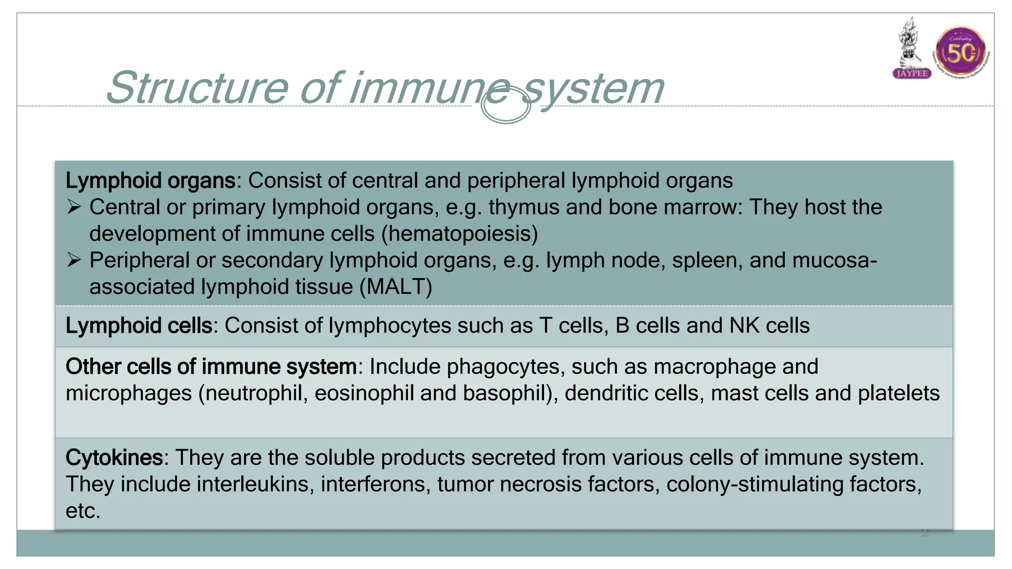
D R S H A R A N K A D A M B A
S R M I C R O B I O L O G Y
H B T M C & D R R N C O O P E R H O S P I T A L
Components of Immune System
Organs, Cells and Products
Structure of immune system
2
Lymphoid organs: Consist of central and peripheral lymphoid organs
 Central or primary lymphoid organs, e.g. thymus and bone marrow: They host the
development of immune cells (hematopoiesis)
 Peripheral or secondary lymphoid organs, e.g. lymph node, spleen, and mucosa-
associated lymphoid tissue (MALT)
Lymphoid cells: Consist of lymphocytes such as T cells, B cells and NK cells
Other cells of immune system: Include phagocytes, such as macrophage and
microphages (neutrophil, eosinophil and basophil), dendritic cells, mast cells and platelets
Cytokines: They are the soluble products secreted from various cells of immune system.
They include interleukins, interferons, tumor necrosis factors, colony-stimulating factors,
etc.
CENTRAL LYMPHOID
ORGANS
3
Bone Marrow
 All the cells in blood are originated from pluripotent hematopoietic
stem cells of bone marrow and the process is called a
hematopoiesis.
 In early fetal life, hematopoiesis occurs in liver; gradually the stem
cells migrate to bone marrow.
 By birth, the stem cells occupy most of the bone marrow space of
large bones. 4
Bone Marrow (Cont..)
 After puberty hematopoiesis is mostly confined to axial bones
such as pelvis, vertebrae, sternum, skull & ribs.
 Progenitor T and B cells originate in bone marrow.
 Further development of B cells occurs in bone marrow itself.
 Progenitor T cells migrate to thymus for further proliferation.
5
Thymus
 Site of proliferation and maturation of T cells.
6
Development
 Developed in the embryonic life
 Highly active at birth, continues to grow for many years, reaches its
peak size at puberty, and then it degenerates.
7
Structure
 Thymus has two lobes surrounded by
a fibrous capsule.
 Septa arising from capsule divide
thymus into lobules, and each lobule
is differentiated into an outer cortex
& an inner medulla
8
Cortex
 Densely populated and contains:
 Thymocytes- Lymphocytes of thymus (immature and many in
number).
 Cortical epithelial cells
 Nurse cells (specialized epithelial cells with long membrane
extensions that surround many thymocytes)
9
Medulla
 Sparsely populated and contains:
 Thymocytes which are relatively more mature and fewer in
number
 Medullary epithelial cells
 Interdigitating dendritic cells
 Hassall’s corpuscles (concentric layers of degenerating epithelial
10
Thymic Hormones
 Produced from the epithelial cells of thymus.
 They are believed to attract the precursor T cells (progenitor T cells)
from bone marrow.
 Thymulin
 Thymopoietin
 Thymosin
11
Maturation of T Cells
 Cell-to-cell interaction between thymocytes and thymic stromal cells
(including epithelial cells, dendritic cells and macrophages) and the
effect of thymic hormones help in maturation of T cells in thymus
12
Central Tolerance
 Only 2-5% of the developing T cells become mature and released
out from thymus.
 Remaining T cells are destroyed as they are either not capable of
recognizing MHC or are believed to be self-reacting in nature.
 Destruction of such self-reacting T cells prevents
development of autoimmunity (immune response against self-
antigens).
 Such tolerance to self-antigens mediated by thymus that occurs in
embryonic life is called as central tolerance.
13
Defect in Thymus
 Leads to defect in maturation of T lymphocytes that in turn results
in severe life threatening cell mediated immunodeficiency
disorders.
 DiGeorge syndrome (immunodeficiency disorder) - congenital
aplasia of thymus.
14
PERIPHERAL LYMPHOID
ORGANS
15
Lymph Node
 Lymph nodes act as physiological barriers - filter the microbial
antigens carried to lymph node by activating the T and B cells.
16
Cortex
 Contains lymphoid follicles that are composed of:
 B cells
 Few special type of dendritic cells (called follicular dendritic
cells)
 Occasional T cells.
 Lymphoid follicles are mainly of two types:
 Primary lymphoid follicles
 Secondary lymphoid follicles 17
Cortex (Cont..)
Primary lymphoid follicles:
 Found before the antigenic stimulus.
 Smaller in size
 Contain the resting B cells.
18
Cortex (Cont..)
Secondary lymphoid follicles:
 Following contact with an antigen - resting B cells starts dividing
and become activated.
 Activated B cells differentiate rapidly into plasma cells and
memory B cells.
 Follicles become larger called secondary lymphoid follicles.
19
Paracortical area
 Present in between cortex and medulla.
 T cell area of lymph node (rich in naive T cells).Contains
macrophages and interdigitating dendritic cells, which trap the
antigens and present to T cells.
20
Medulla
 Innermost area of lymph node.
 Rich in B-lymphocytes (mainly plasma cells).
21
Spleen
 Largest secondary lymphoid organ.
 Acts as physiological barrier similar to lymph node in clearing
the microbial antigens through the antigenic stimulation of T and B
cells.
22
Defect in Spleen
 Increased incidence of bacterial sepsis caused primarily by
capsulated bacteria such as:
 Streptococcus pneumoniae
 Neisseria meningitides
 Haemophilus influenzae.
23
Mucosa-associated Lymphoid Tissue (MALT)
 Defense mechanisms in the mucosal sites to prevent the microbial
entry.
 Group of lymphoid tissues lining these mucosal sites are collectively
known as mucosa associated lymphoid tissue (MALT).
24
Epithelial layer
 Contains few specialized lymphocytes called intraepithelial
lymphocytes (IELs) and modified epithelial cells (called M cells)
 Intraepithelial lymphocytes (IELs) are the γδ T cells.
 M cells
 Secretory IgA: Dimeric IgA antibodies - present in the
submucosa as well as in the lining epithelium.
25
M Cells
 Contains B cells, T cells and macrophages.
 Portal of entry of a number of microbes such as Salmonella,
Shigella, Vibrio, and Poliovirus.
26
M Cells (Cont..)
27
A. MALT; B. Structure of M cell
M Cells (Cont..)
 Invading microbes are taken-up by M cells (by endocytosis) -
transported in a vesicle and are delivered to the basolateral pockets
 B lymphocytes in MALT once activated at a site by antigenic
exposure, migrate to other parts of the intestine, secrete the
dimeric IgA, and thus extend the local immunity
28
Cutaneous-associated Lymphoid Tissue
 Similar to MALT.
 Skin also contains a few loose lymphocytes and specialized antigen
presenting cells in epidermis called Langerhans cells.
29
LYMPHOID CELLS
30
LYMPHOID CELLS
 Lymphocytes are the major components of cells of immune system.
 Approximately 1011 lymphocytes in the body, accounting for 20-40%
of the total white blood cells (WBCs) in blood and 99% of the cells in
the lymph.
31
Distribution of lymphocytes (%) in organs/blood
32
Tissues T cell B cell NK cell
Bone marrow 5-10 80-90 5-10
Thymus 99 <1 <1
Lymph node 70-80 20-30 <1
Spleen 30-40 50-60 1-5
Peripheral
blood
70-80 10-15 10-15
CD molecules (cluster of differentiation)
 Cell surface markers useful for the identification of cells of
immune system.
 Numerous functions, often act as surface receptors or some CD
proteins may help in cell adhesion.
 As of 2015, CD molecules for humans are numbered up to 364,
important examples are CD4 and CD8 molecules—expressed by
helper T cells and cytotoxic T cells respectively. 33
TYPES OF
LYMPHOCYTES
34
TYPES OF LYMPHOCYTES
 Based on function and cell membrane structure:
 T lymphocytes
 B lymphocytes
 NK (natural killer) cells.
 Lymphocytes can also be classified into:
 Naive lymphocytes
 Lymphoblasts.
35
Naive Lymphocytes
 Resting B and T lymphocytes that have not interacted with
antigen (unprimed lymphocytes).
 Also known as small lymphocytes (6µm)
 Short life span (1-3 months).
36
Lymphoblasts
 Naive cells interact with antigen in the presence of certain
cytokines (e.g. interleukin-7) - become activated - transform into
lymphoblasts - eventually differentiate into effector cells or
memory cells.
37
Effector cells
 Function in various ways to eliminate antigen.
 Short life span (few days to few weeks)
 Large lymphocytes (15 µm in size), having wider rim of cytoplasm
with more organelles.
 Effector B cells - Antibody producing plasma cells
 Effector T cells - Helper T cells and cytotoxic T cells.
38
Memory cells
 Remain dormant like naive cells.
 Capable of transforming into effector cells rapidly on subsequent
antigenic challenge.
 Longer life span; providing long term immunity to may pathogens.
39
Differences between naive, effector and
memory cells
40
Naive cell Effector cell Memory cell
Location
(present mostly in)
Secondary
lymphoid organs
Inflamed
tissues & mucosal
surfaces
Both the locations
of naive & effector cell
Cell cycle Dormant
(G0 phase)
Active Dormant
(G0 phase)
Morphology Small lymphocyte Large lymphocyte Small lymphocyte
Life span Short Short Long
Function Transforms to effector
cell on primary exposure
exposure to antigen;
Occurs slow due to lag
period
Eliminate antigen Transforms to effector
cell on secondary
exposure to antigen,
Occurs fast without lag
period
T LYMPHOCYTES
42
T LYMPHOCYTES
 T cells constitute 70–80% of blood lymphocytes.
 Unlike B cells, they do not have microvilli on their surface.
 Bear specialized surface receptors called T cell receptors (TCR).
43
T Cell Receptor
 Main function - antigen recognition.
 Unlike B cell receptor which binds to antigen directly, TCR does not
recognize antigen by itself.
 Can only respond to an antigen which is processed and presented by
the antigen presenting cells such as macrophages.
44
TCR–CD3 Complex
 Most T cell receptors (95%) comprise of two chains (α & β) which
in turn have three regions:
 Extracellular domain
 Transmembrane domain
 Cytoplasmic tail.
 Extracellular domain of each polypeptide chain has 2 regions
(variable and constant region).
45
TCR–CD3 Complex (Cont..)
 TCR is active only when both the
chains (α & β) complexes with CD3
molecule.
46
Types of T Cells - Effector T Cells
 Two types of effector T cells:
 CD4+ helper T cells
 CD8+ cytotoxic T cell.
48
Helper T cells (TH)
 Possess CD4 molecules as surface receptors.
 Recognize the antigenic peptides that are processed by antigen
presenting cells and presented along with MHC-II molecules.
 Following antigenic stimulus, the helper T cells differentiate into
either of the two types of cells- TH1 and TH2 subset.
 Each secrete specific cytokines which modulate the cellular and
humoral immune responses respectively.
49
Helper T cells (TH) (Cont..)
TH17 cells:
 Recently a third subset of T helper cells.
 Produces IL-17 & IL-22 - primarily involved in recruiting
neutrophils which in turn kill the microbes as well as induce
inflammation.
 Contribute to the pathogenesis of many autoimmune inflammatory
diseases. 50
Cytotoxic T cells
 Possess CD8 molecules
 Recognize the intracellular antigens processed by any nucleated cells
and presented along with MHC-I.
 TC cells are involved in destruction of virus infected cells and tumor
cells.
51
Regulatory T cells (TREG cells)
 Subpopulation of T cells which regulate the immune system.
 Provide tolerance to self-antigens (known as peripheral tolerance),
and prevent the development of autoimmune disease.
 Surface markers -TREG cells possess surface markers such as CD4,
CD25 and Foxp3 (a forkhead family transcription factor)
 Deficiency of Foxp3 receptors leads to a severe form of
autoimmune disease known as Immune dysregulation,
Polyendocrinopathy, Enteropathy X-linked (IPEX) syndrome.
52
B LYMPHOCYTES
55
B LYMPHOCYTES
 Mediators of humoral immunity; constitutes 10-15% of blood
lymphocytes.
 Named after their site of maturation (bursa of Fabricius in birds and
bone marrow in human and other mammals).
56
Development of B Lymphocytes
57
Development of B Cells in Peripheral Lymphoid
Organs
 Immature B cells migrate from bone marrow to peripheral lymphoid
organs (lymph node and spleen).
 Transform into mature B cells following contact with appropriate
antigen.
58
Mature or Naive B Cells
 Most mature B cells (95%) belong to the follicular B cell type and
produce surface receptor IgD in addition to IgM.
 Play an important role in humoral immune response.
 Following antigenic stimulus, the mature B cells transform into
activated B cells (lymphoblasts).
 Which further differentiate into either effector B cells i.e. plasma
cells (majority) or memory B cells.
59
Differences between T cell and B cell
60
Property T-cell B-cell
Origin Bone marrow Bone marrow
Maturation Thymus Bone marrow
Peripheral blood 70-80% of total lymphocytes 10-15% of total lymphocytes
Antigen recognition
receptors
T cell receptors complexed
with CD3
B cell receptor-Surface IgM or
IgD complexed with Igα/Igβ
CD markers CD 3,4,8 CD19, 21, 24
Thymus specific Ag Present Absent
Microvilli on the surface Absent Present
NATURAL KILLER
CELLS
61
NATURAL KILLER CELLS
 Large granular lymphocytes.
 Similar to cytotoxic T cells, NK cells also are involved in destruction
of virus infected cells and tumor cells.
62
OTHER CELLS OF
IMMUNE SYSTEM
63
Macrophage
Macrophages:
 When monocytes migrate to tissues, they transform into
macrophages
 Macrophages differ from monocytes in the following:
 5-10 folds larger than monocytes
 Contain more lysozymes and cell organelles
 Produce more lytic enzymes and cytokines
 Possess greater phagocytic activity
 Have a longer life in tissues (months to years) 66
Types of macrophages
67
Body sites Macrophage designation
Peripheral blood Monocytes
Tissues Macrophages
Liver Kupffer cells
Brain Microglial cells
Kidney Mesangial cells
Lungs Alveolar macrophages
Bone Osteoclasts
Secretory products of activated macrophages
68
Secretory Products Examples
Enzymes Lysozyme, acid hydrolases, elastases,
Phosphatases, lipases, collagenases
Free radicals Reactive oxygen intermediates
 Superoxide anion (O2-)
 Hydroxyl radicals (OH)
 Hydrogen peroxide (H2O2)
 Hypochlorite anion(ClO-)
Reactive nitrogen intermediates-
 Nitric oxide (NO)
 Nitrogen dioxide (NO2)
 HNO2(nitrous acid)
Secretory products of activated macrophages
(Cont..)
69
Secretory Products Examples
Cytokines Interferon α,β
Interleukins (IL-1, IL-6, IL-8, IL-12)
Tumor necrosis factor- α (TNF- α)
Growth factors Colony stimulating factors (CSF)
Platelet derived growth factor (PDGF)
Platelet activating factor (PAF)
Transforming growth factor β (TGF-β)
Coagulation factors Factor V,VII,IX,X
Prothrombin
Complement factors C5, C8 properdin, Factor B, D, I
Functions of Macrophage
 Phagocytosis
 Antigen presentation
 Activated macrophages
 Secretory products
70
Phagocytosis
 Macrophages are the principle
cells involved in phagocytosis.
 Steps:
71
Antigen presentation
 Macrophages also promote adaptive immunity, by acting as antigen
presenting cells (APCs).
 Macrophages capture the antigen - process into smaller antigenic
peptides and present the antigenic peptides along with the MHC
class II molecules to the helper T cells.
 Facilitating helper T cell activation.
72
Activated macrophages
 On exposure to cytokines such as interferon-γ, macrophages
become activated  greater phagocytic ability  produce many
cytokines  act against intracellular bacteria, virus infected cells
and tumor cells.
 Also express higher level of MHC class II, hence can act as efficient
APCs.
73
Secretory products
 Interleukin 1 (IL-1):Promotes inflammatory responses, fever and activate
helper T cells.
 IL-6 & TNF-α:Promote innate immunity, (inflammation & fever) and
eliminate the pathogens.
 Interferon α & β -have antiviral activity.
 TNF-α: Lyse the tumor cells (anti-tumor activity)
 Growth factors such as CSF (colony stimulating factor): promote
hematopoiesis.
 Following tissue injury, various mediators are secreted from macrophage;
which help in tissue repair and scar formation. 74
Dendritic Cells
 Possess long membranous cytoplasmic extensions
resembling dendrites of neurons - hence named as
dendritic cells.
 Originate from bone marrow, but the pathway is
uncertain.
 Develop as a separate lineage from stem cells or may
originate from the macrophage lineage. 75
Functions of dendritic cells
 Non-phagocytic in nature.
 Most efficient APCs.
 Main function is to capture, process and present the antigenic
peptides on their cell surface to the helper T cells.
 Carry high level of MHC class II and co-stimulatory B7 molecules.
 Act as messengers between the innate and the adaptive immune
systems. 76
Granulocytic Cells
 Granulocytes are a category of white blood cells characterized by the
presence of granules in their cytoplasm.
 E.g. neutrophils, eosinophils and basophils.
 Differ from each other by cell morphology and cytoplasmic staining
and function.
77
MAJOR
HISTOCOMPATIBILITY
COMPLEX
78
MAJOR HISTOCOMPATIBILITY
COMPLEX
 Complex is a group of genes.
 Coding for a set of host cell surface molecules that bind to peptide
fragments derived from pathogens
 Display them on the host cell surface for recognition by the
appropriate T-cells.
79
MAJOR HISTOCOMPATIBILITY
COMPLEX
(Cont..)
 Present in almost all the human cells, but first discovered on the
surface of leukocytes; hence in humans, the MHC coded proteins are
also called as human leukocyte antigens (HLA).
 Serve as a unique identification marker for every individual.
 Following transplantation of a graft, the recipient mounts an
immune response against the graft’s MHC molecule and vice versa.
80
MAJOR HISTOCOMPATIBILITY
COMPLEX
(Cont..)
 The acceptance or rejection of the graft is directly dependent on the
MHC molecules of the graft and the recipient.
 As the MHC molecules determine the compatibility between the
graft and host tissues, they are named as histocompatibility
antigens.
81
MHC GENES AND
THEIR PRODUCTS
82
MHC GENES AND THEIR PRODUCTS
 Located in short arm of chromosome-6.
 HLA complex extends over 4000kbp length covering >100 genes.
 Genes are clustered in three regions named as MHC region-I, II
and III.
83
Structure of human MHC complex
84
Structure of MHC molecule
85
MHC Class I Molecule
 Association of β2 microglobulin with α chain is necessary for the
expression of MHC I molecules on to the cell surface.
 In Daudi cells (a type of human B cell tumor cell which are not
able to produce β2 microglobulin) - observed that they synthesize
MHC-I but do not express them on cell surface.
86
Structure of MHC Class I molecule
87
Role of MHC Class I Molecules
 Antigen peptide groove of class I MHC molecule (i.e. the site, where
the antigen peptide binds) is formed by the cleft between α1 and α2
domains.
 α3 domain binds to CD8 molecule present on cytotoxic T cells
during antigen presentation.
88
MHC Class II Molecule
 Comprises of one α chain (33 kDa) and one β chain (28 kDa).
 The α and β chains in turn consist of two domains each—(1) α1 and
α2 and (2) β1 and β2, respectively and cytoplasmic tails.
89
Click to add text
Click to add text
MHC Class II Molecule (Cont..)
 The antigen peptide binding groove is formed by the cleft between
α1 and β1 domains
 β2 domain interacts with CD4 molecule of helper T cells during
antigen presentation.
90
Differences between MHC class I and MHC
class II molecules
91
MHC class I MHC class II
Present on All nucleated cells (except
sperms) and platelets
Antigen presenting cells (APCs)
Peptide antigen is presented to
CD8 T cells
presented to
CD4 T cells
Nature of peptide antigen Endogenous or intracellular (viral /
tumor antigen)
Exogenous
Peptide antigen (size) 8-10 amino acid long 13-18 amino acid long
Antigen presentation pathways Cytosolic pathway Endocytic pathway
Peptide-binding site α1/α2 groove α1/β1 groove
CD4 or CD8 binding site α3 binds to CD8 molecules on TC
cells
β2 binds to CD4 on TH cells
Regulation of MHC Expression
 Transcription factors-MHC genes have promoter sequences at
their 5’ end which are regulated by certain transcription factors such
as CIITA, and RFX (both bind to MHC II promoter genes and
increase their transcription). Defects in CIITA, and RFX cause one
of the form of bare lymphocyte syndrome.
92
Regulation of MHC Expression
 Cytokines also influence MHC expression.
 Interferon γ activates both MHC-I & II promoter genes.
 IL-4 increases expression of class II MHC molecules on resting B
cells.
93
Regulation of MHC Expression (Cont..)
 Corticosteroid and prostaglandins decrease the expression of
MHC II molecules.
 In many viral infection, the viral antigens inhibit various
components of MHC-I (e.g., adenovirus proteins inhibit TAP,
cytomegalovirus proteins inhibit β2 microglobulin). As a result,
MHC-I expression is suppressed.
94
Diseases associated with certain HLA alleles
95
HLA allele Associated disease
HLA B27 Ankylosing spondylitis,
Reactive arthritis (Yersinia, Salmonella,
Gonococcus) and
Reiter’s syndrome
DR-2 Multiple sclerosis, Goodpasture’s syndrome,
Narcolepsy
DR-3 Myasthenia gravis, systemic lupus erythematosus
(SLE)
DR-3/DR-4 Insulin-dependent diabetes mellitus
DR-4 Rheumatoid arthritis
A3/B14 Hereditary hemochromatosis
SOLUBLE PRODUCTS
OF
LYMPHOID CELLS
96
CYTOKINES
 Cytokines are chemical substances which serve as messengers,
mediating interaction and communication between the various cells
of immune system.
97
Sources and functions of cytokines
98
Cytokine Cytokine secreting cells Target cells & Functions
Interleukins (IL)
IL-1 Produced by all nucleated cells, but
principal sources are APCs such as-
Macrophages, monocytes
dendritic cell, B cells &
endothelial cell
1. TH cells - IL1 produced by APCs stimulates TH cells
activation & proliferation
 Promotes IL2 secretion by TH cells
 Induces IL-2 receptor expression on TH cells
 Induces ↑MHC-II expression on APCs
1. B cell -Promotes B cell development & maturation
2. Liver- Induces synthesis of acute phase reactant
proteins
3. Hypothalamus- induction of fever
4. Macrophage & neutrophil activation-↑ expression of
ICAM
IL-2 TH 1 cells Induces proliferation activated TH cells, TC cells and some
NK cells
(Previously called as T cell growth factor)
IL-3 TH cell, NK cell, Mast cell 1. Stimulates hematopoiesis (acts as multi-CSF)
Click to add text
Sources and functions of cytokines (Cont..)
99
Cytokine Cytokine secreting cells Target cells & Functions
Interleukins (IL)
IL-4 TH 2 cells 1. TH cells -Promote TH 2 cell activity and inhibit TH 1 cell
2. B cell- Promote B cells activation & proliferation and
induce B cell class switch over to produce IgE, IgG4,
IgG1;previously called as B cell growth factor
3. Macrophage & APCs- Induce ↑MHC-II expression
IL-5 TH 2 cells Promote eosinophil growth and differentiation
IL-6 TH 2 cells, macrophages IL-1 & TNF like effects (synergistic effect)
Promotes B cell proliferation and antibody production
IL-7 Bone marrow/thymic stromal
cells
Serves as a growth factor for T cell and B cell precursors.
Sources and functions of cytokines (Cont..)
100
Cytokine Cytokine secreting cells Target cells & Functions
Interleukins (IL)
IL-8 Macrophages, endothelial
cells
Attracts neutrophils, NK cells, eosinophils and
basophils.
IL-9 TH cells Hematopoietic & thymopoietic effects
IL-10 TH 2 cells Reduces cytokine production by TH1 cell.
IL-11 Bone marrow stromal cells Hematopoietic effect ( B cell and platelet development)
Liver- Induce synthesis of acute phase reactant protein
IL-12 Macrophages Promote TH 1 cell induction and inhibit TH 2 activity;
promotes CMI responses NK cell stimulatory factor
IL-13 TH 2 cells Mimic IL-4 function
IL-17 CD4+ activated memory TH
cell
Initiates and maintains inflammation
Sources and functions of cytokines (Cont..)
101
Cytokine Cytokine secreting cells Target cells & Functions
Interferons (IFN)
IFN-α Leukocytes Antiviral activity
IFN-β Fibroblasts Antiviral activity
IFN-γ TH &TC cells, NK cells 1. Macrophage -Activates the resting macrophages into
activated macrophage
2. B cells -Activate B cells to produce IgG
3. Promotes inflammation of delayed type of
hypersensitivity (along with TNF-β)
4. TH 2 cell - Inhibits TH 2 cell proliferation
Sources and functions of cytokines (Cont..)
102
Cytokine Cytokine secreting cells Target cells & Functions
Tumor necrosis factors (TNF)
TNF-α Macrophage 1. IL-1 like effect
2. Tumor cells- Promote vascular thrombosis and
tumor necrosis
3. Inflammatory cells- Induce cytokine secretion
4. Induces lipolysis, causes extensive weight loss
associated with chronic inflammation
TNF- β TH 1 cell and TC cell Tumor cells- Similar effect like TNF-α
Macrophage-Enhance phagocytic activity
Sources and functions of cytokines (Cont..)
103
Cytokine Cytokine secreting cells Target cells & Functions
Colony stimulating factor(CSF)
GM-CSF Fibroblasts, endothelium,
T cells, macrophages
Macrophage and granulocyte growth stimulation
G-CSF Bone marrow stromal cells,
macrophages
Granulocyte growth stimulation
M-CSF Fibroblasts, endothelium Macrophage growth stimulation
Others
TGF- β Macrophages, mast cells
T and B cells, platelet
1. Inhibit T and B cell proliferation and hematopoiesis:
2. Promote wound healing
3. Promotes class switching of B cells to the IgA class

Components of Immune System Dr Sharan.pptx

  • 1.
    D R SH A R A N K A D A M B A S R M I C R O B I O L O G Y H B T M C & D R R N C O O P E R H O S P I T A L Components of Immune System Organs, Cells and Products
  • 2.
    Structure of immunesystem 2 Lymphoid organs: Consist of central and peripheral lymphoid organs  Central or primary lymphoid organs, e.g. thymus and bone marrow: They host the development of immune cells (hematopoiesis)  Peripheral or secondary lymphoid organs, e.g. lymph node, spleen, and mucosa- associated lymphoid tissue (MALT) Lymphoid cells: Consist of lymphocytes such as T cells, B cells and NK cells Other cells of immune system: Include phagocytes, such as macrophage and microphages (neutrophil, eosinophil and basophil), dendritic cells, mast cells and platelets Cytokines: They are the soluble products secreted from various cells of immune system. They include interleukins, interferons, tumor necrosis factors, colony-stimulating factors, etc.
  • 3.
  • 4.
    Bone Marrow  Allthe cells in blood are originated from pluripotent hematopoietic stem cells of bone marrow and the process is called a hematopoiesis.  In early fetal life, hematopoiesis occurs in liver; gradually the stem cells migrate to bone marrow.  By birth, the stem cells occupy most of the bone marrow space of large bones. 4
  • 5.
    Bone Marrow (Cont..) After puberty hematopoiesis is mostly confined to axial bones such as pelvis, vertebrae, sternum, skull & ribs.  Progenitor T and B cells originate in bone marrow.  Further development of B cells occurs in bone marrow itself.  Progenitor T cells migrate to thymus for further proliferation. 5
  • 6.
    Thymus  Site ofproliferation and maturation of T cells. 6
  • 7.
    Development  Developed inthe embryonic life  Highly active at birth, continues to grow for many years, reaches its peak size at puberty, and then it degenerates. 7
  • 8.
    Structure  Thymus hastwo lobes surrounded by a fibrous capsule.  Septa arising from capsule divide thymus into lobules, and each lobule is differentiated into an outer cortex & an inner medulla 8
  • 9.
    Cortex  Densely populatedand contains:  Thymocytes- Lymphocytes of thymus (immature and many in number).  Cortical epithelial cells  Nurse cells (specialized epithelial cells with long membrane extensions that surround many thymocytes) 9
  • 10.
    Medulla  Sparsely populatedand contains:  Thymocytes which are relatively more mature and fewer in number  Medullary epithelial cells  Interdigitating dendritic cells  Hassall’s corpuscles (concentric layers of degenerating epithelial 10
  • 11.
    Thymic Hormones  Producedfrom the epithelial cells of thymus.  They are believed to attract the precursor T cells (progenitor T cells) from bone marrow.  Thymulin  Thymopoietin  Thymosin 11
  • 12.
    Maturation of TCells  Cell-to-cell interaction between thymocytes and thymic stromal cells (including epithelial cells, dendritic cells and macrophages) and the effect of thymic hormones help in maturation of T cells in thymus 12
  • 13.
    Central Tolerance  Only2-5% of the developing T cells become mature and released out from thymus.  Remaining T cells are destroyed as they are either not capable of recognizing MHC or are believed to be self-reacting in nature.  Destruction of such self-reacting T cells prevents development of autoimmunity (immune response against self- antigens).  Such tolerance to self-antigens mediated by thymus that occurs in embryonic life is called as central tolerance. 13
  • 14.
    Defect in Thymus Leads to defect in maturation of T lymphocytes that in turn results in severe life threatening cell mediated immunodeficiency disorders.  DiGeorge syndrome (immunodeficiency disorder) - congenital aplasia of thymus. 14
  • 15.
  • 16.
    Lymph Node  Lymphnodes act as physiological barriers - filter the microbial antigens carried to lymph node by activating the T and B cells. 16
  • 17.
    Cortex  Contains lymphoidfollicles that are composed of:  B cells  Few special type of dendritic cells (called follicular dendritic cells)  Occasional T cells.  Lymphoid follicles are mainly of two types:  Primary lymphoid follicles  Secondary lymphoid follicles 17
  • 18.
    Cortex (Cont..) Primary lymphoidfollicles:  Found before the antigenic stimulus.  Smaller in size  Contain the resting B cells. 18
  • 19.
    Cortex (Cont..) Secondary lymphoidfollicles:  Following contact with an antigen - resting B cells starts dividing and become activated.  Activated B cells differentiate rapidly into plasma cells and memory B cells.  Follicles become larger called secondary lymphoid follicles. 19
  • 20.
    Paracortical area  Presentin between cortex and medulla.  T cell area of lymph node (rich in naive T cells).Contains macrophages and interdigitating dendritic cells, which trap the antigens and present to T cells. 20
  • 21.
    Medulla  Innermost areaof lymph node.  Rich in B-lymphocytes (mainly plasma cells). 21
  • 22.
    Spleen  Largest secondarylymphoid organ.  Acts as physiological barrier similar to lymph node in clearing the microbial antigens through the antigenic stimulation of T and B cells. 22
  • 23.
    Defect in Spleen Increased incidence of bacterial sepsis caused primarily by capsulated bacteria such as:  Streptococcus pneumoniae  Neisseria meningitides  Haemophilus influenzae. 23
  • 24.
    Mucosa-associated Lymphoid Tissue(MALT)  Defense mechanisms in the mucosal sites to prevent the microbial entry.  Group of lymphoid tissues lining these mucosal sites are collectively known as mucosa associated lymphoid tissue (MALT). 24
  • 25.
    Epithelial layer  Containsfew specialized lymphocytes called intraepithelial lymphocytes (IELs) and modified epithelial cells (called M cells)  Intraepithelial lymphocytes (IELs) are the γδ T cells.  M cells  Secretory IgA: Dimeric IgA antibodies - present in the submucosa as well as in the lining epithelium. 25
  • 26.
    M Cells  ContainsB cells, T cells and macrophages.  Portal of entry of a number of microbes such as Salmonella, Shigella, Vibrio, and Poliovirus. 26
  • 27.
    M Cells (Cont..) 27 A.MALT; B. Structure of M cell
  • 28.
    M Cells (Cont..) Invading microbes are taken-up by M cells (by endocytosis) - transported in a vesicle and are delivered to the basolateral pockets  B lymphocytes in MALT once activated at a site by antigenic exposure, migrate to other parts of the intestine, secrete the dimeric IgA, and thus extend the local immunity 28
  • 29.
    Cutaneous-associated Lymphoid Tissue Similar to MALT.  Skin also contains a few loose lymphocytes and specialized antigen presenting cells in epidermis called Langerhans cells. 29
  • 30.
  • 31.
    LYMPHOID CELLS  Lymphocytesare the major components of cells of immune system.  Approximately 1011 lymphocytes in the body, accounting for 20-40% of the total white blood cells (WBCs) in blood and 99% of the cells in the lymph. 31
  • 32.
    Distribution of lymphocytes(%) in organs/blood 32 Tissues T cell B cell NK cell Bone marrow 5-10 80-90 5-10 Thymus 99 <1 <1 Lymph node 70-80 20-30 <1 Spleen 30-40 50-60 1-5 Peripheral blood 70-80 10-15 10-15
  • 33.
    CD molecules (clusterof differentiation)  Cell surface markers useful for the identification of cells of immune system.  Numerous functions, often act as surface receptors or some CD proteins may help in cell adhesion.  As of 2015, CD molecules for humans are numbered up to 364, important examples are CD4 and CD8 molecules—expressed by helper T cells and cytotoxic T cells respectively. 33
  • 34.
  • 35.
    TYPES OF LYMPHOCYTES Based on function and cell membrane structure:  T lymphocytes  B lymphocytes  NK (natural killer) cells.  Lymphocytes can also be classified into:  Naive lymphocytes  Lymphoblasts. 35
  • 36.
    Naive Lymphocytes  RestingB and T lymphocytes that have not interacted with antigen (unprimed lymphocytes).  Also known as small lymphocytes (6µm)  Short life span (1-3 months). 36
  • 37.
    Lymphoblasts  Naive cellsinteract with antigen in the presence of certain cytokines (e.g. interleukin-7) - become activated - transform into lymphoblasts - eventually differentiate into effector cells or memory cells. 37
  • 38.
    Effector cells  Functionin various ways to eliminate antigen.  Short life span (few days to few weeks)  Large lymphocytes (15 µm in size), having wider rim of cytoplasm with more organelles.  Effector B cells - Antibody producing plasma cells  Effector T cells - Helper T cells and cytotoxic T cells. 38
  • 39.
    Memory cells  Remaindormant like naive cells.  Capable of transforming into effector cells rapidly on subsequent antigenic challenge.  Longer life span; providing long term immunity to may pathogens. 39
  • 40.
    Differences between naive,effector and memory cells 40 Naive cell Effector cell Memory cell Location (present mostly in) Secondary lymphoid organs Inflamed tissues & mucosal surfaces Both the locations of naive & effector cell Cell cycle Dormant (G0 phase) Active Dormant (G0 phase) Morphology Small lymphocyte Large lymphocyte Small lymphocyte Life span Short Short Long Function Transforms to effector cell on primary exposure exposure to antigen; Occurs slow due to lag period Eliminate antigen Transforms to effector cell on secondary exposure to antigen, Occurs fast without lag period
  • 41.
  • 42.
    T LYMPHOCYTES  Tcells constitute 70–80% of blood lymphocytes.  Unlike B cells, they do not have microvilli on their surface.  Bear specialized surface receptors called T cell receptors (TCR). 43
  • 43.
    T Cell Receptor Main function - antigen recognition.  Unlike B cell receptor which binds to antigen directly, TCR does not recognize antigen by itself.  Can only respond to an antigen which is processed and presented by the antigen presenting cells such as macrophages. 44
  • 44.
    TCR–CD3 Complex  MostT cell receptors (95%) comprise of two chains (α & β) which in turn have three regions:  Extracellular domain  Transmembrane domain  Cytoplasmic tail.  Extracellular domain of each polypeptide chain has 2 regions (variable and constant region). 45
  • 45.
    TCR–CD3 Complex (Cont..) TCR is active only when both the chains (α & β) complexes with CD3 molecule. 46
  • 46.
    Types of TCells - Effector T Cells  Two types of effector T cells:  CD4+ helper T cells  CD8+ cytotoxic T cell. 48
  • 47.
    Helper T cells(TH)  Possess CD4 molecules as surface receptors.  Recognize the antigenic peptides that are processed by antigen presenting cells and presented along with MHC-II molecules.  Following antigenic stimulus, the helper T cells differentiate into either of the two types of cells- TH1 and TH2 subset.  Each secrete specific cytokines which modulate the cellular and humoral immune responses respectively. 49
  • 48.
    Helper T cells(TH) (Cont..) TH17 cells:  Recently a third subset of T helper cells.  Produces IL-17 & IL-22 - primarily involved in recruiting neutrophils which in turn kill the microbes as well as induce inflammation.  Contribute to the pathogenesis of many autoimmune inflammatory diseases. 50
  • 49.
    Cytotoxic T cells Possess CD8 molecules  Recognize the intracellular antigens processed by any nucleated cells and presented along with MHC-I.  TC cells are involved in destruction of virus infected cells and tumor cells. 51
  • 50.
    Regulatory T cells(TREG cells)  Subpopulation of T cells which regulate the immune system.  Provide tolerance to self-antigens (known as peripheral tolerance), and prevent the development of autoimmune disease.  Surface markers -TREG cells possess surface markers such as CD4, CD25 and Foxp3 (a forkhead family transcription factor)  Deficiency of Foxp3 receptors leads to a severe form of autoimmune disease known as Immune dysregulation, Polyendocrinopathy, Enteropathy X-linked (IPEX) syndrome. 52
  • 51.
  • 52.
    B LYMPHOCYTES  Mediatorsof humoral immunity; constitutes 10-15% of blood lymphocytes.  Named after their site of maturation (bursa of Fabricius in birds and bone marrow in human and other mammals). 56
  • 53.
    Development of BLymphocytes 57
  • 54.
    Development of BCells in Peripheral Lymphoid Organs  Immature B cells migrate from bone marrow to peripheral lymphoid organs (lymph node and spleen).  Transform into mature B cells following contact with appropriate antigen. 58
  • 55.
    Mature or NaiveB Cells  Most mature B cells (95%) belong to the follicular B cell type and produce surface receptor IgD in addition to IgM.  Play an important role in humoral immune response.  Following antigenic stimulus, the mature B cells transform into activated B cells (lymphoblasts).  Which further differentiate into either effector B cells i.e. plasma cells (majority) or memory B cells. 59
  • 56.
    Differences between Tcell and B cell 60 Property T-cell B-cell Origin Bone marrow Bone marrow Maturation Thymus Bone marrow Peripheral blood 70-80% of total lymphocytes 10-15% of total lymphocytes Antigen recognition receptors T cell receptors complexed with CD3 B cell receptor-Surface IgM or IgD complexed with Igα/Igβ CD markers CD 3,4,8 CD19, 21, 24 Thymus specific Ag Present Absent Microvilli on the surface Absent Present
  • 57.
  • 58.
    NATURAL KILLER CELLS Large granular lymphocytes.  Similar to cytotoxic T cells, NK cells also are involved in destruction of virus infected cells and tumor cells. 62
  • 59.
  • 60.
    Macrophage Macrophages:  When monocytesmigrate to tissues, they transform into macrophages  Macrophages differ from monocytes in the following:  5-10 folds larger than monocytes  Contain more lysozymes and cell organelles  Produce more lytic enzymes and cytokines  Possess greater phagocytic activity  Have a longer life in tissues (months to years) 66
  • 61.
    Types of macrophages 67 Bodysites Macrophage designation Peripheral blood Monocytes Tissues Macrophages Liver Kupffer cells Brain Microglial cells Kidney Mesangial cells Lungs Alveolar macrophages Bone Osteoclasts
  • 62.
    Secretory products ofactivated macrophages 68 Secretory Products Examples Enzymes Lysozyme, acid hydrolases, elastases, Phosphatases, lipases, collagenases Free radicals Reactive oxygen intermediates  Superoxide anion (O2-)  Hydroxyl radicals (OH)  Hydrogen peroxide (H2O2)  Hypochlorite anion(ClO-) Reactive nitrogen intermediates-  Nitric oxide (NO)  Nitrogen dioxide (NO2)  HNO2(nitrous acid)
  • 63.
    Secretory products ofactivated macrophages (Cont..) 69 Secretory Products Examples Cytokines Interferon α,β Interleukins (IL-1, IL-6, IL-8, IL-12) Tumor necrosis factor- α (TNF- α) Growth factors Colony stimulating factors (CSF) Platelet derived growth factor (PDGF) Platelet activating factor (PAF) Transforming growth factor β (TGF-β) Coagulation factors Factor V,VII,IX,X Prothrombin Complement factors C5, C8 properdin, Factor B, D, I
  • 64.
    Functions of Macrophage Phagocytosis  Antigen presentation  Activated macrophages  Secretory products 70
  • 65.
    Phagocytosis  Macrophages arethe principle cells involved in phagocytosis.  Steps: 71
  • 66.
    Antigen presentation  Macrophagesalso promote adaptive immunity, by acting as antigen presenting cells (APCs).  Macrophages capture the antigen - process into smaller antigenic peptides and present the antigenic peptides along with the MHC class II molecules to the helper T cells.  Facilitating helper T cell activation. 72
  • 67.
    Activated macrophages  Onexposure to cytokines such as interferon-γ, macrophages become activated  greater phagocytic ability  produce many cytokines  act against intracellular bacteria, virus infected cells and tumor cells.  Also express higher level of MHC class II, hence can act as efficient APCs. 73
  • 68.
    Secretory products  Interleukin1 (IL-1):Promotes inflammatory responses, fever and activate helper T cells.  IL-6 & TNF-α:Promote innate immunity, (inflammation & fever) and eliminate the pathogens.  Interferon α & β -have antiviral activity.  TNF-α: Lyse the tumor cells (anti-tumor activity)  Growth factors such as CSF (colony stimulating factor): promote hematopoiesis.  Following tissue injury, various mediators are secreted from macrophage; which help in tissue repair and scar formation. 74
  • 69.
    Dendritic Cells  Possesslong membranous cytoplasmic extensions resembling dendrites of neurons - hence named as dendritic cells.  Originate from bone marrow, but the pathway is uncertain.  Develop as a separate lineage from stem cells or may originate from the macrophage lineage. 75
  • 70.
    Functions of dendriticcells  Non-phagocytic in nature.  Most efficient APCs.  Main function is to capture, process and present the antigenic peptides on their cell surface to the helper T cells.  Carry high level of MHC class II and co-stimulatory B7 molecules.  Act as messengers between the innate and the adaptive immune systems. 76
  • 71.
    Granulocytic Cells  Granulocytesare a category of white blood cells characterized by the presence of granules in their cytoplasm.  E.g. neutrophils, eosinophils and basophils.  Differ from each other by cell morphology and cytoplasmic staining and function. 77
  • 72.
  • 73.
    MAJOR HISTOCOMPATIBILITY COMPLEX  Complexis a group of genes.  Coding for a set of host cell surface molecules that bind to peptide fragments derived from pathogens  Display them on the host cell surface for recognition by the appropriate T-cells. 79
  • 74.
    MAJOR HISTOCOMPATIBILITY COMPLEX (Cont..)  Presentin almost all the human cells, but first discovered on the surface of leukocytes; hence in humans, the MHC coded proteins are also called as human leukocyte antigens (HLA).  Serve as a unique identification marker for every individual.  Following transplantation of a graft, the recipient mounts an immune response against the graft’s MHC molecule and vice versa. 80
  • 75.
    MAJOR HISTOCOMPATIBILITY COMPLEX (Cont..)  Theacceptance or rejection of the graft is directly dependent on the MHC molecules of the graft and the recipient.  As the MHC molecules determine the compatibility between the graft and host tissues, they are named as histocompatibility antigens. 81
  • 76.
  • 77.
    MHC GENES ANDTHEIR PRODUCTS  Located in short arm of chromosome-6.  HLA complex extends over 4000kbp length covering >100 genes.  Genes are clustered in three regions named as MHC region-I, II and III. 83
  • 78.
    Structure of humanMHC complex 84
  • 79.
    Structure of MHCmolecule 85
  • 80.
    MHC Class IMolecule  Association of β2 microglobulin with α chain is necessary for the expression of MHC I molecules on to the cell surface.  In Daudi cells (a type of human B cell tumor cell which are not able to produce β2 microglobulin) - observed that they synthesize MHC-I but do not express them on cell surface. 86
  • 81.
    Structure of MHCClass I molecule 87
  • 82.
    Role of MHCClass I Molecules  Antigen peptide groove of class I MHC molecule (i.e. the site, where the antigen peptide binds) is formed by the cleft between α1 and α2 domains.  α3 domain binds to CD8 molecule present on cytotoxic T cells during antigen presentation. 88
  • 83.
    MHC Class IIMolecule  Comprises of one α chain (33 kDa) and one β chain (28 kDa).  The α and β chains in turn consist of two domains each—(1) α1 and α2 and (2) β1 and β2, respectively and cytoplasmic tails. 89 Click to add text Click to add text
  • 84.
    MHC Class IIMolecule (Cont..)  The antigen peptide binding groove is formed by the cleft between α1 and β1 domains  β2 domain interacts with CD4 molecule of helper T cells during antigen presentation. 90
  • 85.
    Differences between MHCclass I and MHC class II molecules 91 MHC class I MHC class II Present on All nucleated cells (except sperms) and platelets Antigen presenting cells (APCs) Peptide antigen is presented to CD8 T cells presented to CD4 T cells Nature of peptide antigen Endogenous or intracellular (viral / tumor antigen) Exogenous Peptide antigen (size) 8-10 amino acid long 13-18 amino acid long Antigen presentation pathways Cytosolic pathway Endocytic pathway Peptide-binding site α1/α2 groove α1/β1 groove CD4 or CD8 binding site α3 binds to CD8 molecules on TC cells β2 binds to CD4 on TH cells
  • 86.
    Regulation of MHCExpression  Transcription factors-MHC genes have promoter sequences at their 5’ end which are regulated by certain transcription factors such as CIITA, and RFX (both bind to MHC II promoter genes and increase their transcription). Defects in CIITA, and RFX cause one of the form of bare lymphocyte syndrome. 92
  • 87.
    Regulation of MHCExpression  Cytokines also influence MHC expression.  Interferon γ activates both MHC-I & II promoter genes.  IL-4 increases expression of class II MHC molecules on resting B cells. 93
  • 88.
    Regulation of MHCExpression (Cont..)  Corticosteroid and prostaglandins decrease the expression of MHC II molecules.  In many viral infection, the viral antigens inhibit various components of MHC-I (e.g., adenovirus proteins inhibit TAP, cytomegalovirus proteins inhibit β2 microglobulin). As a result, MHC-I expression is suppressed. 94
  • 89.
    Diseases associated withcertain HLA alleles 95 HLA allele Associated disease HLA B27 Ankylosing spondylitis, Reactive arthritis (Yersinia, Salmonella, Gonococcus) and Reiter’s syndrome DR-2 Multiple sclerosis, Goodpasture’s syndrome, Narcolepsy DR-3 Myasthenia gravis, systemic lupus erythematosus (SLE) DR-3/DR-4 Insulin-dependent diabetes mellitus DR-4 Rheumatoid arthritis A3/B14 Hereditary hemochromatosis
  • 90.
  • 91.
    CYTOKINES  Cytokines arechemical substances which serve as messengers, mediating interaction and communication between the various cells of immune system. 97
  • 92.
    Sources and functionsof cytokines 98 Cytokine Cytokine secreting cells Target cells & Functions Interleukins (IL) IL-1 Produced by all nucleated cells, but principal sources are APCs such as- Macrophages, monocytes dendritic cell, B cells & endothelial cell 1. TH cells - IL1 produced by APCs stimulates TH cells activation & proliferation  Promotes IL2 secretion by TH cells  Induces IL-2 receptor expression on TH cells  Induces ↑MHC-II expression on APCs 1. B cell -Promotes B cell development & maturation 2. Liver- Induces synthesis of acute phase reactant proteins 3. Hypothalamus- induction of fever 4. Macrophage & neutrophil activation-↑ expression of ICAM IL-2 TH 1 cells Induces proliferation activated TH cells, TC cells and some NK cells (Previously called as T cell growth factor) IL-3 TH cell, NK cell, Mast cell 1. Stimulates hematopoiesis (acts as multi-CSF) Click to add text
  • 93.
    Sources and functionsof cytokines (Cont..) 99 Cytokine Cytokine secreting cells Target cells & Functions Interleukins (IL) IL-4 TH 2 cells 1. TH cells -Promote TH 2 cell activity and inhibit TH 1 cell 2. B cell- Promote B cells activation & proliferation and induce B cell class switch over to produce IgE, IgG4, IgG1;previously called as B cell growth factor 3. Macrophage & APCs- Induce ↑MHC-II expression IL-5 TH 2 cells Promote eosinophil growth and differentiation IL-6 TH 2 cells, macrophages IL-1 & TNF like effects (synergistic effect) Promotes B cell proliferation and antibody production IL-7 Bone marrow/thymic stromal cells Serves as a growth factor for T cell and B cell precursors.
  • 94.
    Sources and functionsof cytokines (Cont..) 100 Cytokine Cytokine secreting cells Target cells & Functions Interleukins (IL) IL-8 Macrophages, endothelial cells Attracts neutrophils, NK cells, eosinophils and basophils. IL-9 TH cells Hematopoietic & thymopoietic effects IL-10 TH 2 cells Reduces cytokine production by TH1 cell. IL-11 Bone marrow stromal cells Hematopoietic effect ( B cell and platelet development) Liver- Induce synthesis of acute phase reactant protein IL-12 Macrophages Promote TH 1 cell induction and inhibit TH 2 activity; promotes CMI responses NK cell stimulatory factor IL-13 TH 2 cells Mimic IL-4 function IL-17 CD4+ activated memory TH cell Initiates and maintains inflammation
  • 95.
    Sources and functionsof cytokines (Cont..) 101 Cytokine Cytokine secreting cells Target cells & Functions Interferons (IFN) IFN-α Leukocytes Antiviral activity IFN-β Fibroblasts Antiviral activity IFN-γ TH &TC cells, NK cells 1. Macrophage -Activates the resting macrophages into activated macrophage 2. B cells -Activate B cells to produce IgG 3. Promotes inflammation of delayed type of hypersensitivity (along with TNF-β) 4. TH 2 cell - Inhibits TH 2 cell proliferation
  • 96.
    Sources and functionsof cytokines (Cont..) 102 Cytokine Cytokine secreting cells Target cells & Functions Tumor necrosis factors (TNF) TNF-α Macrophage 1. IL-1 like effect 2. Tumor cells- Promote vascular thrombosis and tumor necrosis 3. Inflammatory cells- Induce cytokine secretion 4. Induces lipolysis, causes extensive weight loss associated with chronic inflammation TNF- β TH 1 cell and TC cell Tumor cells- Similar effect like TNF-α Macrophage-Enhance phagocytic activity
  • 97.
    Sources and functionsof cytokines (Cont..) 103 Cytokine Cytokine secreting cells Target cells & Functions Colony stimulating factor(CSF) GM-CSF Fibroblasts, endothelium, T cells, macrophages Macrophage and granulocyte growth stimulation G-CSF Bone marrow stromal cells, macrophages Granulocyte growth stimulation M-CSF Fibroblasts, endothelium Macrophage growth stimulation Others TGF- β Macrophages, mast cells T and B cells, platelet 1. Inhibit T and B cell proliferation and hematopoiesis: 2. Promote wound healing 3. Promotes class switching of B cells to the IgA class