Download to read offline
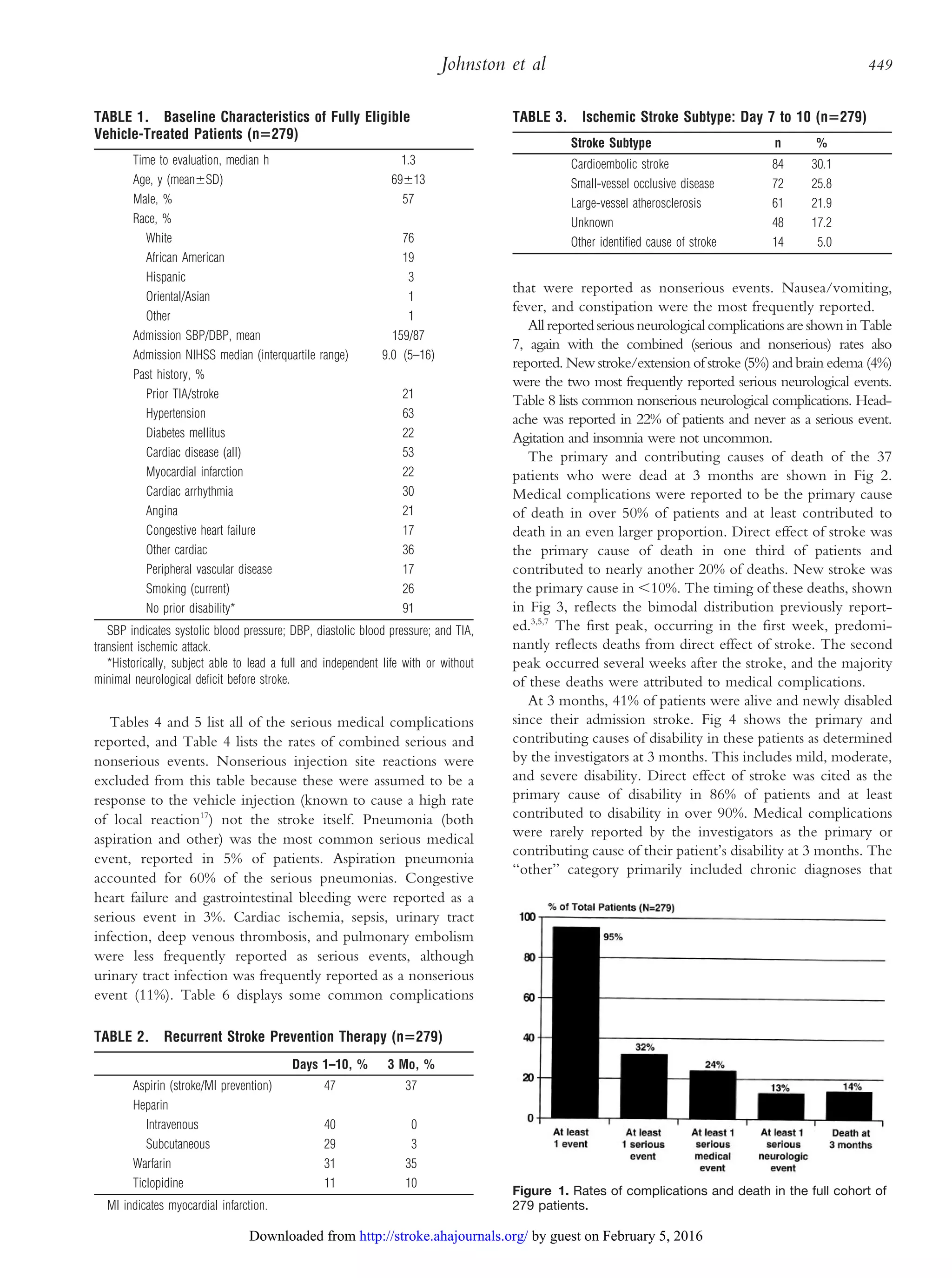
![Medical and Neurological Complications
of Ischemic Stroke
Experience From the RANTTAS Trial
Karen C. Johnston, MD; Jiang Y. Li, MS; Patrick D. Lyden, MD; Sandra K. Hanson, MD;
Thomas E. Feasby, MD; Robert J. Adams, MD; R. Edward Faught, Jr, MD; E. Clarke Haley, Jr, MD;
for the RANTTAS InvestigatorsV
Background and Purpose—Medical and neurological complications after acute ischemic stroke may adversely impact outcome and
in some cases may be preventable. Limited data exist regarding the frequency of such complications occurring in the first days
after the ictus and the relationship of these complications to outcome. Our objective was to identify the types, severity, and
frequency of medical and neurological complications following acute ischemic stroke and to determine their role in mortality
and functional outcome.
Methods—Rates of serious (life-threatening) and nonserious medical and neurological complications and mortality were derived
from the placebo limb of the Randomized Trial of Tirilazad Mesylate in Acute Stroke (RANTTAS) database (nϭ279).
Complications were correlated with clinical outcome using logistic regression techniques.
Results—Of all patients, 95% had at least one complication. The most common serious medical complication was pneumonia (5%),
and the most common serious neurological complication was new cerebral infarction or extension of the admission infarction
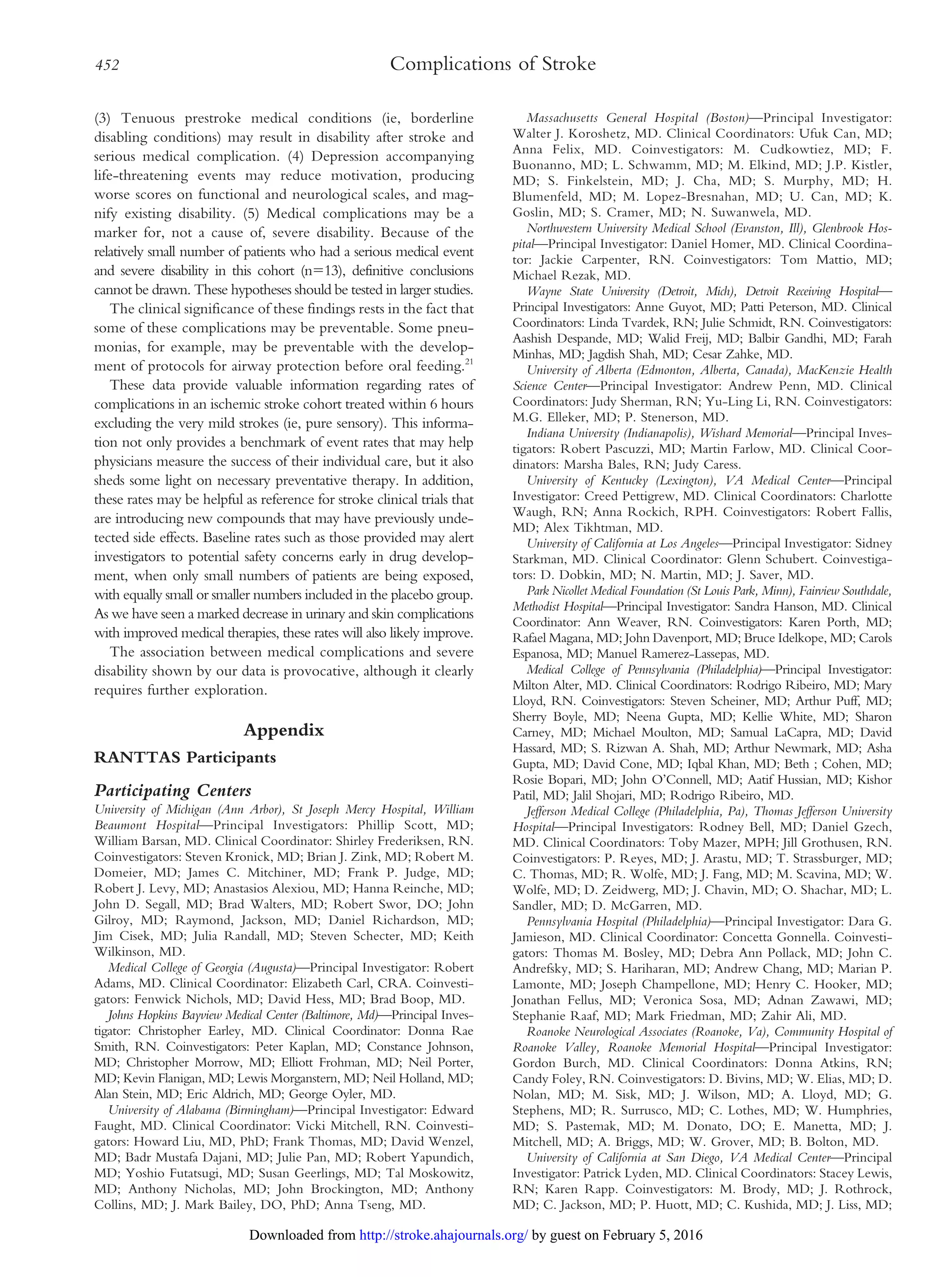
(5%). The 3-month mortality was 14%; 51% of these deaths were attributed primarily to medical complications. Outcome was
significantly worse in patients with serious medical complications, after adjustment for baseline imbalances, as measured by the
Barthel Index (odds ratio [OR], 6.1; 95% confidence interval [CI], 2.5 to 15.1) and by the Glasgow Outcome Scale (OR, 11.6;
95% CI, 4.3 to 30.9). After death was discounted, serious medical complications were associated with severe disability at 3
months as determined by the Glasgow Outcome Scale (OR, 4.4; 95% CI, 1.3 to 14.8).
Conclusions—Medical complications that follow ischemic stroke not only influence mortality but may influence functional
outcome. (Stroke. 1998;29:447-453.)
Key Words: complications Ⅲ stroke Ⅲ stroke, acute Ⅲ stroke outcome
Ischemic stroke remains the third leading cause of death after
heart disease and cancer in the United States.1
Mortality rates
have declined over the last several decades1
and are now consis-
tently reported to be approximately 20%,2–5
although this rate
varies from 15% to 58% depending on stroke subtype.3
Medical
complications are also known to be common in stroke patients,
although the implications of these complications have less fre-
quently been studied. Davenport et al6
retrospectively reported
complication rates in 597 stroke patients (ischemic and hemor-
rhagic). They found 59% had complications and 23% died in the
hospital. Silver et al3
reported that approximately 40% of deaths
were from medical complications in a series of nearly 1000
ischemic stroke patients. They also noted that while most deaths
occurring in the first week were due to brain edema associated
with stroke, most deaths in the second and third weeks after stroke
could be attributed to medical complications.3
In a retrospective
autopsy review from 1966 to 1975, Bounds et al7
found that
Ͼ50% of deaths occurred secondary to medical complications.
Kalra et al8
reported medical complications occurring in 60% of
the 245 patients involved in a stroke rehabilitation program.
The relationship between serious medical complications and
functional outcome has not been systematically examined. The
purpose of this study was to describe the rates of medical and
neurological complications in an acute ischemic stroke cohort
and then to investigate the impact of these complications on
functional outcome and mortality.
Subjects and Methods
Our study population included the 279 fully eligible, vehicle-treated
stroke patients from the Randomized Trial of Tirilazad Mesylate in
Received September 22, 1997; final revision received November 18, 1997; accepted December 1, 1997.
From the Departments of Neurology (K.C.J., E.C.H.) and Neurosurgery (J.Y.L., E.C.H.), University of Virginia (Charlottesville); the Department of
Neurology, University of California at San Diego (P.D.L.); Park Nicollet Medical Foundation, St Louis Park, Minn (S.K.H.); Foothills Hospital, Calgary,
Alberta, Canada (T.E.F.); the Department of Neurology, Medical College of Georgia (Augusta) (R.J.A.); and the Department of Neurology, University of
Alabama (Birmingham) (R.E.F.).
V
The investigators and participating centers are listed in the Appendix.
This work was presented in part at the 49th Annual Meeting of the American Academy of Neurology, April 17, 1997.
Correspondence to Karen C. Johnston, MD, Department of Neurology, Box 394, University of Virginia Health Sciences Center, Charlottesville, VA 22908.
E-mail kj4v@virginia.edu
© 1998 American Heart Association, Inc.
447 by guest on February 5, 2016http://stroke.ahajournals.org/Downloaded from](https://image.slidesharecdn.com/complicationsofstroke-200706071952/75/Complications-of-stroke-1-2048.jpg)
![Patients with Acute Stroke (RANTTAS).9
This was a multicenter,
randomized, double-blinded, vehicle-controlled trial that evaluated
the efficacy and safety of intravenous tirilazad mesylate in patients with
acute ischemic stroke. Patients were selected based on eligibility
criteria (see below) from all patients with acute ischemic stroke
admitted to each of 27 participating North American centers from
May 1993 through December 1994. The protocol was approved by
each center’s institutional review board.
Eligible patients included those who had a serious neurological
deficit due to focal ischemia, could be treated within 6 hours of stroke
onset, were 18 years of age or older, were not pregnant if female, and
from whom informed consent could be obtained (patient and/or
representative). Subjects were excluded if they had sensory loss, ataxia,
or dysarthria alone; coma due to mass effect by CT scan; severe
hypertension (mean arterial pressure of Ͼ160 mm Hg); seizure at
onset; stroke as a complication of a medical or surgical procedure (not
including cardiac catheterization or cerebral angiography); hemor-
rhage on initial CT scan; or severe concomitant medical, neurological,
or psychiatric illness. The most frequent reasons for exclusion included
stroke onset Ͼ6 hours (36%) and transient deficit (12%). Further
details of the study population have been described previously.9
Each
patient who met eligibility criteria was randomly assigned to receive
either tirilazad mesylate or vehicle (sodium citrate) diluted with
sodium chloride (recommended concentration, 0.9% or 0.45%) for a
total volume of 250 mL administered as a rapid intravenous infusion
over 10 to 30 minutes. A head CT scan without contrast was required
before administration of subsequent doses. If each component of the
complete evaluation (including laboratory work, CT scan, and clinical
examination) obtained before the second dose met the eligibility
criteria, then the patient was allowed to continue participation in the
study as a fully eligible patient. Only the fully eligible patients received
subsequent doses (250 mL per dose or an alternatively lower volume
[based on weight] for patients with central lines or who could not
tolerate the total volume) of study drug or vehicle every 6 hours for
11 additional doses (12 doses total).
Patients in the RANTTAS study were managed in an acute-care area
with current 1990s medical therapy according to standard practice in each
center. Treatment with corticosteroids and the calcium channel blockers
nicardipine and nimodipine, or any experimental stroke therapy (includ-
ing thrombolytic therapy at that time), was prohibited. Allowed medical
therapies included heparin, antiplatelet agents, volume expansion, vaso-
pressors, mannitol, anticonvulsants, and all antihypertensive agents except
for nicardipine and nimodipine. Emergency surgery was also allowed if
judged to be clinically indicated.
The frequency of complications and death was monitored continu-
ously and prospectively by the investigators and coordinators at each site
during the trial. A standardized medical event coding guide based on the
COSTART medical dictionary was used to code complication events.10
This dictionary includes both symptoms and diagnoses. Specific diagnoses
are used when known; however, the system allows a default to a symptom
when the diagnosis is unknown. Adverse events were only reported if
clinically significant. Data on serious complications (ie, those that were
immediately life-threatening, prolonged or resulted in hospitalization, or
resulted in death) were collected by investigators and coordinators at each
site for 3 months. Data on nonserious complications were collected for
the first 7 to 10 days only. Neurological complications included those that
occurred in the nervous system, whereas medical complications were all
other events reported. Clinical outcome was measured at 7 to 10 days,
discharge, and at 3 months according to the BI,11
the GOS,12
and the
NIHSS.13
Primary and contributing causes of death and disability were
designated by the treating investigator based on clinical judgment. New
infarction and extension of infarction were combined for the analysis due
to the somewhat arbitrary assignment of the event in some cases of limited
information.
Statistical Analysis
Poor outcome was defined as severe disability (a BI score of Ͻ6014
or a
GOS rating of severe disability or vegetative survival) or death at 3
months. For patients who were dead at 3 months, the worst possible score
(BIϭ0, GOSϭ5) was assigned. Logistic regression analysis15
was used to
evaluate the association between (1) serious events and poor outcome
(severe disability and death) and (2) serious events and severe disability.
Each analysis was adjusted for admission NIHSS score, patient age, and
the presence of diabetes mellitus, since each of these factors was indepen-
dently associated with outcome in the RANTTAS trial.9
Results
From May 1993 through December 1994, trial subjects were
selected by eligibility criteria from a total of 3853 patients admitted
within 12 hours of stroke onset registered in the combined stroke
logs of the centers. A total of 660 patients (329 vehicle-treated and
331 tirilazad-treated) were randomized. Of those who received
treatment, 556 (280 vehicle and 276 tirilazad) were fully eligible
and 104 (49 vehicle and 55 tirilazad) were subsequently excluded.
Of the 280 fully eligible, vehicle-treated group, 1 patient was
determined to not have a stroke but to have a conversion disorder,
and this patient was excluded from this analysis. The fully eligible,
vehicle-treated stroke group used for this analysis included 279
patients. Patients in the tirilazad-treated group were excluded
because this is not considered standard stroke care, and the
possibility that some of the events were drug related could not be
excluded. Patient characteristics are summarized in Table 1. The
median time from onset of symptoms to admission to the
emergency department was slightly more than 1 hour. The mean
age was 69 years, most patients were white with a slight male
preponderance, and the median NIHSS score reflected a moder-
ate deficit with a minimum NIHSS score of 1 and a maximum of
30. Over 60% of patients had a history of hypertension, over 50%
had a history of cardiac disease, and less than one quarter had a
history of prior transient ischemic attack or stroke. Over 90% of
patients were able to lead a full and independent life before the
presenting stroke. The rates of antiplatelet and anticoagulant
treatment prescribed for these patients, both in the first 10 days
and at the 3-month follow-up are reflected in Table 2. Nearly half
of patients were treated with aspirin in the first 10 days, and a
slightly lower proportion were taking aspirin at 3 months.
Intravenous heparin was used in 40% of patients in the first 10
days. Warfarin therapy was prescribed in approximately one third
of patients in the first 10 days and at 3 months.
Table 3 shows the ischemic stroke subtypes as determined by the
investigator at 7 to 10 days using the Trial of Org 10172 in Acute
Stroke Treatment (TOAST) criteria.16
Cardioembolic stroke was the
most common subtype, accounting for nearly a third of strokes.
Small-vessel occlusive disease and large-vessel atherosclerosis each
accounted for approximately one quarter of the strokes.
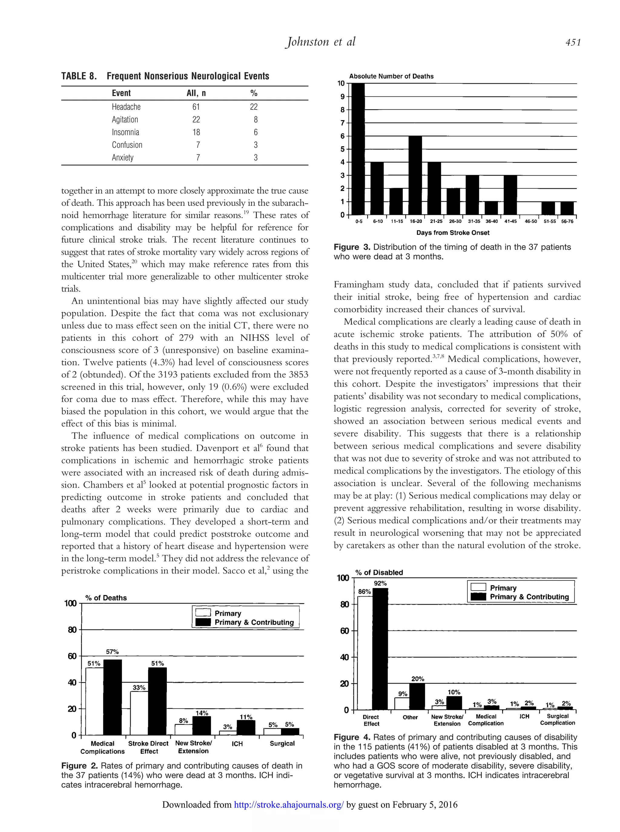
Fig 1 displays the rates of complications and death in the full
cohort of 279 patients. Ninety-five percent of patients had at
least one event, 32% had at least one serious event, and 14% of
patients were dead at 3 months.
Selected Abbreviations and Acronyms
BI ϭ Barthel Index
CI ϭ confidence interval
GOS ϭ Glasgow Outcome Scale
NIHSS ϭ National Institutes of Health Stroke Scale
OR ϭ odds ratio
RANTTAS ϭ Randomized Trial of Tirilazad Mesylate in Acute
Stroke
448 Complications of Stroke
by guest on February 5, 2016http://stroke.ahajournals.org/Downloaded from](https://image.slidesharecdn.com/complicationsofstroke-200706071952/75/Complications-of-stroke-2-2048.jpg)


This document summarizes a study examining medical and neurological complications in 279 patients with acute ischemic stroke. The study found that 95% of patients experienced at least one complication. The most common serious medical complication was pneumonia (5%) and the most common serious neurological complication was new or extended cerebral infarction (5%). Medical complications contributed to 51% of deaths within 3 months. Patients with serious medical complications had significantly worse outcomes on functional scales even after accounting for baseline differences.


