More Related Content
Similar to A novel coronavirus from patients with pneumonia in china, 2019
Similar to A novel coronavirus from patients with pneumonia in china, 2019 (20)
More from MANUELPERALTA33
More from MANUELPERALTA33 (7)
A novel coronavirus from patients with pneumonia in china, 2019
- 1. The new engl and jour nal of medicine
n engl j med nejm.org 1
Brief Report
Summary
In December 2019, a cluster of patients with pneumonia of unknown cause was
linked to a seafood wholesale market in Wuhan, China. A previously unknown
betacoronavirus was discovered through the use of unbiased sequencing in sam-
ples from patients with pneumonia. Human airway epithelial cells were used to
isolate a novel coronavirus, named 2019-nCoV, which formed another clade within
the subgenus sarbecovirus, Orthocoronavirinae subfamily. Different from both
MERS-CoV and SARS-CoV, 2019-nCoV is the seventh member of the family of
coronaviruses that infect humans. Enhanced surveillance and further investigation
are ongoing. (Funded by the National Key Research and Development Program of
China and the National Major Project for Control and Prevention of Infectious
Disease in China.)
E
merging and reemerging pathogens are global challenges for
public health.1
Coronaviruses are enveloped RNA viruses that are distributed
broadly among humans, other mammals, and birds and that cause respira-
tory, enteric, hepatic, and neurologic diseases.2,3
Six coronavirus species are known
to cause human disease.4
Four viruses — 229E, OC43, NL63, and HKU1 — are
prevalent and typically cause common cold symptoms in immunocompetent indi-
viduals.4
The two other strains — severe acute respiratory syndrome coronavirus
(SARS-CoV) and Middle East respiratory syndrome coronavirus (MERS-CoV) — are
zoonotic in origin and have been linked to sometimes fatal illness.5
SARS-CoV was
the causal agent of the severe acute respiratory syndrome outbreaks in 2002 and
2003 in Guangdong Province, China.6-8
MERS-CoV was the pathogen responsible
for severe respiratory disease outbreaks in 2012 in the Middle East.9
Given the high
prevalence and wide distribution of coronaviruses, the large genetic diversity and
frequent recombination of their genomes, and increasing human–animal interface
activities, novel coronaviruses are likely to emerge periodically in humans owing
to frequent cross-species infections and occasional spillover events.5,10
In late December 2019, several local health facilities reported clusters of pa-
tients with pneumonia of unknown cause that were epidemiologically linked to a
seafood and wet animal wholesale market in Wuhan, Hubei Province, China.11
On
December 31, 2019, the Chinese Center for Disease Control and Prevention (China
CDC) dispatched a rapid response team to accompany Hubei provincial and Wuhan
city health authorities and to conduct an epidemiologic and etiologic investigation.
From the MHC Key Laboratory of Bio-
safety, National Institute for Viral Dis-
ease Control and Prevention, Chinese
Center for Disease Control and Preven-
tion (N.Z., W.W., J.S., X.Z., B.H., R.L.,
P.N., X.M., D.W., W.X., G.W., G.F.G.,
W.T.), and the Department of Infectious
Diseases, Beijing Ditan Hospital, Capital
Medical University (X.L.) — both in Bei-
jing; Wuhan Jinyintan Hospital (D.Z.),
the Division for Viral Disease Detection,
Hubei Provincial Center for Disease Con-
trol and Prevention (B.Y., F.Z.), and the
Center for Biosafety Mega-Science, Chi-
nese Academy of Sciences (W.T.) — all in
Wuhan; and the Shandong First Medical
University and Shandong Academy of
Medical Sciences, Jinan, China (W.S.).
Address reprint requests to Dr. Tan at the
NHC Key Laboratory of Biosafety, Nation-
al Institute for Viral Disease Control and
Prevention, China CDC, 155 Changbai
Road, Changping District, Beijing 102206,
China; or at tanwj@ivdc.chinacdc.cn, Dr.
Gao at the National Institute for Viral
Disease Control and Prevention, China
CDC, Beijing 102206, China, or at gaof@
im.ac.cn, or Dr. Wu at the NHC Key Labo-
ratory of Biosafety, National Institute for
Viral Disease Control and Prevention,
China CDC, Beijing 102206, China, or at
wugz@ivdc.chinacdc.cn.
Drs. Zhu, Zhang, W. Wang, Li, and Yang
contributed equally to this article.
This article was published on January 24,
2020, at NEJM.org.
DOI: 10.1056/NEJMoa2001017
Copyright © 2020 Massachusetts Medical Society.
A Novel Coronavirus from Patients with
Pneumonia in China, 2019
Na Zhu, Ph.D., Dingyu Zhang, M.D., Wenling Wang, Ph.D., Xinwang Li, M.D.,
Bo Yang, M.S., Jingdong Song, Ph.D., Xiang Zhao, Ph.D., Baoying Huang, Ph.D.,
Weifeng Shi, Ph.D., Roujian Lu, M.D., Peihua Niu, Ph.D., Faxian Zhan, Ph.D.,
Xuejun Ma, Ph.D., Dayan Wang, Ph.D., Wenbo Xu, M.D., Guizhen Wu, M.D.,
George F. Gao, D.Phil., and Wenjie Tan, M.D., Ph.D., for the China Novel
Coronavirus Investigating and Research Team
The New England Journal of Medicine
Downloaded from nejm.org by Alex Ortiz on January 24, 2020. For personal use only. No other uses without permission.
Copyright © 2020 Massachusetts Medical Society. All rights reserved.
- 2. n engl j med nejm.org2
The new engl and jour nal of medicine
We report the results of this investigation, iden-
tifying the source of the pneumonia clusters,
and describe a novel coronavirus detected in
patients with pneumonia whose specimens were
tested by the China CDC at an early stage of the
outbreak. We also describe clinical features of
the pneumonia in two of these patients.
Methods
Viral Diagnostic Methods
Four lower respiratory tract samples, including
bronchoalveolar-lavage fluid, were collected from
patients with pneumonia of unknown cause who
were identified in Wuhan on December 21, 2019,
or later and who had been present at the Huanan
Seafood Market close to the time of their clinical
presentation. Seven bronchoalveolar-lavage fluid
specimens were collected from patients in Beijing
hospitals with pneumonia of known cause to
serve as control samples. Extraction of nucleic
acids from clinical samples (including uninfect-
ed cultures that served as negative controls) was
performed with a High Pure Viral Nucleic Acid
Kit, as described by the manufacturer (Roche).
Extracted nucleic acid samples were tested for
viruses and bacteria by polymerase chain reac-
tion (PCR), using the RespiFinderSmart22kit
(PathoFinder BV) and the LightCycler 480 real-
time PCR system, in accordance with manufac-
turer instructions.12
Samples were analyzed for
22 pathogens (18 viruses and 4 bacteria) as de-
tailed in the Supplementary Appendix. In addition,
unbiased, high-throughput sequencing, described
previously,13
was used to discover microbial se-
quences not identifiable by the means described
above. A real-time reverse transcription PCR (RT-
PCR) assay was used to detect viral RNA by tar-
geting a consensus RdRp region of pan β-CoV, as
described in the Supplementary Appendix.
Isolation of Virus
Bronchoalveolar-lavage fluid samples were col-
lected in sterile cups to which virus transport
medium was added. Samples were then centri-
fuged to remove cellular debris. The supernatant
was inoculated on human airway epithelial cells,14
which had been obtained from airway specimens
resected from patients undergoing surgery for
lung cancer and were confirmed to be special-
pathogen-free by NGS.13
Human airway epithelial cells were expanded
on plastic substrate to generate passage-1 cells
and were subsequently plated at a density of
2.5×10⁵
cells per well on permeable Transwell-
COL (12-mm diameter) supports. Human airway
epithelial cell cultures were generated in an
air–liquid interface for 4 to 6 weeks to form
well-differentiated, polarized cultures resem-
bling in vivo pseudostratified mucociliary epi-
thelium.13
Prior to infection, apical surfaces of the hu-
man airway epithelial cells were washed three
times with phosphate-buffered saline; 150 μl of
supernatant from bronchoalveolar-lavage fluid
samples was inoculated onto the apical surface
of the cell cultures. After a 2-hour incubation at
37°C, unbound virus was removed by washing with
500 μl of phosphate-buffered saline for 10 min-
utes; human airway epithelial cells were main-
tained in an air–liquid interface incubated at
37°C with 5% carbon dioxide. Every 48 hours,
150 μl of phosphate-buffered saline was applied
to the apical surfaces of the human airway epi-
thelial cells, and after 10 minutes of incubation
at 37°C the samples were harvested. Pseudostrat-
ified mucociliary epithelium cells were main-
tained in this environment; apical samples were
passaged in a 1:3 diluted vial stock to new cells.
The cells were monitored daily with light micros-
copy, for cytopathic effects, and with RT-PCR, for
the presence of viral nucleic acid in the superna-
tant. After three passages, apical samples and
human airway epithelial cells were prepared for
transmission electron microscopy.
Transmission Electron Microscopy
Supernatant from human airway epithelial cell
cultures that showed cytopathic effects was col-
lected, inactivated with 2% paraformaldehyde
for at least 2 hours, and ultracentrifuged to
sediment virus particles. The enriched superna-
tant was negatively stained on film-coated grids
for examination. Human airway epithelial cells
showing cytopathic effects were collected and
fixed with 2% paraformaldehyde–2.5% glutaral-
dehyde and were then fixed with 1% osmium
tetroxide dehydrated with grade ethanol embed-
ded with PON812 resin. Sections (80 nm) were
cut from resin block and stained with uranyl
acetate and lead citrate, separately. The negative
stained grids and ultrathin sections were ob-
served under transmission electron microscopy.
The New England Journal of Medicine
Downloaded from nejm.org by Alex Ortiz on January 24, 2020. For personal use only. No other uses without permission.
Copyright © 2020 Massachusetts Medical Society. All rights reserved.
- 3. n engl j med nejm.org 3
Brief Report
Viral Genome Sequencing
RNA extracted from bronchoalveolar-lavage flu-
id and culture supernatants was used as a tem-
plate to clone and sequence the genome. We
used a combination of Illumina sequencing and
nanopore sequencing to characterize the virus
genome. Sequence reads were assembled into
contig maps (a set of overlapping DNA segments)
with the use of CLC Genomics software, version
4.6.1 (CLC Bio). Specific primers were subse-
quently designed for PCR, and 5′- or 3′- RACE
(rapid amplification of cDNA ends) was used to
fill genome gaps from conventional Sanger se-
quencing. These PCR products were purified
from gels and sequenced with a BigDye Termi-
nator v3.1 Cycle Sequencing Kit and a 3130XL
Genetic Analyzer, in accordance with the manu-
facturers’ instructions.
Multiple-sequence alignment of the 2019-
nCoV and reference sequences was performed
with the use of Muscle. Phylogenetic analysis of
the complete genomes was performed with
RAxML (13) with 1000 bootstrap replicates and
a general time-reversible model used as the nu-
cleotide substitution model.
Results
Patients
Three adult patients presented with severe pneu-
monia and were admitted to a hospital in Wu-
han on December 27, 2019. Patient 1 was a
49-year-old woman, Patient 2 was a 61-year-old
man, and Patient 3 was a 32-year-old man.
Clinical profiles were available for Patients 1 and
2. Patient 1 reported having no underlying
chronic medical conditions but reported fever
(temperature, 37°C to 38°C) and cough with
chest discomfort on December 23, 2019. Four
days after the onset of illness, her cough and
chest discomfort worsened, but the fever was
reduced; a diagnosis of pneumonia was based
on computed tomographic (CT) scan. Her occu-
pation was retailer in the seafood wholesale
market. Patient 2 initially reported fever and
cough on December 20, 2019; respiratory dis-
tress developed 7 days after the onset of illness
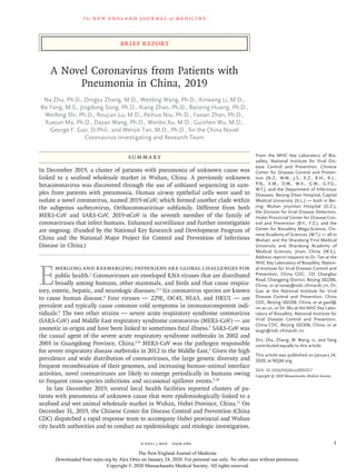
and worsened over the next 2 days (see chest
radiographs, Fig. 1), at which time mechanical
ventilation was started. He had been a frequent
visitor to the seafood wholesale market. Patients
1 and 3 recovered and were discharged from the
hospital on January 16, 2020. Patient 2 died on
January 9, 2020. No biopsy specimens were ob-
tained.
Figure 1. Chest Radiographs.
Shown are chest radiographs from Patient 2 on days 8
and 11 after the onset of illness. The trachea was intu-
bated and mechanical ventilation instituted in the peri-
od between the acquisition of the two images. Bilateral
fluffy opacities are present in both images but are in-
creased in density, profusion, and confluence in the
second image; these changes are most marked in the
lower lung fields. Changes consistent with the accu-
mulation of pleural liquid are also visible in the second
image.
A
B
The New England Journal of Medicine
Downloaded from nejm.org by Alex Ortiz on January 24, 2020. For personal use only. No other uses without permission.
Copyright © 2020 Massachusetts Medical Society. All rights reserved.
- 4. n engl j med nejm.org4
The new engl and jour nal of medicine
Detection and Isolation of a Novel
Coronavirus
Three bronchoalveolar-lavage samples were col-
lected from Wuhan Jinyintan Hospital on De-
cember 30, 2019. No specific pathogens (includ-
ing HCoV-229E, HCoV-NL63, HCoV-OC43, and
HCoV-HKU1) were detected in clinical specimens
from these patients by the RespiFinderSmart-
22kit. RNA extracted from bronchoalveolar-la-
vage fluid from the patients was used as a tem-
plate to clone and sequence a genome using a
combination of Illumina sequencing and nano-
pore sequencing. More than 20,000 viral reads
from individual specimens were obtained, and
most contigs matched to the genome from lin-
eage B of the genus betacoronavirus — showing
more than 85% identity with a bat SARS-like
CoV (bat-SL-CoVZC45, MG772933.1) genome
published previously. Positive results were also
obtained with use of a real-time RT-PCR assay
for RNA targeting to a consensus RdRp region
of pan β-CoV (although the cycle threshold value
was higher than 34 for detected samples). Virus
isolation from the clinical specimens was per-
formed with human airway epithelial cells and
Vero E6 and Huh-7 cell lines. The isolated virus
was named 2019-nCoV.
To determine whether virus particles could be
visualized in 2019-nCoV–infected human airway
epithelial cells, mock-infected and 2019-nCoV–
infected human airway epithelial cultures were
examined with light microscopy daily and with
transmission electron microscopy 6 days after
inoculation. Cytopathic effects were observed 96
hours after inoculation on surface layers of hu-
man airway epithelial cells; a lack of cilium
beating was seen with light microcopy in the
center of the focus (Fig. 2). No specific cyto-
pathic effects were observed in the Vero E6 and
Huh-7 cell lines until 6 days after inoculation.
Electron micrographs of negative-stained
2019-nCoV particles were generally spherical
with some pleomorphism (Fig. 3). Diameter var-
ied from about 60 to 140 nm. Virus particles had
quite distinctive spikes, about 9 to 12 nm, and
gave virions the appearance of a solar corona.
Extracellular free virus particles and inclusion
bodies filled with virus particles in membrane-
bound vesicles in cytoplasm were found in the
human airway epithelial ultrathin sections. This
observed morphology is consistent with the
Coronaviridae family.
To further characterize the virus, de novo se-
quences of 2019-nCoV genome from clinical spec-
imens (bronchoalveolar-lavage fluid) and human
airway epithelial cell virus isolates were obtained
by Illumina and nanopore sequencing. The novel
coronavirus was identified from all three patients.
Two nearly full-length coronavirus sequences
were obtained from bronchoalveolar-lavage fluid
(BetaCoV/Wuhan/IVDC-HB-04/2020, BetaCoV/
Wuhan/IVDC-HB-05/2020|EPI_ISL_402121), and
one full-length sequence was obtained from a
virus isolated from a patient (BetaCoV/Wuhan/
IVDC-HB-01/2020|EPI_ISL_402119). Complete ge-
nome sequences of the three novel coronaviruses
were submitted to GASAID (BetaCoV/Wuhan/
IVDC-HB-01/2019,accessionID:EPI_ISL_402119;
BetaCoV/Wuhan/IVDC-HB-04/2020, accession ID:
EPI_ISL_402120;BetaCoV/Wuhan/IVDC-HB-05/2019,
Figure 2. Cytopathic Effects in Human Airway Epithelial Cell Cultures after Inoculation with 2019-nCoV.
A BMock HAE-CPE
100 µm
The New England Journal of Medicine
Downloaded from nejm.org by Alex Ortiz on January 24, 2020. For personal use only. No other uses without permission.
Copyright © 2020 Massachusetts Medical Society. All rights reserved.
- 5. n engl j med nejm.org 5
Brief Report
accession ID: EPI_ISL_402121) and have a 86.9%
nucleotide sequence identity to a previously pub-
lished bat SARS-like CoV (bat-SL-CoVZC45,
MG772933.1) genome. The three 2019-nCoV ge-
nomes clustered together and formed an inde-
pendent subclade within the sarbecovirus subge-
nus, which shows the typical betacoronavirus
organization: a 5′ untranslated region (UTR),
replicase complex (orf1ab), S gene, E gene, M
gene, N gene, 3′ UTR, and several unidentified
nonstructural open reading frames.
Although 2019-nCoV is similar to some beta-
coronaviruses detected in bats (Fig. 4), it is dis-
tinct from SARS-CoV and MERS-CoV. The three
2019-nCoV coronaviruses from Wuhan, together
with two bat-derived SARS-like strains, ZC45
and ZXC21, form a distinct clade in lineage B of
the subgenus sarbecovirus. SARS-CoV strains
from humans and genetically similar SARS-like
coronaviruses from bats collected from south-
western China formed another clade within the
subgenus sarbecovirus. Since the sequence iden-
tity in conserved replicase domains (ORF 1ab) is
less than 90% between 2019-nCoV and other
members of betacoronavirus, the 2019-nCoV —
the likely causative agent of the viral pneumonia
in Wuhan — is a novel betacoronavirus belonging
to the sarbecovirus subgenus of Coronaviridae
family.
Discussion
We report a novel CoV (2019-nCoV) that was
identified in hospitalized patients in Wuhan,
China, in December 2019 and January 2020. Evi-
dence for the presence of this virus includes
identification in bronchoalveolar-lavage fluid in
three patients by whole-genome sequencing, di-
rect PCR, and culture. The illness likely to have
been caused by this CoV was named “novel coro-
navirus-infected pneumonia” (NCIP). Complete
genomes were submitted to GASAID. Phyloge-
netic analysis revealed that 2019-nCoV falls into
the genus betacoronavirus, which includes coro-
naviruses (SARS-CoV, bat SARS-like CoV, and
others) discovered in humans, bats, and other
wild animals.15
We report isolation of the virus
and the initial description of its specific cyto-
pathic effects and morphology.
Molecular techniques have been used suc-
Figure 3. Visualization of 2019-nCoV with Transmission Electron Microscopy.
Negative-stained 2019-nCoV particles are shown in Panel A, and 2019-nCoV particles in the human airway epithelial
cell ultrathin sections are shown in Panel B.
A
100 nm 1 µm
B
The New England Journal of Medicine
Downloaded from nejm.org by Alex Ortiz on January 24, 2020. For personal use only. No other uses without permission.
Copyright © 2020 Massachusetts Medical Society. All rights reserved.
- 6. n engl j med nejm.org6
The new engl and jour nal of medicine
cessfully to identify infectious agents for many
years. Unbiased, high-throughput sequencing is
a powerful tool for the discovery of patho-
gens.14,16
Next-generation sequencing and bioin-
formatics are changing the way we can respond
to infectious disease outbreaks, improving our
understanding of disease occurrence and trans-
mission, accelerating the identification of patho-
gens, and promoting data sharing. We describe
in this report the use of molecular techniques
and unbiased DNA sequencing to discover a
novel betacoronavirus that is likely to have been
the cause of severe pneumonia in three patients
in Wuhan, China.
Although establishing human airway epithe-
lial cell cultures is labor intensive, they appear
to be a valuable research tool for analysis of hu-
man respiratory pathogens.14
Our study showed
that initial propagation of human respiratory se-
cretions onto human airway epithelial cell cul-
tures, followed by transmission electron micros-
copy and whole genome sequencing of culture
supernatant, was successfully used for visualiza-
tion and detection of new human coronavirus
that can possibly elude identification by tradi-
tional approaches.
Further development of accurate and rapid
methods to identify unknown respiratory patho-
gens is still needed. On the basis of analysis of
three complete genomes obtained in this study,
we designed several specific and sensitive assays
targeting ORF1ab, N, and E regions of the 2019-
nCoV genome to detect viral RNA in clinical
specimens. The primer sets and standard oper-
ating procedures have been shared with the
World Health Organization and are intended for
surveillance and detection of 2019-nCoV infec-
tion globally and in China. More recent data
show 2019-nCoV detection in 830 persons in
China.17
Although our study does not fulfill Koch’s
postulates, our analyses provide evidence impli-
cating 2019-nCoV in the Wuhan outbreak. Ad-
ditional evidence to confirm the etiologic sig-
Figure 4. Phylogenetic Analysis of 2019-nCoV and Other Betacoronavirus Genomes in the Orthocoronavirinae Subfamily.
AY508724 SARS_coronavirus_NS-1
AY485277 SARS_coronavirus_Sino1-11
AY390556 SARS_coronavirus_GZ02
AY278489 SARS_coronavirus_GD01
KT444582 SARS-like_coronavirus_WIV16
KY417146 Bat_SARS-like_coronavirus_Rs4231
KY417151 Bat_SARS-like_coronavirus_Rs7327
KY417152 Bat_SARS-like_coronavirus_Rs9401
MK211376 Coronavirus_BtRs-BetaCoV/YN2018B
MK211377 Coronavirus_BtRs-BetaCoV/YN2018C
MK211374 Coronavirus_BtRl-BetaCoV/SC2018
BetaCoV-Wuhan-IVDC-HB-04-2020
BetaCoV/Wuhan/IVDC-HB-01/2019 EPI_ISL_402119
BetaCoV-WuhanI-VDC-HB-05-2019EPI_ISL_402121
KY770859 Bat_coronavirus Anlong-112
KJ473816 BtRs_BetaCoV/YN2013
KJ473815 BtRs_BetaCoV/GX2013 BtRs-GX2013
JX993988 Bat_coronavirus_Cp/Yunnan2011
KY417145 Bat_SARS-like_coronavirus_Rf4092
KY417142 Bat_SARS-like_coronavirus As6526
KY417148 Bat_SARS-like_coronavirus Rs4247
MG772934 Bat_SARS-like_coronavirus bat-SL-CoVZXC21
KF636752 Bat_Hp-betacoronavirus/Zhejiang2013
EF065513 Bat_coronavirus_HKU9-1 BF_005I
EF065505 Bat_coronavirus_HKU4-1 B04f
KU762338 Rousettus_bat_coronavirus GCCDC1_356
EF065509 Bat_coronavirus_HKU5-1 LMH03f
JX869059 Human_betacoronavirus_2c_EMC/2012 HCoV-EMC
KC545386 Betacoronavirus_Erinaceus/VMC/DEU/2012 ErinaceusCoV/2012-216/GER/2012
KM349744 Betacoronavirus_HKU24 HKU24-R05010l
AY391777 Human_coronavirus_OC43 ATCC_VR-759
MK167038 Human_coronavirus_HKU1 SC2521
FJ647223 Murine_coronavirus_MHV-1 MHV-1
MG772933 Bat_SARS-like_coronavirus bat-SL-CoVZC45
SARS-CoV
MERS-CoV
Sarbecovirus
Hibecovirus
Nobecovirus
Merbecovirus
Embevovirus
0.2
100
100
100
100
83
66
84
60
90
94
100
100
100
100
100
100
100
100
100
96
99
100
The New England Journal of Medicine
Downloaded from nejm.org by Alex Ortiz on January 24, 2020. For personal use only. No other uses without permission.
Copyright © 2020 Massachusetts Medical Society. All rights reserved.
- 7. n engl j med nejm.org 7
Brief Report
nificance of 2019-nCoV in the Wuhan outbreak
include identification of a 2019-nCoV antigen in
the lung tissue of patients by immunohisto-
chemical analysis, detection of IgM and IgG
antiviral antibodies in the serum samples from
a patient at two time points to demonstrate se-
roconversion, and animal (monkey) experiments
to provide evidence of pathogenicity. Of critical
importance are epidemiologic investigations to
characterize transmission modes, reproduction in-
terval, and clinical spectrum resulting from infec-
tion to inform and refine strategies that can pre-
vent, control, and stop the spread of 2019-nCoV.
This work was supported by grants from the National Key
Research and Development Program of China (2016YFD0500301)
and the National Major Project for Control and Prevention of
Infectious Disease in China (2018ZX10101002).
Disclosure forms provided by the authors are available with
the full text of this article at NEJM.org.
We thank Dr. Zhongjie Li, Dr. Guangxue He, Dr. Lance Rode-
wald, Yu Li, Fei Ye, Li Zhao, Weimin Zhou, Jun Liu, Yao Meng,
Huijuan Wang, and many staff members at the China CDC for
their contributions and assistance in this preparation and sub-
mission of an earlier version of the manuscript.
References
1. Gao GF. From “A”IV to “Z”IKV: attacks
from emerging and re-emerging patho-
gens. Cell 2018;172:1157-9.
2. Weiss SR, Leibowitz JL. Coronavirus
pathogenesis. Adv Virus Res 2011;81:85-
164.
3. Masters PS, Perlman S. Coronaviridae.
In:Knipe DM, Howley PM, eds. Fields vi-
rology. 6th ed. Lippincott Williams &
Wilkins, 2013:825-58.
4. Su S, Wong G, Shi W, et al. Epidemiol-
ogy, genetic recombination, and patho-
genesis of coronaviruses. Trends Micro-
biol 2016;24:490-502.
5. Cui J, Li F, Shi ZL. Origin and evolu-
tion of pathogenic coronaviruses. Nat Rev
Microbiol 2019;17:181-92.
6. Zhong NS, Zheng BJ, Li YM, et al. Epi-
demiology and cause of severe acute respi-
ratory syndrome (SARS) in Guangdong,
People’s Republic of China, in February,
2003. Lancet 2003;362:1353-8.
7. Ksiazek TG, Erdman D, Goldsmith
CS, et al. A novel coronavirus associated
with severe acute respiratory syndrome.
N Engl J Med 2003;348:1953-66.
8. Drosten C, Günther S, Preiser W, et
al. Identification of a novel coronavirus
in patients with severe acute respiratory
syndrome. N Engl J Med 2003;348:1967-
76.
9. Zaki AM, van Boheemen S, Bestebroer
TM, Osterhaus AD, Fouchier RA. Isola-
tion of a novel coronavirus from a man
with pneumonia in Saudi Arabia. N Engl J
Med 2012;367:1814-20.
10. Wong G, Liu W, Liu Y, Zhou B, Bi Y,
Gao GF. MERS, SARS, and Ebola: the role
of super-spreaders in infectious disease.
Cell Host Microbe 2015;18:398-401.
11. Report of clustering pneumonia of
unknown etiology in Wuhan City. Wuhan
Municipal Health Commission, 2019.
(http://wjw.wuhan.gov.cn/front/web/
showDetail/2019123108989).
12. Liu GS, Li H, Zhao SC, Lu RJ, Niu PH,
Tan WJ. Viral and bacterial etiology of
acute febrile respiratory syndrome among
patients in Qinghai, China. Biomed Envi-
ron Sci 2019;32:438-45.
13. Jonsdottir HR, Dijkman R. Coronavi-
ruses and the human airway: a universal
system for virus-host interaction studies.
Virol J 2016;13:24.
14. Palacios G, Druce J, Du L, et al. A new
arenavirus in a cluster of fatal transplant-
associated diseases. N Engl J Med 2008;
358:991-8.
15. Tan WJ, Zhao X, Ma XJ, et al. A novel
coronavirus genome identified in a clus-
ter of pneumonia cases — Wuhan, China
2019−2020. China CDC Weekly 2020;2:
61-2.
16. Armstrong GL, MacCannell DR, Tay-
lor J, et al. Pathogen genomics in public
health. N Engl J Med 2019;381:2569-80.
17. Report of novel coronavirus-infected
pneumonia in Wuhan City. Wuhan Mu-
nicipal Health Commission, 2020 (http://
wjw.wuhan.gov.cn/front/web/showDetail/
2020012009077).
Copyright © 2020 Massachusetts Medical Society.
The New England Journal of Medicine
Downloaded from nejm.org by Alex Ortiz on January 24, 2020. For personal use only. No other uses without permission.
Copyright © 2020 Massachusetts Medical Society. All rights reserved.