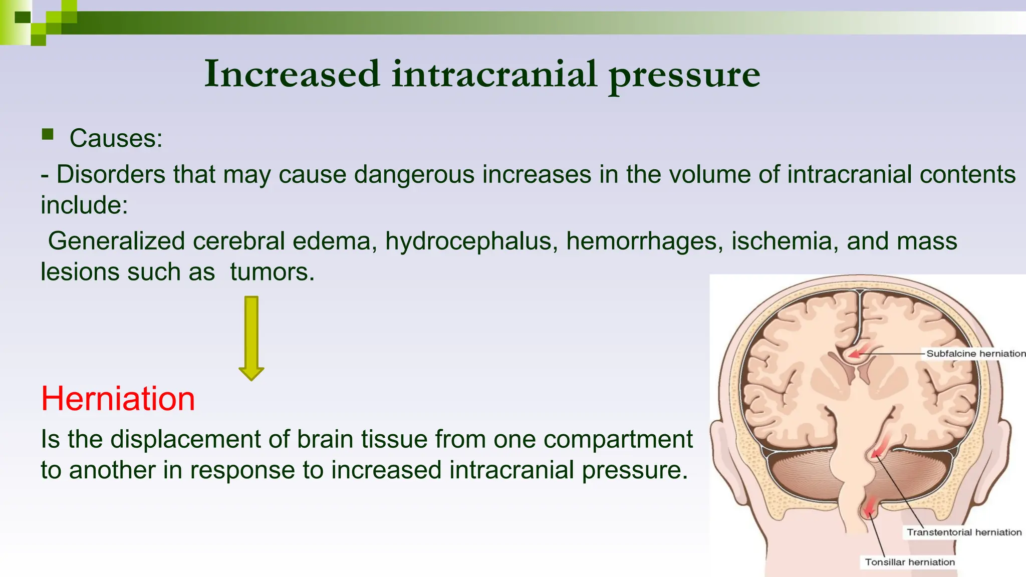
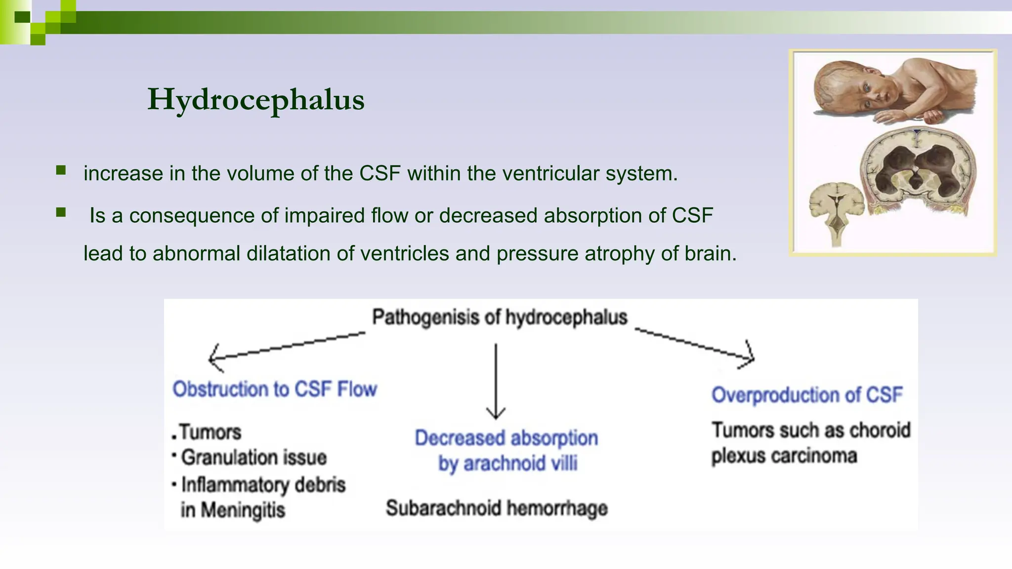
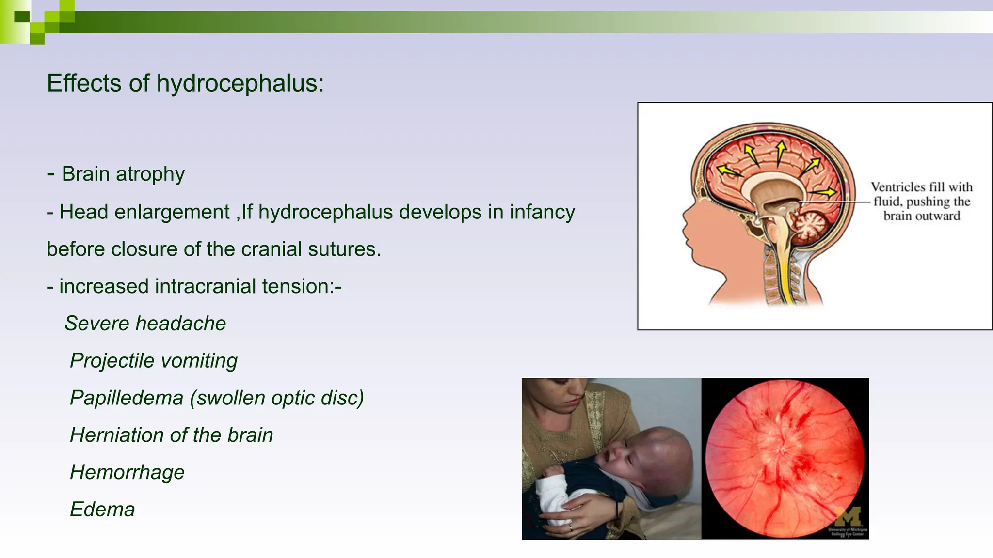
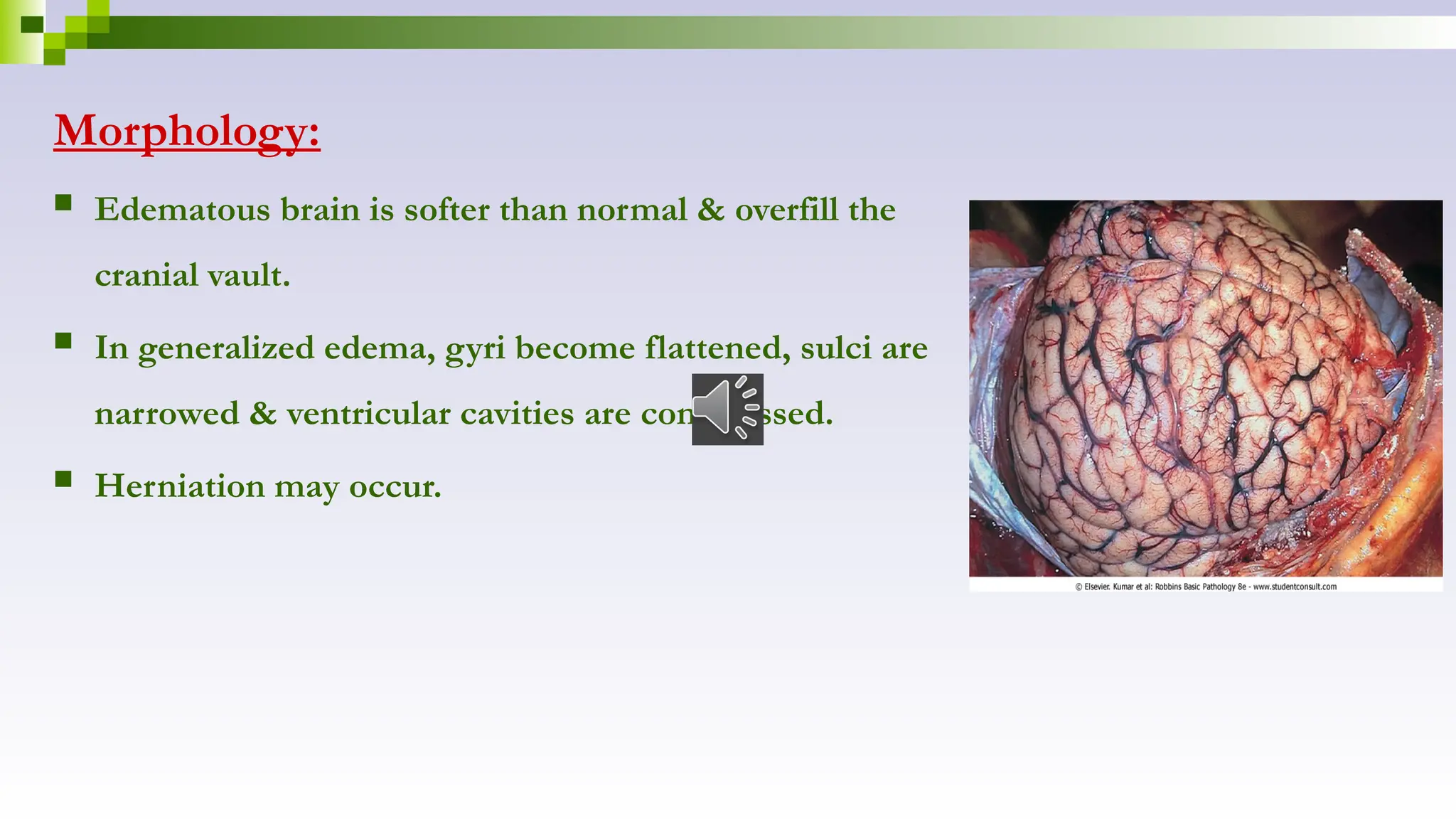
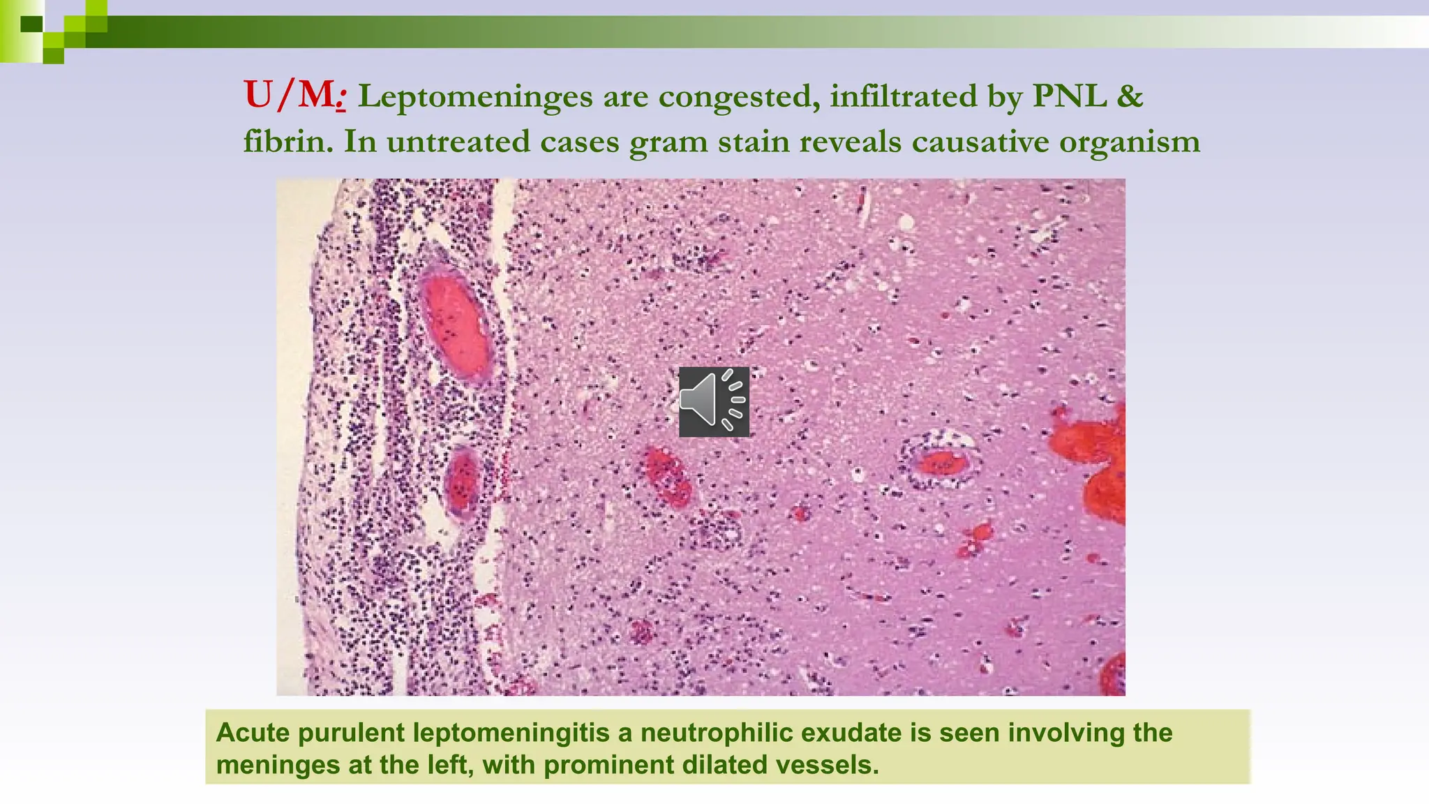
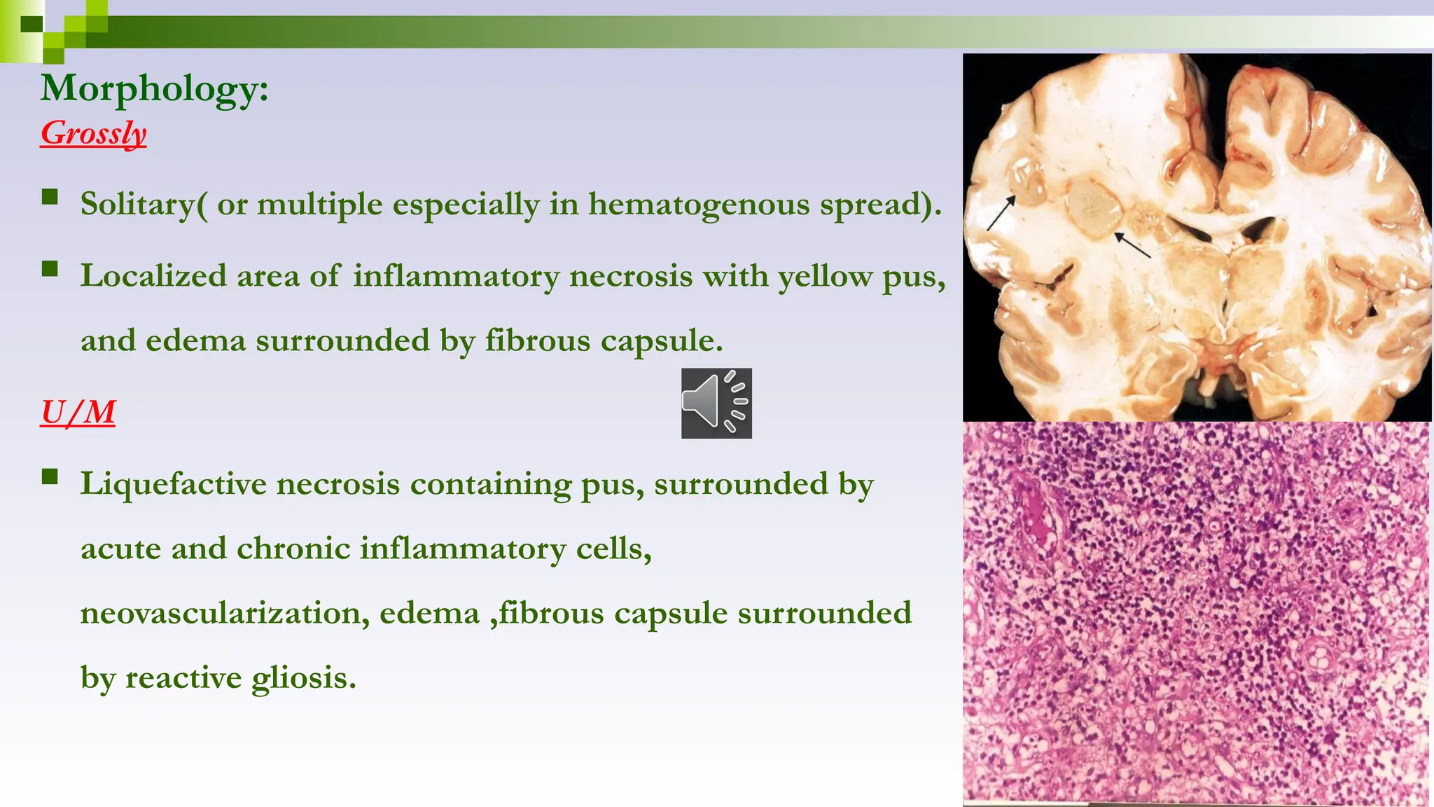
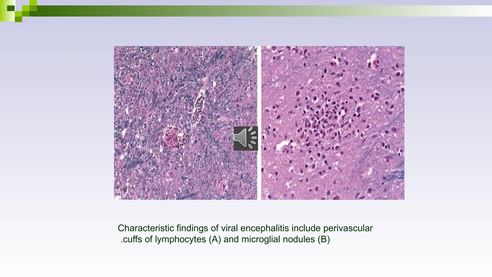
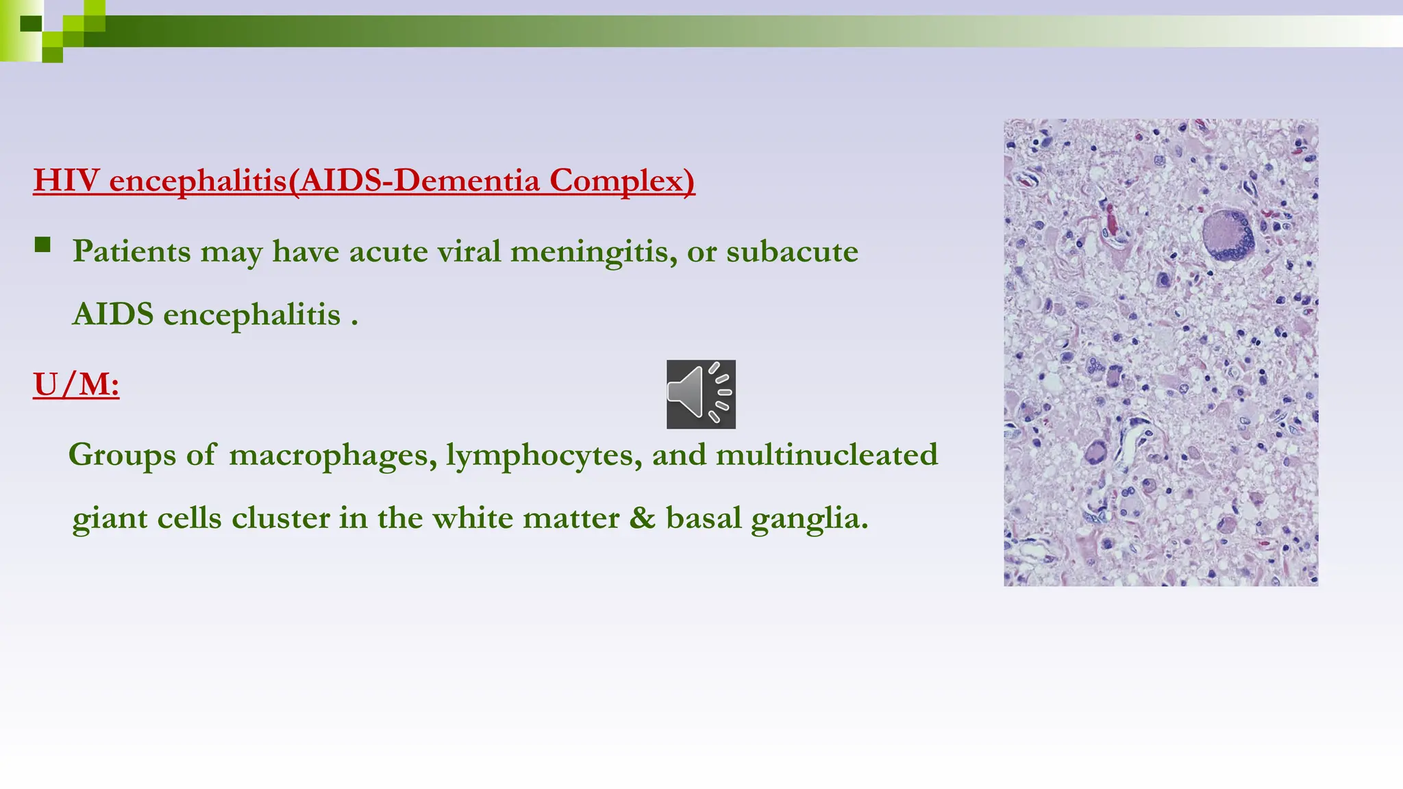
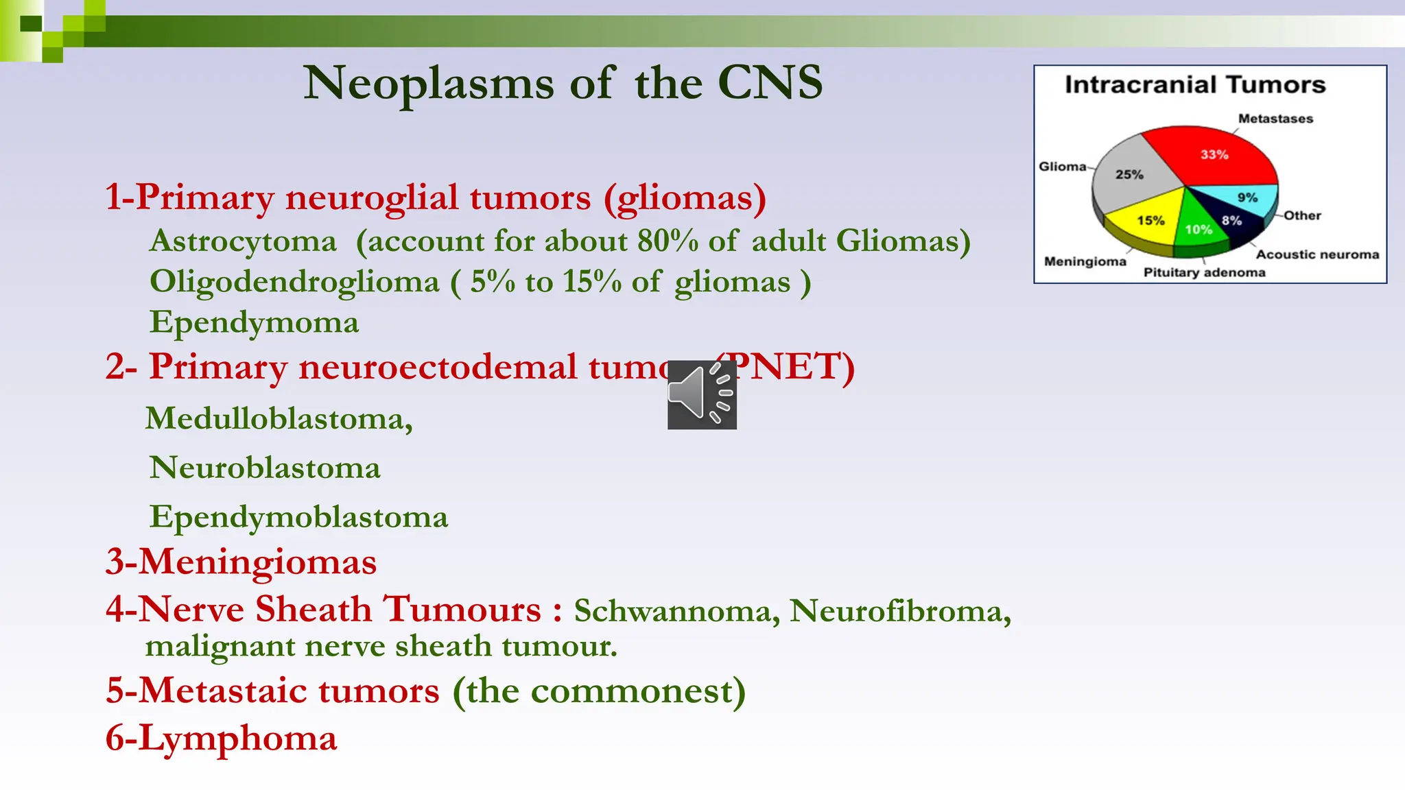
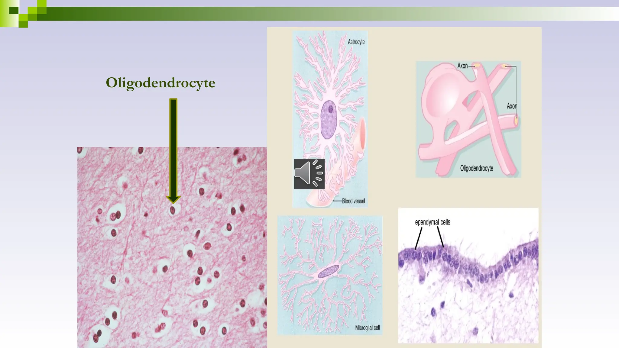
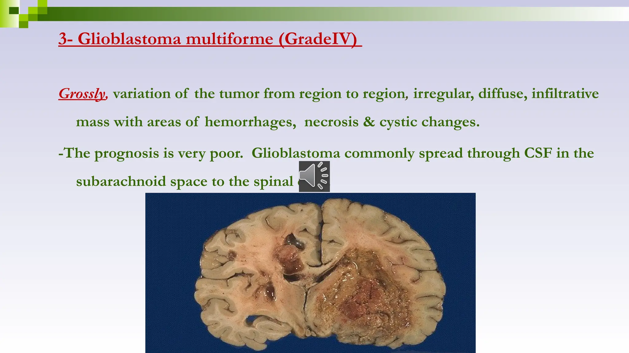
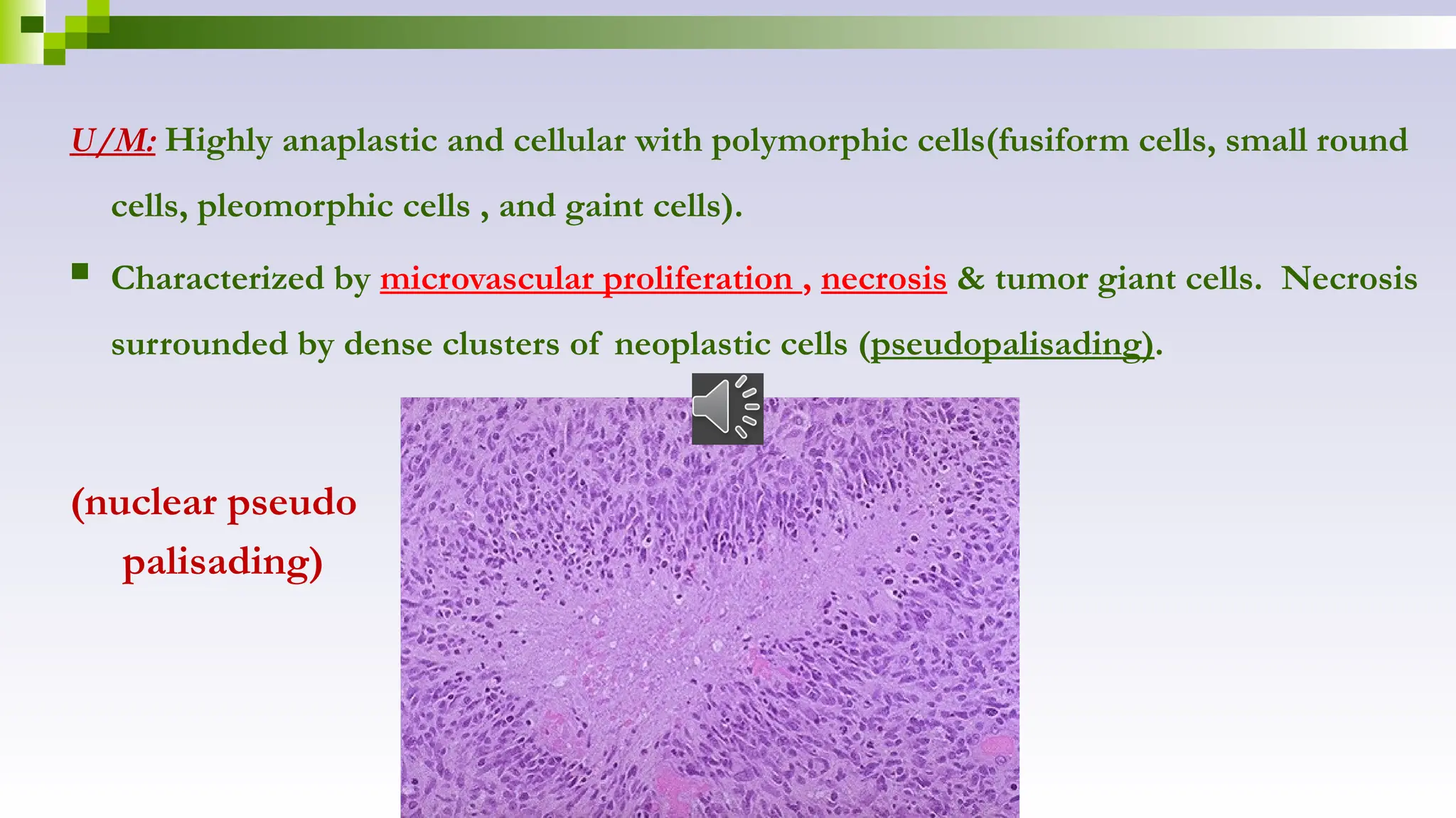
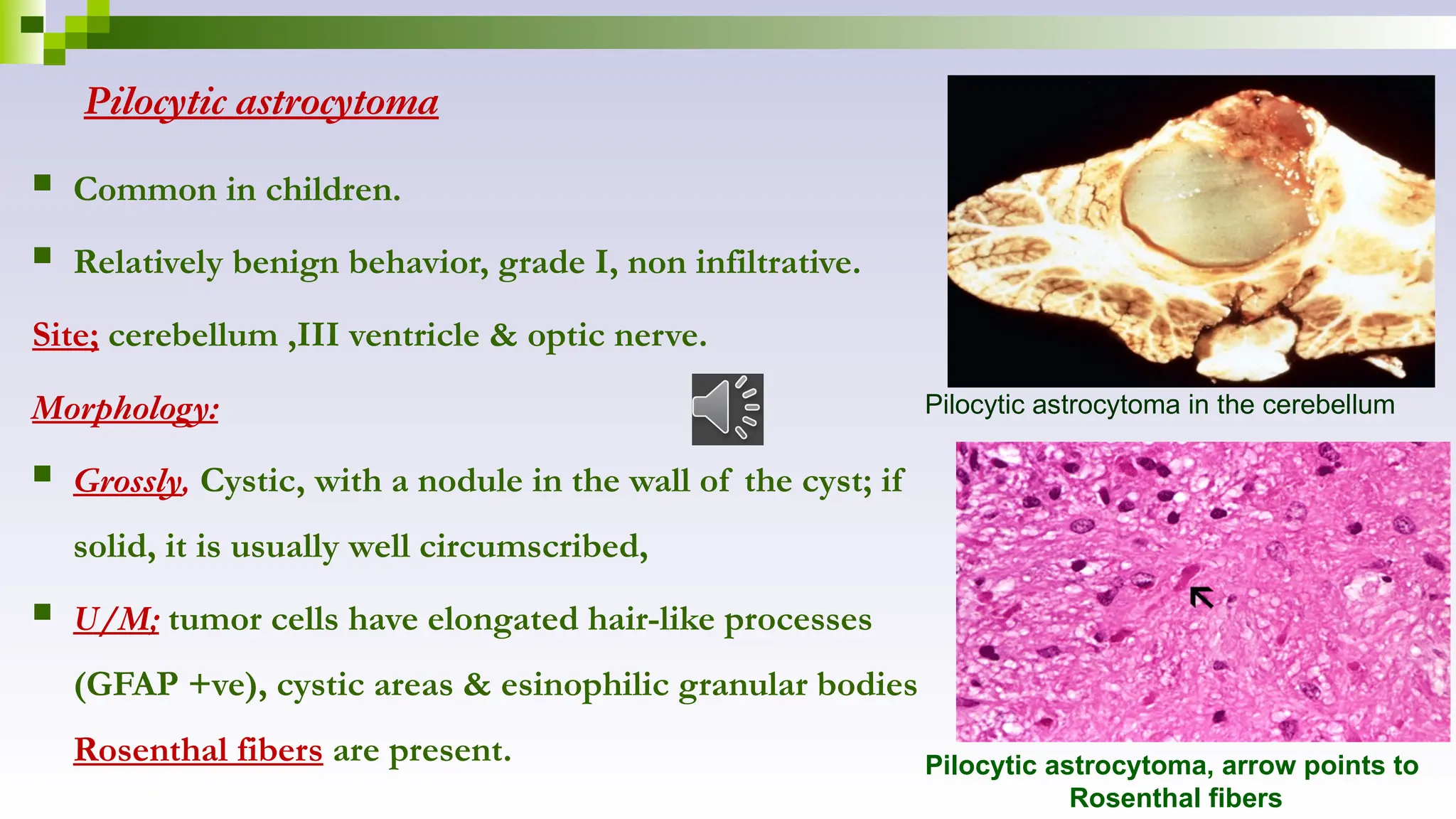
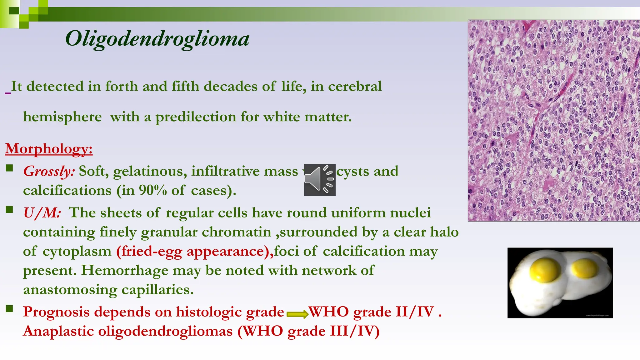
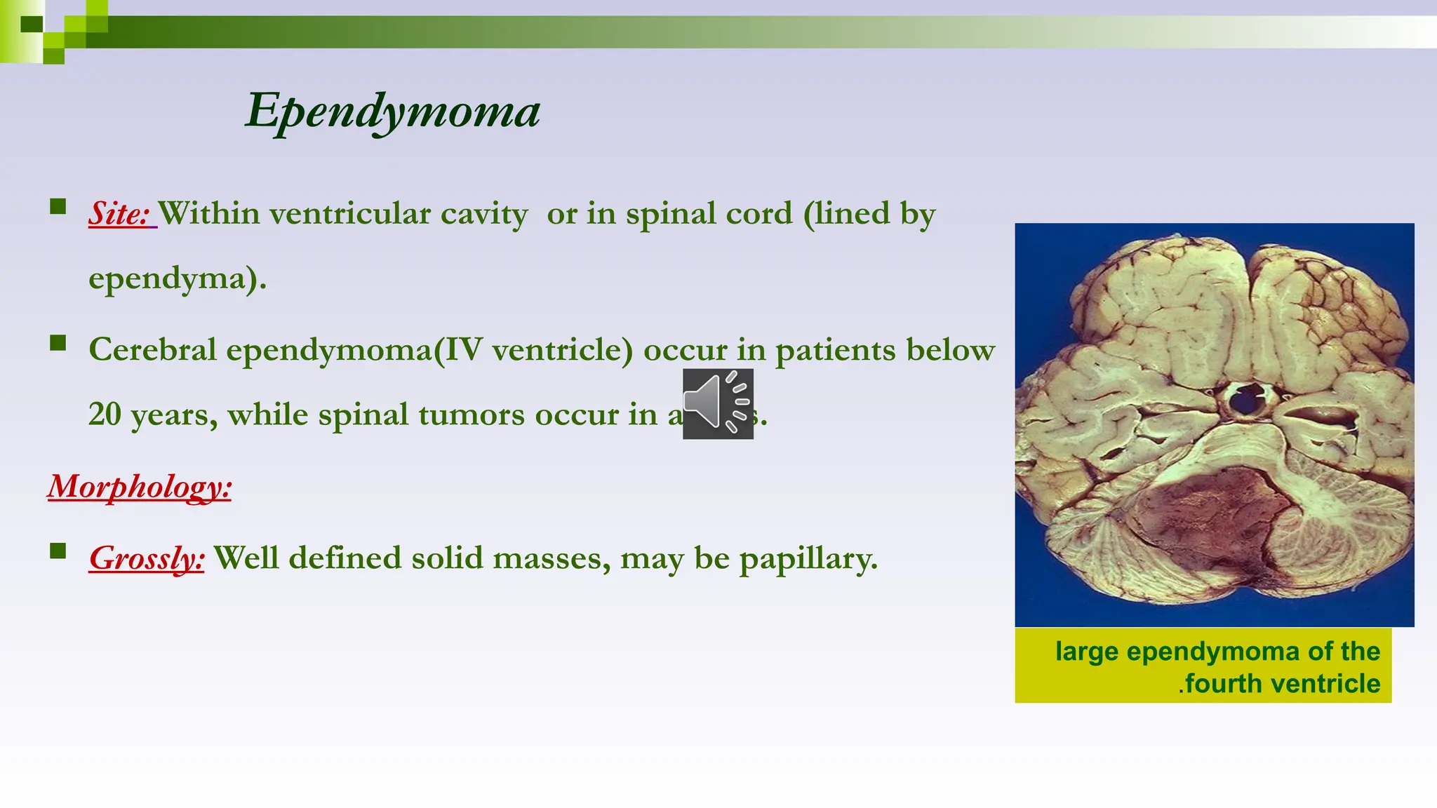
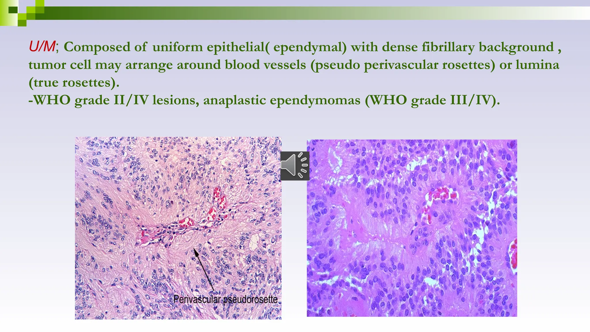
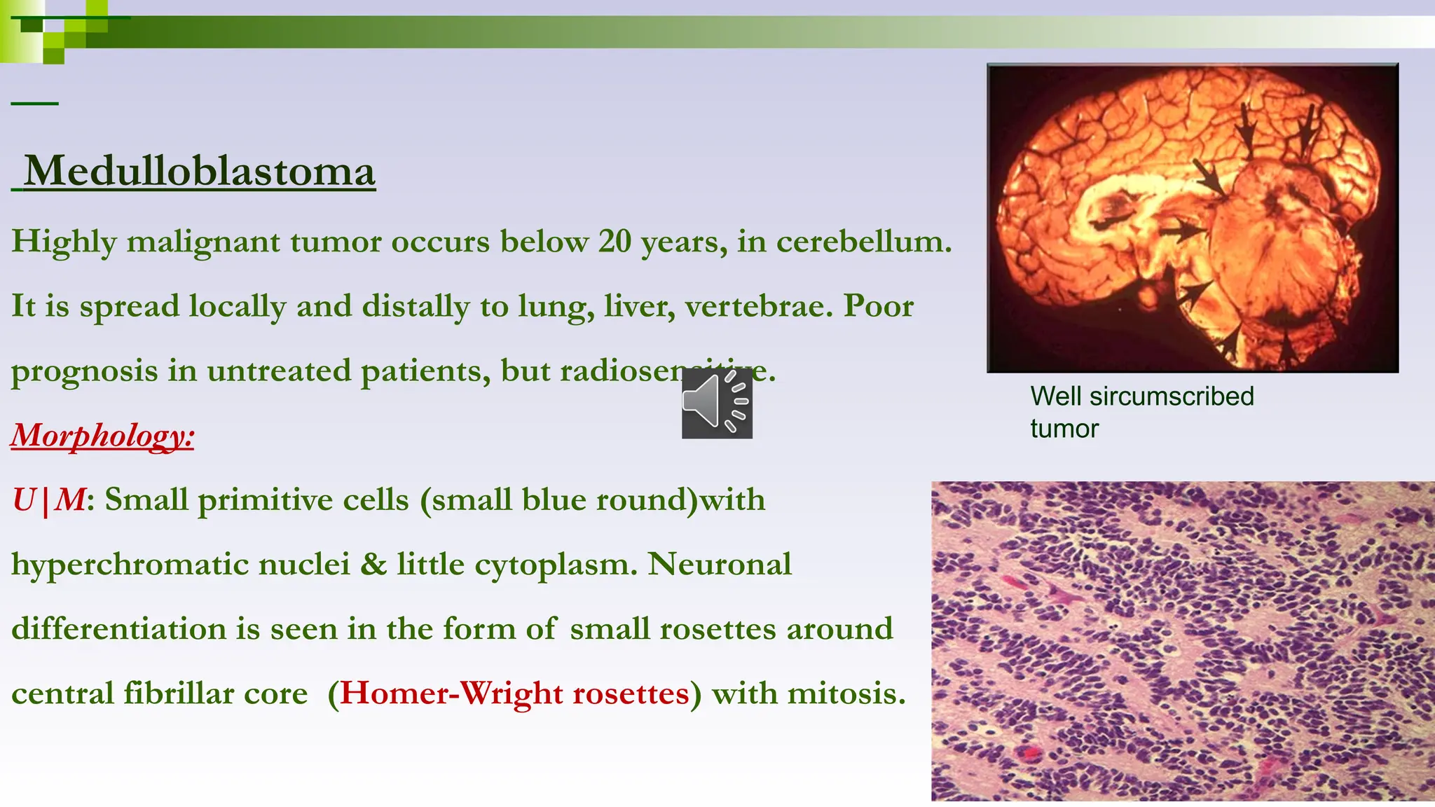
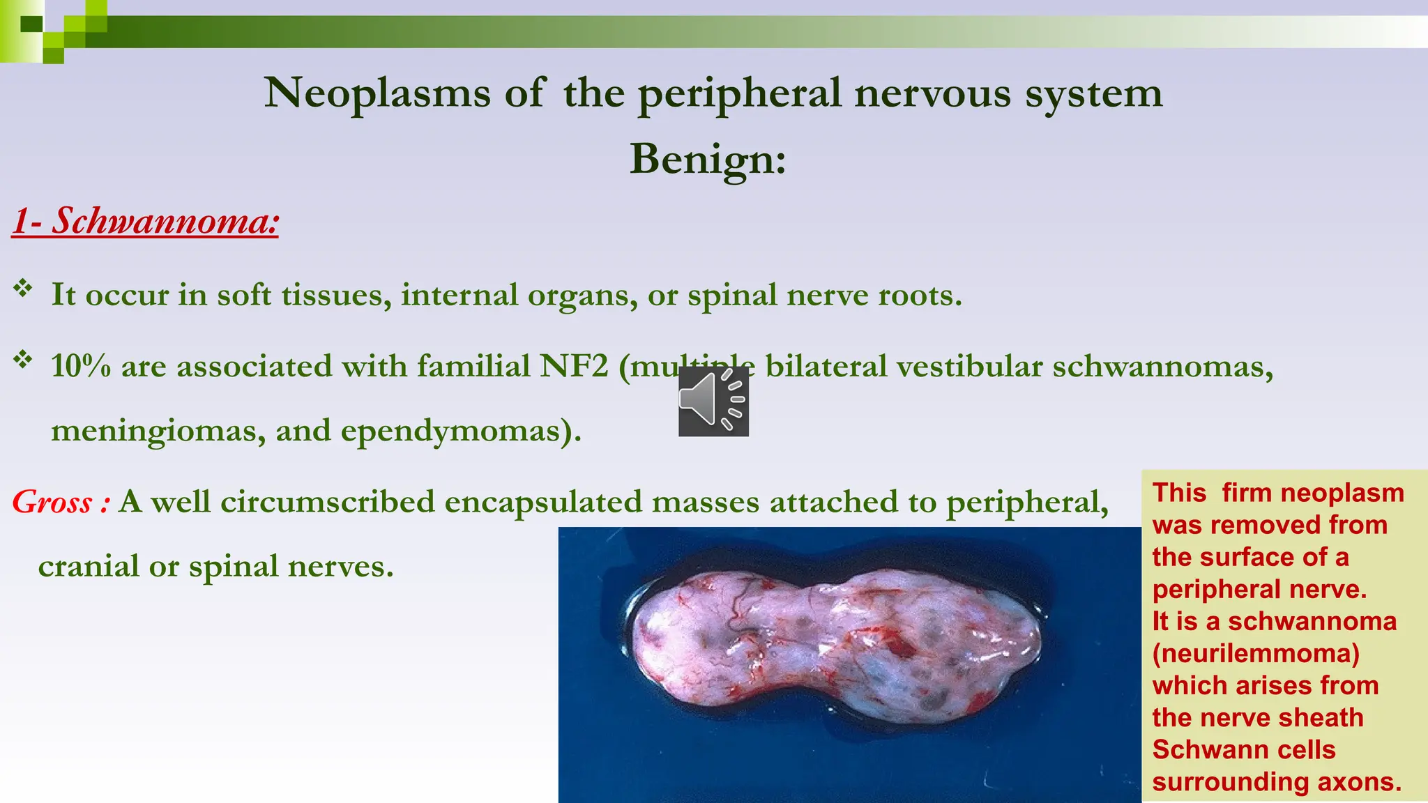
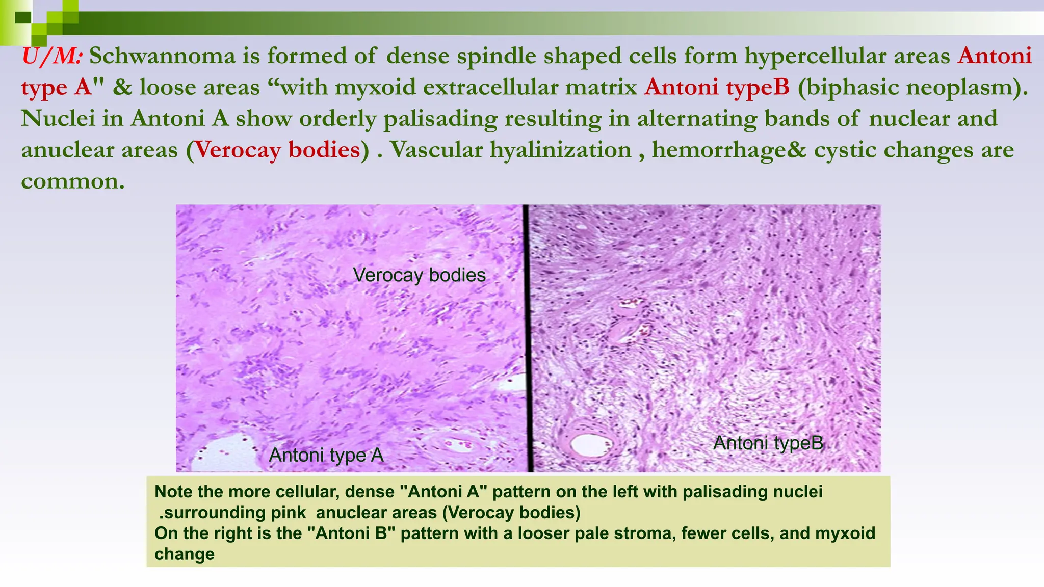
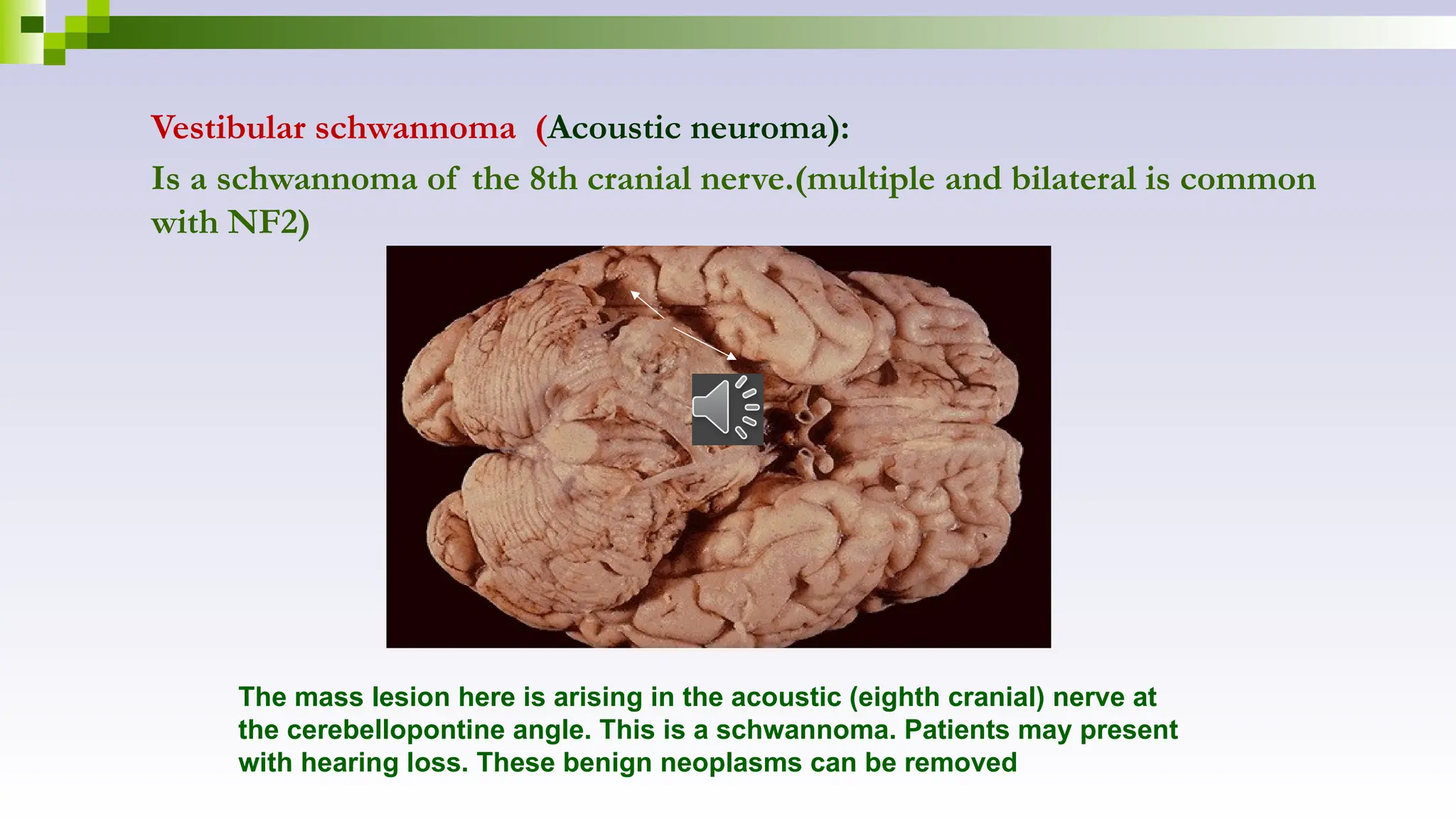
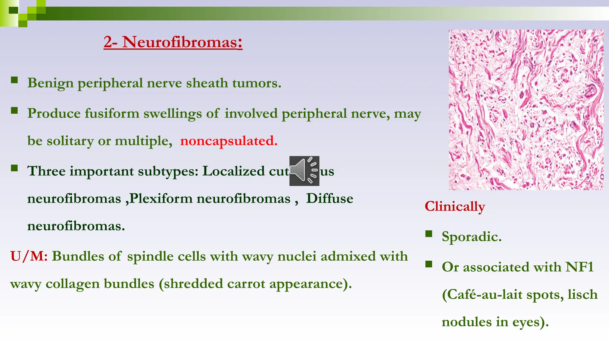
Diseases of the CNS
Dr.Mai Inbiek
Subjects:
 Increased intracranial pressure
 Hydrocephalus
 Cerebral edema
 Vascular diseases
 Vascular malformation
 CNS trauma
 Infections of CNS
 Neoplasms of CNS
 Neoplasms of peripheral nervous system
Increased intracranial pressure
 Causes:
- Disorders that may cause dangerous increases in the volume of intracranial contents
include:
Generalized cerebral edema, hydrocephalus, hemorrhages, ischemia, and mass
lesions such as tumors.
Herniation
Is the displacement of brain tissue from one compartment
to another in response to increased intracranial pressure.
Hydrocephalus
 increase in the volume of the CSF within the ventricular system.
 Is a consequence of impaired flow or decreased absorption of CSF
lead to abnormal dilatation of ventricles and pressure atrophy of brain.
Effects of hydrocephalus:
- Brain atrophy
- Head enlargement ,If hydrocephalus develops in infancy
before closure of the cranial sutures.
- increased intracranial tension:-
Severe headache
Projectile vomiting
Papilledema (swollen optic disc)
Herniation of the brain
Hemorrhage
Edema
Cerebral edema
 It indicates accumulation of excess fluid within the brain parenchyma.
There are two types,
1-Vasogenic edema:
 Due to disruption of integrity of normal blood brain barrier, so fluid escape from
blood vessels to interstitial spaces of brain.
 (intravascular extracellular).
 The absence of significant lymphatic drainage impairs reabsorption of excess water.
 A- localized in cases of abscess or tumors(due to increased vascular permeability).
 B- generalized in cases of sever head trauma, malignant hypertension.
2-Cytotoxic edema:
Due to increased intracellular fluid secondary to neuronal and glial cell
injury in generalized hypoxic-ischemic insult or exposure to certain toxins.
---► abnormalities in ion transport ---► increased amount of water within
the cell Intracellular odema.
 The two types of odema, often occur together, particularly after
generalized injury.
Morphology:
 Edematous brain is softer than normal & overfill the
cranial vault.
 In generalized edema, gyri become flattened, sulci are
narrowed & ventricular cavities are compressed.
 Herniation may occur.
Cerebrovascular diseases.
I- Ischemic brain damage:
a. Global hypoxic, ischemic- encephalopathy.
b. Focal : infarcts. (70-80% of cerebro-vascular accidents)
II-Intracranial hemorrhages: Primary intracerebral haemorrhage and subarachnoid
haemorrhage
I-Ischemic brain damage
a. Global hypoxic, ischemic- encephalopathy:
Causes: Generalized reduction in blood flow due to decrease of blood pressure below 50
mmHg ----► severe hypoxemia ,inadequate autoregulation , results in rapid fall in
cerebral perfusion as in cardiac arrest, shock, sever systemic hypotension .
 Neurons are highly susceptible to hypoxic damage, more than glial cells.
 The resulting deficits range from mild , transient neurologic disturbances, to brain death
(diffuse cortical injury) brain autolysis so called respirator brain.
Morphology:
Gross: After 48hr , soft, swollen brain with wide gyri and narrowed sulci and the cut
surface shows poor demarcation between gray matter and white matter.
-Areas of watershed infarct (border zone) wedge-shaped necrosis (occur in regions of
the brain and spinal cord that lie at the most distal portions of arterial territories).
U/M
:
-
Early changes-12-24hr acute neuronal cell
change in form of eosinphilic cytoplasm and
pyknotic nuclei (red neurons)
.
-
Subacute changes, after 24-48 hours ---
►infiltration by neutrophil, necrosis of tissue,
macrophage influx
.
-
Repair seen after 2weeks characterized by
reactive gliosis
.
b-Cerebral infarction (focal cerebral ischemia)
 Common in old male, 70 years, due to local vascular obstruction.
Causes: Cerebral arterial occlusion leads first to focal ischemia and then to infarction in the
distribution of the compromised vessel.
1-2-Embolic infarction: (commonest cause) , cardiac origin, and commonly affecting
middle cerebral artery.
2-Thrombotic occlusions: Commonly due to atherosclerosis, common sites are the carotid
bifurcation, the origin of the middle cerebral artery, and either end of the basilar artery.
3- Vasculitis & trauma.
 Morphology:
 Gross: Two types:
-Hemorrhagic infarct (due to reperfusion of
ischemic tissue, either through collaterals or after
dissolution of emboli).
Manifest as multiple, petechial hemorrhages.
-Non hemorrhagic infarct (due to acute vascular
occlusions ):
-By 48h,the tissue becomes pale, soft ,and swollen.
-From days 2 to 10, the injured brain turns
gelatinous and friable.
-From day 10 to week 3 ,the tissue liquefies leaving
fluid filled cavity.
This is an infarct in the distribution of
the middle cerebral artery
.
Histopathologic
progression of
CNS infarcts
B- “RED” NEURONS
(eosinophilic cytoplasm,
and nuclear pyknosis
and karyorrhexis)
D- MONO’s
(MACs)
E- GLIOSIS
A- Odema
C- POLYs
U/M:
 Cell death occurs within minutes, but morphologic changes appears after
8-12 hours ischemic necrosis (red neurons), PNL infiltrate, edema & hemorrhage.
 3rd day macrophages appear to phagocytose necrotic debris with vascular
proliferation.
 By one month liquefaction of infarct irregular cavities(old cystic infarcts) with
peripheral fibrillary gliosis (astrocytes at the edges of the lesion enlarge, divide
a prominent network of cytoplasmic extensions dense network of glial fibers
admixed with new capillaries and a few perivascular connective tissue fibers)
(Note: Lacunar infarcts: When thrombotic occlusions causing small infarcts of only a few
millimeters in diameter , usually from long-standing hypertension).
II- Intracranial hemorrhages:
a-primary haemorrhage in the brain parenchyma
( intracerebral haemorrhage).
b- Haemorrhage in the subarachnoid space ( subarachnoid haemorrhage
most commonly due to rupture of aneurysm).
A- Primary parenchymal hemorrhages (intracerebral hemorrhage)
 Second most common cause of stroke (after atherosclerosis).
 Common in mid to late adult life, with a peak incidence at about 60 years of age.
Causes:
1- Hypertension accounts for 50% of cases.
 Hypertension ---► Accelerated atherosclerosis , hyaline arteriolosclerosis ,proliferative
changes and necrosis of arterioles ---► rupture ---► hemorrhage (the common sites
are the region of the basal ganglia, ,thalamus, pons and the cerebellum).
2-Cerebral amyloid angiopathy(CAA), cagulation disorders, vascular malformations, tumor.
Morphology:
 Gross: Hemorrhage may extend to brain
parenchyma (compression) and later form a well
localized hematoma, that is absorbed over time
leaving a fluid filled cavity with brown
rim(apopleptic cyst).
 Microscopic: Clotted blood surrounded by
edematous brain tissue.
 The cellular events follow the same time course
observed after cerebral infarction.
Hemorrhages involving the basal ganglia
area tend to be non-traumatic and caused
by hypertension, which damages and
weakens the small penetrating arteries. A
mass effect with midline shift, often with
secondary edema, may lead to herniation.
Vascular malformations:
 Are important cause of intracranial hemorrhage,
specially in young patients.
Types:
 Arteriovenous malformations (most common)
 Capillary telangiectases.
 Venous & cavernous angiomas
The intraventricular and
intracerebral hemorrhage seen
here was due to a ruptured
vascular malformation.
b- Subarachnoid heamorrhage & saccular aneurysm :
 Haemorrhage caused by rupture of saccular aneurysm. Saccular
(berry aneurysms) occur in 2% of population.
 80% occur in anterior circulation at the bifurcation in the
branches of the internal carotid artery (between anterior cerebral
& anterior communicating arteries), and 20% occur with
branches of posterior circulation (vertebrobasilar).
 There is congenital defect in the media of arteries at the
branching points.
 Etiology unknown, sporadic, genatic common in patients with
polycystic kidney disease, coarcatation of the aorta &
arteriovenous malformation.
Morphology:
 Saccular aneurysms are usually small, less than 3mm,
multiple in 30% of cases.
 The wall of the aneurysm shows no media, and lined
only by thickened intima ,lumen may contain laminated
thrombus.
Rupture ---► releasing blood into subarachnoid space or
brain substance ,or both. Healing by scar may obstruct
CSF hydrocephalus.
 rupture of a saccular aneurysm occurs at the time of
an acute increase in intracranial pressure causing
sudden sever headache(the most important sign),and
death in 25% and 50% of affected individuals .
The subarachnoid hemorrhage
from a ruptured berry aneurysm
carry a risk for ischemic injury from
vasospasm of other vessels.
Central nervous system trauma
Traumatic Vascular Injury
1-Epidural hematoma: Accumulation of blood between the dura and the skull.
Cause:
 Traumatic tear of middle meningeal artery in skull fracture (or easily deformable skull
in infants) (arterial cause).
A blood clot is seen over the external surface of the dura. Thus,
this is an epidural hematoma. Such a location for hemorrhage is
virtually always the result of trauma that causes a tear in the
middle meningeal artery
Effects:
 Epidural hematoma strip the dura from the inner skull surface and compresses the brain
surface.
 If not immediately drained, herniation effects & brain stem compression & death may occur.
 Many patients ---► lucid interval followed by loss of consciousness .
2-Subdural hematoma: Accumulation of blood between dura and
subarachnoid.
 Caused by disruption of bridging veins that extend from surface of brain to
dural sinuses(vinous cause),elderly persons with brain atrophy &infants are
more susceptible.
a- Acute; Appear as large fresh hematoma,and underlying brain is flattened.
-About 1 week ---►hematomas organize by lysis of the clot.
 -2 weeks growth of granulation tissue and, in 1–3months fibrosis.
-Slow symptoms than epidural hemorrhage.
b- Chronic ; rebleeding from the thin- walled vessels of granulation tissue
---► altered mental state & focal neurologic deficits(surgical removal).
The dura has been reflected above to
reveal the bridging veins that extend
across to the superior aspect of the
cerebral hemispheres. These can be
torn with trauma, particularly if there is
significant cerebral atrophy that
exposes these veins even more.
The dura has been reflected back
(with a small portion visible at the
lower right) to reveal a subdural
hematoma. Such a blood clot is
usually the result of trauma with
tearing of the bridging veins
Infections of the CNS
Meningitis (lepto and pachy) ,encephalitis (cerebritis, cerebral
abscess).
Meningitis (Leptominigitis)
Acute purulent leptomeningitis: (bacterial)
Aseptic (viral) meningitis
Chronic meningitis
Acute purulent leptomeningitis: (bacterial)
It is acute infection of the pia-arachnoid.
Causative organisms:
 Neonates: Beta-streptococci, E-coli, listeria. (BEL)
 Children older than 2 years: H.influenza, strept.pneumonia.
 Neisseria meningitides in older children, adolescence & young adults.
 Strept. pneumonia in older adults(most common cause )
 Listeria& klebsiella in immunocompromised patients.
Route of entry: 1-Hematogenous spread, as in bacterial endocarditis, lung abscess,
bronchiectasis, pneumonia.
2-Contiguous spread from adjacent focus (sinusitis, otitis media).
3-Direct implantation of organisms in trauma.
.
Morphology:
Grossly:
 Meninges are congested , thick and opaque with
presence of creamy purulent exudates in
subarachnoid space.
 CSF turbid to frankly purulent, contain PNL,
increase in protein content, decrease in glucose
content, bacteria are present.
 Fulminant miningitis may cause focal cerebritis,
brain abscesses,or ventriculitis.
A thick layer of suppurative exudate covers the
brainstem and cerebellum and thickens the
leptomeninges
U/M: Leptomeninges are congested, infiltrated by PNL &
fibrin. In untreated cases gram stain reveals causative organism
Acute purulent leptomeningitis a neutrophilic exudate is seen involving the
meninges at the left, with prominent dilated vessels.
Aseptic meningitis (viral meningitis )
Self- limited disease and has better prognosis than suppurative type and symptoms
are less severe than bacterial.
Causes:
 Echovirus, Coxsackie's virus, mumps virus, HIV.
CSF shows:
● Mild increase in protein content .
● Normal glucose.
● Lymphocytosis.
Chronic meningitis: TB, spirochetal infections, cryptococcal (with AIDS).
Encephalitis:
Refers to inflammation of the brain parenchyma.
It is may be the result of bacterial, viral, fungal and protozal infection.
 Brain abscess(localized ,capsulated), cerebritis(localized, without capsule).
A- Brain abscess:
Causative organism:
 Streptococcal, staph,…
Route of entry: -Hematogenous spread, contiguous spread from adjacent focus
(sinusitis, otitis media), direct implantation of organisms in trauma.
Morphology:
Grossly
 Solitary( or multiple especially in hematogenous spread).
 Localized area of inflammatory necrosis with yellow pus,
and edema surrounded by fibrous capsule.
U/M
 Liquefactive necrosis containing pus, surrounded by
acute and chronic inflammatory cells,
neovascularization, edema ,fibrous capsule surrounded
by reactive gliosis.
Chronic Brain Abscess
Smooth wall
(Fibrous Tissue)
Acute Brain Abscess
Liquifactive center filled with pus
Shaggy irregular necrotic wall
Surrounded by a zone of congestion
Clinically:
 Fever& neurologic symptoms.
 CSF ---► White cell count and protein levels are high, but normal glucose.
Complications:
 Increased intracranial pressure(ICP), brain herniation(fatal).
 Rupture of abscess into ventricles or subarachnoid space can lead to ventriculitis, and
meningitis.
B- Tuberculous encephalitis:
 Complicate pulmonary TB.
 Affect meninges or brain substance with single or multiple tuberculomata.
C- Toxoplasmosis:
 Common in AIDS patients.
 Areas of necrosis & mononuclear inflammation.
D- Viral encephalitis
-It is the most common cause of encephalitis. most sever
-Rabies virus, Poliovirus, Arbovirus, HSV, CMV, HIV,….
-It is usually associated with meningitis (meningoencephalitis).
U/M
 Multiple foci of necrosis of gray and white matter are found.
 Perivascular infiltrate by lymphocytes, plasma cells & macrophages.
 Viral inclusion bodies within infected cells as Negri bodies in rabies.
 Microglial nodules Microglial cells form small aggregates around foci of necrosis .
 Neurophagia (single-cell neuronal necrosis with phagocytosis of the debris ).
Characteristic findings of viral encephalitis include perivascular
cuffs of lymphocytes (A) and microglial nodules (B)
.
HIV encephalitis(AIDS-Dementia Complex)
 Patients may have acute viral meningitis, or subacute
AIDS encephalitis .
U/M:
Groups of macrophages, lymphocytes, and multinucleated
giant cells cluster in the white matter & basal ganglia.
Neoplasms of the CNS
1-Primary neuroglial tumors (gliomas)
Astrocytoma (account for about 80% of adult Gliomas)
Oligodendroglioma ( 5% to 15% of gliomas )
Ependymoma
2- Primary neuroectodemal tumors(PNET)
Medulloblastoma,
Neuroblastoma
Ependymoblastoma
3-Meningiomas
4-Nerve Sheath Tumours : Schwannoma, Neurofibroma,
malignant nerve sheath tumour.
5-Metastaic tumors (the commonest)
6-Lymphoma
Oligodendrocyte
Gliomas :-
 All tumors arising from neuroglia.
 Most common primary CNS tumors.
 Rarely metastasize outside the CNS (even the most highly malignant Gliomas).
 Even low-grade lesions may infiltrate large regions of the brain.
 The anatomic site of the neoplasm can influence outcome independent of
histologic classification due to local effects.
 The subarachnoid space provides a pathway for spread (seeding along the brain
and spinal cord can occur in highly anaplastic as well as in well-differentiated
neoplasms that extend into the CSF).
Astrocytoma
Common in adults, in cerebral hemispheres (80%of adult
brain tumors).
Histologic types:
1-Diffuse astrocytoma (Grade II )
Grossly, Poorly defined, infiltrative lesions distorts the
underlying brain tissue.
U/M:
 low-grade neoplasia ,with increased number of irregularly
distributed astrocytes.
 Nuclei are mildly atypical.
 Stroma is fibrillary due to an intervening network of fine,
glial fibrillary acidic protein (GFAP positive) astrocytic
cell processes with microcystic changes.
This astrocytoma demonstrates
increased cellularity and mild
pleomorphism, as compared to
normal brain ,with fibrillary
background.
2- Anaplastic astrocytoma(Grade III)
U/M:
 There is increased cellularity, nuclear pleomorphism and hyperchromatism, mitotic
activity and proliferation of vascular endothelium.
3- Glioblastoma multiforme (GradeIV)
Grossly, variation of the tumor from region to region, irregular, diffuse, infiltrative
mass with areas of hemorrhages, necrosis & cystic changes.
-The prognosis is very poor. Glioblastoma commonly spread through CSF in the
subarachnoid space to the spinal cord.
U/M: Highly anaplastic and cellular with polymorphic cells(fusiform cells, small round
cells, pleomorphic cells , and gaint cells).
 Characterized by microvascular proliferation , necrosis & tumor giant cells. Necrosis
surrounded by dense clusters of neoplastic cells (pseudopalisading).
(nuclear pseudo
palisading)
Pilocytic astrocytoma
 Common in children.
 Relatively benign behavior, grade I, non infiltrative.
Site; cerebellum ,III ventricle & optic nerve.
Morphology:
 Grossly, Cystic, with a nodule in the wall of the cyst; if
solid, it is usually well circumscribed,
 U/M; tumor cells have elongated hair-like processes
(GFAP +ve), cystic areas & esinophilic granular bodies
Rosenthal fibers are present. Pilocytic astrocytoma, arrow points to
Rosenthal fibers
Pilocytic astrocytoma in the cerebellum
Oligodendroglioma
It detected in forth and fifth decades of life, in cerebral
hemisphere with a predilection for white matter.
Morphology:
 Grossly: Soft, gelatinous, infiltrative mass with cysts and
calcifications (in 90% of cases).
 U/M: The sheets of regular cells have round uniform nuclei
containing finely granular chromatin ,surrounded by a clear halo
of cytoplasm (fried-egg appearance),foci of calcification may
present. Hemorrhage may be noted with network of
anastomosing capillaries.
 Prognosis depends on histologic grade WHO grade II/IV .
Anaplastic oligodendrogliomas (WHO grade III/IV)
Ependymoma
 Site: Within ventricular cavity or in spinal cord (lined by
ependyma).
 Cerebral ependymoma(IV ventricle) occur in patients below
20 years, while spinal tumors occur in adults.
Morphology:
 Grossly: Well defined solid masses, may be papillary.
large ependymoma of the
fourth ventricle
.
U/M; Composed of uniform epithelial( ependymal) with dense fibrillary background ,
tumor cell may arrange around blood vessels (pseudo perivascular rosettes) or lumina
(true rosettes).
-WHO grade II/IV lesions, anaplastic ependymomas (WHO grade III/IV).
Primitive neuroectodermal tumors (PNET)
 PNET; refers to neoplasms composed of primitive embryonal small round cells that
may show neuronal, glial or mesenchymal differentiation.
 In CNS:
- Medulloblastoma(20% of pediatric brain tumor).
-Neuroblastoma &
-Ependymoblastoma.
Medulloblastoma
Highly malignant tumor occurs below 20 years, in cerebellum.
It is spread locally and distally to lung, liver, vertebrae. Poor
prognosis in untreated patients, but radiosensitive.
Morphology:
U|M: Small primitive cells (small blue round)with
hyperchromatic nuclei & little cytoplasm. Neuronal
differentiation is seen in the form of small rosettes around
central fibrillar core (Homer-Wright rosettes) with mitosis.
Well sircumscribed
tumor
Other intracranial tumors
 Lymphomas (in immunosuppression).
 Germ cell tumors (in suprasellar & pineal region,
as germinoma & teratoma).
 Hemangioblastoma in cerebellum.
Meningioma
 Benign, arise from arachnoid meningiothelial cells.
 Common in adults, females , specially in patients with neurofibromatosis 2 (multiple
meningiomas+ vestibular schwannomas or glial tumors=NF2).
Morphology:
Grossly: Smooth firm lobulated mass attached to dura with compression of underlying
brain( may cause cardiorespiratory arrest from compression of the medulla).
A meningioma is composed of whorled
nests of oval, spindle cells.
The histological types: grade I
meningothelial, fibroblastic,
transitional, psammomatous, secratory,
or grade II (atypical),and grade
III(malignant type).
psammoma bodies
Spammoma bodies is a calcium deposit
Metastatic neoplasms:
 40-50% of brain tumors are metastatic.
 brain is a common site for metastasis, from(carcinoma) cancer lung, breast, malignant
melanoma, kidney and GIT.
 Typically occur at the junction between grey and white matter.
 Metastatic lesions are multicentric with well-demarcated margins both grossly and
microscopically.
 Carcinoma may metastasize to leptomeninges ----► cranial nerve palsies.
Lung Carcinoma
Brain
Neoplasms of the peripheral nervous system
Benign:
1- Schwannoma:
 It occur in soft tissues, internal organs, or spinal nerve roots.
 10% are associated with familial NF2 (multiple bilateral vestibular schwannomas,
meningiomas, and ependymomas).
Gross : A well circumscribed encapsulated masses attached to peripheral,
cranial or spinal nerves.
This firm neoplasm
was removed from
the surface of a
peripheral nerve.
It is a schwannoma
(neurilemmoma)
which arises from
the nerve sheath
Schwann cells
surrounding axons.
U/M: Schwannoma is formed of dense spindle shaped cells form hypercellular areas Antoni
type A" & loose areas “with myxoid extracellular matrix Antoni typeB (biphasic neoplasm).
Nuclei in Antoni A show orderly palisading resulting in alternating bands of nuclear and
anuclear areas (Verocay bodies) . Vascular hyalinization , hemorrhage& cystic changes are
common.
Note the more cellular, dense "Antoni A" pattern on the left with palisading nuclei
surrounding pink anuclear areas (Verocay bodies)
.
On the right is the "Antoni B" pattern with a looser pale stroma, fewer cells, and myxoid
change
Antoni type A
Verocay bodies
Antoni typeB
Vestibular schwannoma (Acoustic neuroma):
Is a schwannoma of the 8th cranial nerve.(multiple and bilateral is common
with NF2)
The mass lesion here is arising in the acoustic (eighth cranial) nerve at
the cerebellopontine angle. This is a schwannoma. Patients may present
with hearing loss. These benign neoplasms can be removed
2- Neurofibromas:
Clinically
 Sporadic.
 Or associated with NF1
(Café-au-lait spots, lisch
nodules in eyes).
 Benign peripheral nerve sheath tumors.
 Produce fusiform swellings of involved peripheral nerve, may
be solitary or multiple, noncapsulated.
 Three important subtypes: Localized cutaneous
neurofibromas ,Plexiform neurofibromas , Diffuse
neurofibromas.
U/M: Bundles of spindle cells with wavy nuclei admixed with
wavy collagen bundles (shredded carrot appearance).
Leptomeninges (pia, arachnoid). Pachy
meninges(dura)

CNS lecture Dr.Mai.pptxhttps://www.slideshare.net/slideshow/embolism-154443210/154443210

  • 1.
    Diseases of theCNS Dr.Mai Inbiek
  • 2.
    Subjects:  Increased intracranialpressure  Hydrocephalus  Cerebral edema  Vascular diseases  Vascular malformation  CNS trauma  Infections of CNS  Neoplasms of CNS  Neoplasms of peripheral nervous system
  • 3.
    Increased intracranial pressure Causes: - Disorders that may cause dangerous increases in the volume of intracranial contents include: Generalized cerebral edema, hydrocephalus, hemorrhages, ischemia, and mass lesions such as tumors. Herniation Is the displacement of brain tissue from one compartment to another in response to increased intracranial pressure.
  • 4.
    Hydrocephalus  increase inthe volume of the CSF within the ventricular system.  Is a consequence of impaired flow or decreased absorption of CSF lead to abnormal dilatation of ventricles and pressure atrophy of brain.
  • 5.
    Effects of hydrocephalus: -Brain atrophy - Head enlargement ,If hydrocephalus develops in infancy before closure of the cranial sutures. - increased intracranial tension:- Severe headache Projectile vomiting Papilledema (swollen optic disc) Herniation of the brain Hemorrhage Edema
  • 6.
    Cerebral edema  Itindicates accumulation of excess fluid within the brain parenchyma. There are two types, 1-Vasogenic edema:  Due to disruption of integrity of normal blood brain barrier, so fluid escape from blood vessels to interstitial spaces of brain.  (intravascular extracellular).  The absence of significant lymphatic drainage impairs reabsorption of excess water.  A- localized in cases of abscess or tumors(due to increased vascular permeability).  B- generalized in cases of sever head trauma, malignant hypertension.
  • 7.
    2-Cytotoxic edema: Due toincreased intracellular fluid secondary to neuronal and glial cell injury in generalized hypoxic-ischemic insult or exposure to certain toxins. ---► abnormalities in ion transport ---► increased amount of water within the cell Intracellular odema.  The two types of odema, often occur together, particularly after generalized injury.
  • 8.
    Morphology:  Edematous brainis softer than normal & overfill the cranial vault.  In generalized edema, gyri become flattened, sulci are narrowed & ventricular cavities are compressed.  Herniation may occur.
  • 9.
    Cerebrovascular diseases. I- Ischemicbrain damage: a. Global hypoxic, ischemic- encephalopathy. b. Focal : infarcts. (70-80% of cerebro-vascular accidents) II-Intracranial hemorrhages: Primary intracerebral haemorrhage and subarachnoid haemorrhage I-Ischemic brain damage a. Global hypoxic, ischemic- encephalopathy: Causes: Generalized reduction in blood flow due to decrease of blood pressure below 50 mmHg ----► severe hypoxemia ,inadequate autoregulation , results in rapid fall in cerebral perfusion as in cardiac arrest, shock, sever systemic hypotension .  Neurons are highly susceptible to hypoxic damage, more than glial cells.
  • 10.
     The resultingdeficits range from mild , transient neurologic disturbances, to brain death (diffuse cortical injury) brain autolysis so called respirator brain. Morphology: Gross: After 48hr , soft, swollen brain with wide gyri and narrowed sulci and the cut surface shows poor demarcation between gray matter and white matter. -Areas of watershed infarct (border zone) wedge-shaped necrosis (occur in regions of the brain and spinal cord that lie at the most distal portions of arterial territories).
  • 11.
    U/M : - Early changes-12-24hr acuteneuronal cell change in form of eosinphilic cytoplasm and pyknotic nuclei (red neurons) . - Subacute changes, after 24-48 hours --- ►infiltration by neutrophil, necrosis of tissue, macrophage influx . - Repair seen after 2weeks characterized by reactive gliosis .
  • 12.
    b-Cerebral infarction (focalcerebral ischemia)  Common in old male, 70 years, due to local vascular obstruction. Causes: Cerebral arterial occlusion leads first to focal ischemia and then to infarction in the distribution of the compromised vessel. 1-2-Embolic infarction: (commonest cause) , cardiac origin, and commonly affecting middle cerebral artery. 2-Thrombotic occlusions: Commonly due to atherosclerosis, common sites are the carotid bifurcation, the origin of the middle cerebral artery, and either end of the basilar artery. 3- Vasculitis & trauma.
  • 13.
     Morphology:  Gross:Two types: -Hemorrhagic infarct (due to reperfusion of ischemic tissue, either through collaterals or after dissolution of emboli). Manifest as multiple, petechial hemorrhages. -Non hemorrhagic infarct (due to acute vascular occlusions ): -By 48h,the tissue becomes pale, soft ,and swollen. -From days 2 to 10, the injured brain turns gelatinous and friable. -From day 10 to week 3 ,the tissue liquefies leaving fluid filled cavity. This is an infarct in the distribution of the middle cerebral artery .
  • 14.
    Histopathologic progression of CNS infarcts B-“RED” NEURONS (eosinophilic cytoplasm, and nuclear pyknosis and karyorrhexis) D- MONO’s (MACs) E- GLIOSIS A- Odema C- POLYs
  • 15.
    U/M:  Cell deathoccurs within minutes, but morphologic changes appears after 8-12 hours ischemic necrosis (red neurons), PNL infiltrate, edema & hemorrhage.  3rd day macrophages appear to phagocytose necrotic debris with vascular proliferation.  By one month liquefaction of infarct irregular cavities(old cystic infarcts) with peripheral fibrillary gliosis (astrocytes at the edges of the lesion enlarge, divide a prominent network of cytoplasmic extensions dense network of glial fibers admixed with new capillaries and a few perivascular connective tissue fibers) (Note: Lacunar infarcts: When thrombotic occlusions causing small infarcts of only a few millimeters in diameter , usually from long-standing hypertension).
  • 16.
    II- Intracranial hemorrhages: a-primaryhaemorrhage in the brain parenchyma ( intracerebral haemorrhage). b- Haemorrhage in the subarachnoid space ( subarachnoid haemorrhage most commonly due to rupture of aneurysm).
  • 17.
    A- Primary parenchymalhemorrhages (intracerebral hemorrhage)  Second most common cause of stroke (after atherosclerosis).  Common in mid to late adult life, with a peak incidence at about 60 years of age. Causes: 1- Hypertension accounts for 50% of cases.  Hypertension ---► Accelerated atherosclerosis , hyaline arteriolosclerosis ,proliferative changes and necrosis of arterioles ---► rupture ---► hemorrhage (the common sites are the region of the basal ganglia, ,thalamus, pons and the cerebellum). 2-Cerebral amyloid angiopathy(CAA), cagulation disorders, vascular malformations, tumor.
  • 18.
    Morphology:  Gross: Hemorrhagemay extend to brain parenchyma (compression) and later form a well localized hematoma, that is absorbed over time leaving a fluid filled cavity with brown rim(apopleptic cyst).  Microscopic: Clotted blood surrounded by edematous brain tissue.  The cellular events follow the same time course observed after cerebral infarction. Hemorrhages involving the basal ganglia area tend to be non-traumatic and caused by hypertension, which damages and weakens the small penetrating arteries. A mass effect with midline shift, often with secondary edema, may lead to herniation.
  • 19.
    Vascular malformations:  Areimportant cause of intracranial hemorrhage, specially in young patients. Types:  Arteriovenous malformations (most common)  Capillary telangiectases.  Venous & cavernous angiomas The intraventricular and intracerebral hemorrhage seen here was due to a ruptured vascular malformation.
  • 20.
    b- Subarachnoid heamorrhage& saccular aneurysm :  Haemorrhage caused by rupture of saccular aneurysm. Saccular (berry aneurysms) occur in 2% of population.  80% occur in anterior circulation at the bifurcation in the branches of the internal carotid artery (between anterior cerebral & anterior communicating arteries), and 20% occur with branches of posterior circulation (vertebrobasilar).  There is congenital defect in the media of arteries at the branching points.  Etiology unknown, sporadic, genatic common in patients with polycystic kidney disease, coarcatation of the aorta & arteriovenous malformation.
  • 21.
    Morphology:  Saccular aneurysmsare usually small, less than 3mm, multiple in 30% of cases.  The wall of the aneurysm shows no media, and lined only by thickened intima ,lumen may contain laminated thrombus. Rupture ---► releasing blood into subarachnoid space or brain substance ,or both. Healing by scar may obstruct CSF hydrocephalus.  rupture of a saccular aneurysm occurs at the time of an acute increase in intracranial pressure causing sudden sever headache(the most important sign),and death in 25% and 50% of affected individuals . The subarachnoid hemorrhage from a ruptured berry aneurysm carry a risk for ischemic injury from vasospasm of other vessels.
  • 22.
    Central nervous systemtrauma Traumatic Vascular Injury 1-Epidural hematoma: Accumulation of blood between the dura and the skull. Cause:  Traumatic tear of middle meningeal artery in skull fracture (or easily deformable skull in infants) (arterial cause). A blood clot is seen over the external surface of the dura. Thus, this is an epidural hematoma. Such a location for hemorrhage is virtually always the result of trauma that causes a tear in the middle meningeal artery
  • 23.
    Effects:  Epidural hematomastrip the dura from the inner skull surface and compresses the brain surface.  If not immediately drained, herniation effects & brain stem compression & death may occur.  Many patients ---► lucid interval followed by loss of consciousness .
  • 24.
    2-Subdural hematoma: Accumulationof blood between dura and subarachnoid.  Caused by disruption of bridging veins that extend from surface of brain to dural sinuses(vinous cause),elderly persons with brain atrophy &infants are more susceptible. a- Acute; Appear as large fresh hematoma,and underlying brain is flattened. -About 1 week ---►hematomas organize by lysis of the clot.  -2 weeks growth of granulation tissue and, in 1–3months fibrosis. -Slow symptoms than epidural hemorrhage. b- Chronic ; rebleeding from the thin- walled vessels of granulation tissue ---► altered mental state & focal neurologic deficits(surgical removal).
  • 25.
    The dura hasbeen reflected above to reveal the bridging veins that extend across to the superior aspect of the cerebral hemispheres. These can be torn with trauma, particularly if there is significant cerebral atrophy that exposes these veins even more. The dura has been reflected back (with a small portion visible at the lower right) to reveal a subdural hematoma. Such a blood clot is usually the result of trauma with tearing of the bridging veins
  • 26.
    Infections of theCNS Meningitis (lepto and pachy) ,encephalitis (cerebritis, cerebral abscess). Meningitis (Leptominigitis) Acute purulent leptomeningitis: (bacterial) Aseptic (viral) meningitis Chronic meningitis
  • 27.
    Acute purulent leptomeningitis:(bacterial) It is acute infection of the pia-arachnoid. Causative organisms:  Neonates: Beta-streptococci, E-coli, listeria. (BEL)  Children older than 2 years: H.influenza, strept.pneumonia.  Neisseria meningitides in older children, adolescence & young adults.  Strept. pneumonia in older adults(most common cause )  Listeria& klebsiella in immunocompromised patients. Route of entry: 1-Hematogenous spread, as in bacterial endocarditis, lung abscess, bronchiectasis, pneumonia. 2-Contiguous spread from adjacent focus (sinusitis, otitis media). 3-Direct implantation of organisms in trauma. .
  • 28.
    Morphology: Grossly:  Meninges arecongested , thick and opaque with presence of creamy purulent exudates in subarachnoid space.  CSF turbid to frankly purulent, contain PNL, increase in protein content, decrease in glucose content, bacteria are present.  Fulminant miningitis may cause focal cerebritis, brain abscesses,or ventriculitis. A thick layer of suppurative exudate covers the brainstem and cerebellum and thickens the leptomeninges
  • 29.
    U/M: Leptomeninges arecongested, infiltrated by PNL & fibrin. In untreated cases gram stain reveals causative organism Acute purulent leptomeningitis a neutrophilic exudate is seen involving the meninges at the left, with prominent dilated vessels.
  • 30.
    Aseptic meningitis (viralmeningitis ) Self- limited disease and has better prognosis than suppurative type and symptoms are less severe than bacterial. Causes:  Echovirus, Coxsackie's virus, mumps virus, HIV. CSF shows: ● Mild increase in protein content . ● Normal glucose. ● Lymphocytosis. Chronic meningitis: TB, spirochetal infections, cryptococcal (with AIDS).
  • 31.
    Encephalitis: Refers to inflammationof the brain parenchyma. It is may be the result of bacterial, viral, fungal and protozal infection.  Brain abscess(localized ,capsulated), cerebritis(localized, without capsule). A- Brain abscess: Causative organism:  Streptococcal, staph,… Route of entry: -Hematogenous spread, contiguous spread from adjacent focus (sinusitis, otitis media), direct implantation of organisms in trauma.
  • 32.
    Morphology: Grossly  Solitary( ormultiple especially in hematogenous spread).  Localized area of inflammatory necrosis with yellow pus, and edema surrounded by fibrous capsule. U/M  Liquefactive necrosis containing pus, surrounded by acute and chronic inflammatory cells, neovascularization, edema ,fibrous capsule surrounded by reactive gliosis.
  • 33.
    Chronic Brain Abscess Smoothwall (Fibrous Tissue) Acute Brain Abscess Liquifactive center filled with pus Shaggy irregular necrotic wall Surrounded by a zone of congestion
  • 34.
    Clinically:  Fever& neurologicsymptoms.  CSF ---► White cell count and protein levels are high, but normal glucose. Complications:  Increased intracranial pressure(ICP), brain herniation(fatal).  Rupture of abscess into ventricles or subarachnoid space can lead to ventriculitis, and meningitis.
  • 35.
    B- Tuberculous encephalitis: Complicate pulmonary TB.  Affect meninges or brain substance with single or multiple tuberculomata. C- Toxoplasmosis:  Common in AIDS patients.  Areas of necrosis & mononuclear inflammation.
  • 36.
    D- Viral encephalitis -Itis the most common cause of encephalitis. most sever -Rabies virus, Poliovirus, Arbovirus, HSV, CMV, HIV,…. -It is usually associated with meningitis (meningoencephalitis). U/M  Multiple foci of necrosis of gray and white matter are found.  Perivascular infiltrate by lymphocytes, plasma cells & macrophages.  Viral inclusion bodies within infected cells as Negri bodies in rabies.  Microglial nodules Microglial cells form small aggregates around foci of necrosis .  Neurophagia (single-cell neuronal necrosis with phagocytosis of the debris ).
  • 37.
    Characteristic findings ofviral encephalitis include perivascular cuffs of lymphocytes (A) and microglial nodules (B) .
  • 38.
    HIV encephalitis(AIDS-Dementia Complex) Patients may have acute viral meningitis, or subacute AIDS encephalitis . U/M: Groups of macrophages, lymphocytes, and multinucleated giant cells cluster in the white matter & basal ganglia.
  • 39.
    Neoplasms of theCNS 1-Primary neuroglial tumors (gliomas) Astrocytoma (account for about 80% of adult Gliomas) Oligodendroglioma ( 5% to 15% of gliomas ) Ependymoma 2- Primary neuroectodemal tumors(PNET) Medulloblastoma, Neuroblastoma Ependymoblastoma 3-Meningiomas 4-Nerve Sheath Tumours : Schwannoma, Neurofibroma, malignant nerve sheath tumour. 5-Metastaic tumors (the commonest) 6-Lymphoma
  • 40.
  • 41.
    Gliomas :-  Alltumors arising from neuroglia.  Most common primary CNS tumors.  Rarely metastasize outside the CNS (even the most highly malignant Gliomas).  Even low-grade lesions may infiltrate large regions of the brain.  The anatomic site of the neoplasm can influence outcome independent of histologic classification due to local effects.  The subarachnoid space provides a pathway for spread (seeding along the brain and spinal cord can occur in highly anaplastic as well as in well-differentiated neoplasms that extend into the CSF).
  • 42.
    Astrocytoma Common in adults,in cerebral hemispheres (80%of adult brain tumors). Histologic types: 1-Diffuse astrocytoma (Grade II ) Grossly, Poorly defined, infiltrative lesions distorts the underlying brain tissue.
  • 43.
    U/M:  low-grade neoplasia,with increased number of irregularly distributed astrocytes.  Nuclei are mildly atypical.  Stroma is fibrillary due to an intervening network of fine, glial fibrillary acidic protein (GFAP positive) astrocytic cell processes with microcystic changes. This astrocytoma demonstrates increased cellularity and mild pleomorphism, as compared to normal brain ,with fibrillary background.
  • 44.
    2- Anaplastic astrocytoma(GradeIII) U/M:  There is increased cellularity, nuclear pleomorphism and hyperchromatism, mitotic activity and proliferation of vascular endothelium.
  • 45.
    3- Glioblastoma multiforme(GradeIV) Grossly, variation of the tumor from region to region, irregular, diffuse, infiltrative mass with areas of hemorrhages, necrosis & cystic changes. -The prognosis is very poor. Glioblastoma commonly spread through CSF in the subarachnoid space to the spinal cord.
  • 46.
    U/M: Highly anaplasticand cellular with polymorphic cells(fusiform cells, small round cells, pleomorphic cells , and gaint cells).  Characterized by microvascular proliferation , necrosis & tumor giant cells. Necrosis surrounded by dense clusters of neoplastic cells (pseudopalisading). (nuclear pseudo palisading)
  • 47.
    Pilocytic astrocytoma  Commonin children.  Relatively benign behavior, grade I, non infiltrative. Site; cerebellum ,III ventricle & optic nerve. Morphology:  Grossly, Cystic, with a nodule in the wall of the cyst; if solid, it is usually well circumscribed,  U/M; tumor cells have elongated hair-like processes (GFAP +ve), cystic areas & esinophilic granular bodies Rosenthal fibers are present. Pilocytic astrocytoma, arrow points to Rosenthal fibers Pilocytic astrocytoma in the cerebellum
  • 48.
    Oligodendroglioma It detected inforth and fifth decades of life, in cerebral hemisphere with a predilection for white matter. Morphology:  Grossly: Soft, gelatinous, infiltrative mass with cysts and calcifications (in 90% of cases).  U/M: The sheets of regular cells have round uniform nuclei containing finely granular chromatin ,surrounded by a clear halo of cytoplasm (fried-egg appearance),foci of calcification may present. Hemorrhage may be noted with network of anastomosing capillaries.  Prognosis depends on histologic grade WHO grade II/IV . Anaplastic oligodendrogliomas (WHO grade III/IV)
  • 49.
    Ependymoma  Site: Withinventricular cavity or in spinal cord (lined by ependyma).  Cerebral ependymoma(IV ventricle) occur in patients below 20 years, while spinal tumors occur in adults. Morphology:  Grossly: Well defined solid masses, may be papillary. large ependymoma of the fourth ventricle .
  • 50.
    U/M; Composed ofuniform epithelial( ependymal) with dense fibrillary background , tumor cell may arrange around blood vessels (pseudo perivascular rosettes) or lumina (true rosettes). -WHO grade II/IV lesions, anaplastic ependymomas (WHO grade III/IV).
  • 51.
    Primitive neuroectodermal tumors(PNET)  PNET; refers to neoplasms composed of primitive embryonal small round cells that may show neuronal, glial or mesenchymal differentiation.  In CNS: - Medulloblastoma(20% of pediatric brain tumor). -Neuroblastoma & -Ependymoblastoma.
  • 52.
    Medulloblastoma Highly malignant tumoroccurs below 20 years, in cerebellum. It is spread locally and distally to lung, liver, vertebrae. Poor prognosis in untreated patients, but radiosensitive. Morphology: U|M: Small primitive cells (small blue round)with hyperchromatic nuclei & little cytoplasm. Neuronal differentiation is seen in the form of small rosettes around central fibrillar core (Homer-Wright rosettes) with mitosis. Well sircumscribed tumor
  • 53.
    Other intracranial tumors Lymphomas (in immunosuppression).  Germ cell tumors (in suprasellar & pineal region, as germinoma & teratoma).  Hemangioblastoma in cerebellum.
  • 54.
    Meningioma  Benign, arisefrom arachnoid meningiothelial cells.  Common in adults, females , specially in patients with neurofibromatosis 2 (multiple meningiomas+ vestibular schwannomas or glial tumors=NF2). Morphology: Grossly: Smooth firm lobulated mass attached to dura with compression of underlying brain( may cause cardiorespiratory arrest from compression of the medulla).
  • 55.
    A meningioma iscomposed of whorled nests of oval, spindle cells. The histological types: grade I meningothelial, fibroblastic, transitional, psammomatous, secratory, or grade II (atypical),and grade III(malignant type). psammoma bodies Spammoma bodies is a calcium deposit
  • 56.
    Metastatic neoplasms:  40-50%of brain tumors are metastatic.  brain is a common site for metastasis, from(carcinoma) cancer lung, breast, malignant melanoma, kidney and GIT.  Typically occur at the junction between grey and white matter.  Metastatic lesions are multicentric with well-demarcated margins both grossly and microscopically.  Carcinoma may metastasize to leptomeninges ----► cranial nerve palsies. Lung Carcinoma Brain
  • 57.
    Neoplasms of theperipheral nervous system Benign: 1- Schwannoma:  It occur in soft tissues, internal organs, or spinal nerve roots.  10% are associated with familial NF2 (multiple bilateral vestibular schwannomas, meningiomas, and ependymomas). Gross : A well circumscribed encapsulated masses attached to peripheral, cranial or spinal nerves. This firm neoplasm was removed from the surface of a peripheral nerve. It is a schwannoma (neurilemmoma) which arises from the nerve sheath Schwann cells surrounding axons.
  • 58.
    U/M: Schwannoma isformed of dense spindle shaped cells form hypercellular areas Antoni type A" & loose areas “with myxoid extracellular matrix Antoni typeB (biphasic neoplasm). Nuclei in Antoni A show orderly palisading resulting in alternating bands of nuclear and anuclear areas (Verocay bodies) . Vascular hyalinization , hemorrhage& cystic changes are common. Note the more cellular, dense "Antoni A" pattern on the left with palisading nuclei surrounding pink anuclear areas (Verocay bodies) . On the right is the "Antoni B" pattern with a looser pale stroma, fewer cells, and myxoid change Antoni type A Verocay bodies Antoni typeB
  • 59.
    Vestibular schwannoma (Acousticneuroma): Is a schwannoma of the 8th cranial nerve.(multiple and bilateral is common with NF2) The mass lesion here is arising in the acoustic (eighth cranial) nerve at the cerebellopontine angle. This is a schwannoma. Patients may present with hearing loss. These benign neoplasms can be removed
  • 60.
    2- Neurofibromas: Clinically  Sporadic. Or associated with NF1 (Café-au-lait spots, lisch nodules in eyes).  Benign peripheral nerve sheath tumors.  Produce fusiform swellings of involved peripheral nerve, may be solitary or multiple, noncapsulated.  Three important subtypes: Localized cutaneous neurofibromas ,Plexiform neurofibromas , Diffuse neurofibromas. U/M: Bundles of spindle cells with wavy nuclei admixed with wavy collagen bundles (shredded carrot appearance).
  • 62.
    Leptomeninges (pia, arachnoid).Pachy meninges(dura)

Editor's Notes

  • #9 Cerebrovascular diseases are brain disorders caused by pathologic processes involving blood vessels
  • #10 “brain death mean all voluntary and reflex brain and brain stem function is absent , watershed infarct caused by reduction of blood flow in arterial boundary zones eg hypotension
  • #12 Non- occlusive causes of cerebral infarction:- compression of the cerebral arteries from outside e.g. herniation
  • #14 Histopathologic progression of CNS infarcts, parallels the general cellular progression of events in inflammation. No exception here. The only exception is brain infarcts do NOT usually result in fibrosis.
  • #15 Hem
  • #16 iHemorrhagic (embolic), H
  • #20 Hemorrhage into the subarachnoid space also may result from vascular malformation, trauma, rupture of an intracerebral hemorrhage into the ventricular system, coagulopathies, and tumors.
  • #21 25% and 50% of affected individuals die from the first bleed, and recurrent bleeds are common in survivors. Nonsaccular aneurysms usually manifest as cerebral infarction due to vascular occlusion instead of subarachnoid hemorrhage.
  • #22  epidural, subdural, subarachnoid, or intraparenchymal hg
  • #26 Lepto :inflam of arachnoid and pia pachy :inflame of dura
  • #28 Csf low glucose due to bacteria consumption and low glucose go to brain, high protein due to increase bact replication and inc. immunoglobulin
  • #40 Various glia
  • #41 Because of their infitrative behavior, it is often not feasible to resect glial neoplasms completely without compromising neurologic function….benign meningioma, by compressing the medulla, causes cardiorespiratory arrest
  • #48 Negative for astrocyte marker
  • #49  first
  • #54 a benign meningioma may cause cardiorespiratory arrest from compression of the medulla
  • #62 Leptominiges are arachnoid and pia mean dilecate