2. Introduction
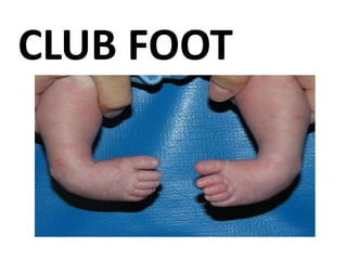
Clubfoot is a condition in which one or both feet are
twisted into an abnormal position at birth.
Common birth defect
Other terms congenital talipes aquinovarus (CTEV)
The condition is also known as talipes. It is a general
term used to describe a range of unusual positions of
the foot.
Present at birth and affects the foot and/or ankle.
3. • It is a common birth defect, occurring in about one in
every 1,000 live births.
• Approximately 50% of cases of clubfoot are
bilateral.
• This occurs in males more often than in females by a
ratio of 2:1.
• Main cause is the result of arrested or anomalous
development in utero.
5. • Clubfoot is a condition in which one or both
feet are twisted into an abnormal position at
birth. The condition is also known as talipes or
talipes equinovarus.
6. Causes
Family history of clubfoot.
Position of the baby in the uterus.
Increased occurrences in those children with
neuromuscular disorders, such as cerebral palsy
and spina bifida.
Amniotic Band Syndrome
Oligohydramnios
7. Clinical types
• There are four variations of clubfoot:
(1) talipes varus: the most common form of
clubfoot, the foot generally turns inward so that
the leg and foot look somewhat like the letter J ).
(2) talipes valgus, the foot rotates outward like the
letter L.
(3) talipes equinus, the foot points downward,
similar to that of a toe dancer.
(4) talipes calcaneus, the foot points upward, with
the heel pointing down.
Slide 7
8.
9.
10. pathophysiology
Predisposing Factors:
Family history of clubfoot.
Position of the baby in the uterus.
Increased occurrences in those
children with neuromuscular
disorders, such as cerebral palsy and
spina bifida.
Amniotic Band Syndrome
Oligohydramnios
Distal limb amniotic
banding
Amnion forms
constrictive bands
around a limb in utero
Cutting off the
circulation to the limb
Defective cartiliganious
anlage of the talus
Resulting in further
abnormal or arrested
development
Arrest of the fetal
development in the
fibular stage
11. Diagnostic Procedures
CT-scan
Ante-natal ultrasound scan.
After birth it can be detected by means of looking at
the shape and position of the foot.
X-ray
14. Treatment
It consist 3 stages
Correct the deformity
Maintenance of correction until muscle balance
developed
Follow up observation to detect reoccurance
19. Nursing Responsibilities
Review the pathology, prognosis and future
expectations to mothers to provideknowledge base
from which parents can make informed choice.
Discuss deformity and expected treatment in terms
the parents can understandto rule out misconceptions
and to provide information about the deformity.
Encourage parents to hold and play with child and
participate in care to promotebonding.
20. Assess and teach parent to assess for signs of
excessive pressure on
skin,redness, excoriation because these signs require i
mmediate evaluation andintervention.
Elevate the extremity to promote venous return and
prevents edema.
Check the toes every 1-2 hours for temperature, color,
sensation, motion, andcapillary refill time.
21. Stimulate movement of toes to promote circulation.
Insert plastic petals over the top edges of a new cast
while it is still wet to keep urine from soaking and
softening the cast.
Provide comfort measures such as soft music,
pacifier, teething ring, or rocking to promote
relaxation and may enhance patients coping abilities
by refocusing attention.
22. When the Kite casting method is being used,
check circulatory status frequently. Circulation
maybe impaired because of increased pressure
on tissues and blood
vessels. The equines correction specially places consi
derable strain on ligaments, blood vessels, and
tendons.
Discuss the importance of physical therapist to
enhance mobility
23. Nursing Diagnosis
1. Risk for disproportionate growth related to congenital
disorders.
2. Impaired physical mobility related to musculoskeletal
impairment.
3. Impaired skin integrity related to musculoskeletal impairment.
4. Disturbed body image related to developmental changes.
5. Social isolation related to alterations in physical appearance