Clinical correlation angina folate
•Download as PPT, PDF•
0 likes•225 views

Clinical Correlation angina folate

Recommended
More Related Content
What's hot
What's hot (20)
Viewers also liked
Viewers also liked (11)
Similar to Clinical correlation angina folate
Similar to Clinical correlation angina folate (20)
More from Mario Sanchez
More from Mario Sanchez (20)
Recently uploaded
Recently uploaded (20)
Clinical correlation angina folate
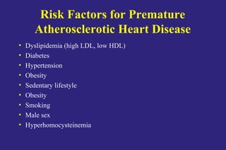
- 1. Risk Factors for Premature Atherosclerotic Heart Disease • Dyslipidemia (high LDL, low HDL) • Diabetes • Hypertension • Obesity • Sedentary lifestyle • Obesity • Smoking • Male sex • Hyperhomocysteinemia
- 2. Homocysteine • Non-protein-forming, sulfur-containing amino acid • Formed exclusively by demethylation of methionine • Eliminated through one of two vitamin-dependent pathways, in addition to an alternate vitamin- independent pathway in liver
- 3. Hyperhomocysteinemia • Independent risk factor for atherosclerotic and thromboembolic disease • A 5 µM increase in serum level confers a 80% increased risk to women and a 60% increased risk to men for atherosclerotic vascular disease • In patients with coronary artery disease, serum homocysteine levels increase with the number of stenosed coronary vessels • Hyperhomocysteinemia may reflect: – Genetic defects – Folate (most common), pyridoxine (vitamin B6), or cobalamin (vitamin B12) deficiencies – Renal failure • Serum levels of homocysteine may be lowered by supplementation with folate, vitamin B6, and vitamin B12
- 4. QuickTime™ and a GIF decompressor are needed to see this picture. Hajjar KA, J Clin Invest 107:663, 2001 Homocysteine Metabolism and Vascular Dysfunction
- 5. Case #1 • 45 year-old African-American male • Presents with acute onset of chest pain • Undergoes cardiac catheterization and is found to have blockage in 4 coronary arteries and is scheduled for bypass surgery • Past medical history is unremarkable • No current medications
- 6. Case #1 • Activity – Works as a bank teller. Very little vigorous physical activity. Walks for 30 min three times per week. • Social – Divorced, lives alone – Smokes 1/2 pack cigarettes/day – Denies alcohol/substance abuse
- 7. Case #1 • Physical Exam – BP 138/90 mm Hg right arm sitting (normal 140/90) – Ht 180 cm – Wt 77 kg – BMI (wt/ht2 ) 23.8 (normal < 25) – Rest of physical exam normal
- 8. Case #1 54 yo hypertension MI 62 yo hypertension stroke 72 yo hypertension 69 yo cancer 70 yo hypertension CH 236 TG 120 HDL 42 69 yo obese CH 204 TG 204 HDL 42 49 yo stroke 40 yo healthy CH ? 42 yo obese CH 210 TG 201 HDL 38 45 yo borderline hypertension CAD CH 230 TG 120 HDL 40 hyperhomocysteinemia
- 9. Case #1 • Diet – No breakfast – Takes lunch: meat sandwich and fruit – Dinner: eats alone, usually fast food or frozen dinner – Analysis of 3-day food diary • Average 2230 kcal/day • Diet composition (% of total calories) – Protein 20% – Fat 38% (desirable < 30%) – Carbohydrate 42% • Cholesterol: 340 mg/day (desirable < 300 mg/day) • Folic acid: 212 mcg/day (desirable > 240 mcg/day)
- 10. Case #1 • Cardiovascular Risk Profile – Lipids • Total cholesterol 230 mg/dL (normal < 200 mg/dL) • Triglycerides 120 mg/dL (normal < 200 mg/dL) • HDL cholesterol 40 mg/dL (normal > 35 mg/dL) • LDL cholesterol 166 mg/dL (normal < 130 mg/dL) – Homocysteine 16 µmol/L (normal < 12 µg/L
- 11. Dietary Sources of Folate and Vitamins B6 and B12 • Foods Rich in Folate – Leafy vegetables, liver, whole-grain cereals, legumes, and some fruits, such as oranges • Foods Rich in Vitamin B6 – Fish, poultry, meat, and wheat • Foods Rich in Vitamin B12 – Animal products
- 13. Case #2 • 4 hr old infant boy born to 25 yo G1P1 Caucasian • Pregnancy notable only for UTI at 5mths • Term by dates • Uncomplicated vaginal delivery • Family history negative for birth defects
- 15. Folic Acid and Birth Defects • About 2,500 neural tube defects per year in US • Occur at 26-28 days post-conception • 95% are spontaneous with no family history • 1991 UK study showed 71% risk reduction in recurrences (4mg dose) • In 1999 (Nov NEJM) 85% reduction in risk for primary prevention (0.4mg dose) • Lifetime costs estimated to be $330.000 (1998)
- 16. Case # 3 • 38 year old female pediatrician • Normal fast food diet, social drinker • Routine Ob/Gyn visit • Mass noted in left breast • Biopsy c/w breast cancer (BRCA negative)
- 17. Folate and Cancer • Increased risk for breast cancer in women who consume 15g/day of ethanol and < 300mcg folate/day (RR=1.32) • Reduced risk for colon cancer in women who consume daily folate suplements for 15 years or more (RR=0.25) • Reduced risk for colon cancer in men who consumed supplements for 10 years (RR=0.75)
- 18. Mechanisms of benefit • Cardiovascular disease – -thrombogenic factors, decreased nitric oxide, increased inflammation • Neural tube defects – ???? • Cancer – Hypomethylation of DNA, low CH2THF leading to uracil for thymidine
Editor's Notes
- Patients, particularly adults, rarely have just one risk factor. As we go along, see how many risk factors you can identify for this patient.
- Iatrogenic causes of elevated homocysteine include methotrexate therapy, theophylline, L-dopa and the co-administration of niacin and bile acid sequestrants. Increased risks of atherosclerotic disease based on micromolar increases in homocysteine were derived from a metanalysis of 27 studies (JAMA 1995;274:1049-57)
- Homocysteine (HC) is formed by demethylation of methionine via S-adenosylmethionine (AdoMet) and S-adenosylhomocysteine (AdoHC). HC is eliminated in the methionine cycle by remethylation through the action of methionine synthase (MS), a vitamin B12-dependent enzyme. In this reaction, methyltetrahydrofolate (CH3THF) serves as the methyl donor, and is formed from methylene tetrahydrofolate (CH2THF) through the action of methyltetrahydrofolate reductase (MTHFR). CH2THF is formed from tetrahydrofolate (THF), a folate derivative. Alternatively, HC can participate in transsulfuration in which it condenses with with serine through the action of the vitamin B6-dependent enzyme, cystathionine--synthase (CBS). Cystathionine then splits into cysteine and -ketobutyrate. HC can modify vascular cell function by forming a direct disulfide protein derivative, as in the case of the fibrinolytic receptor, annexin II; by inducing a prothrombotic phenotype through largely unknown mechanisms; or by undergoing auto-oxidation, generating superoxide radicals (redox stress), leading to depletion of nitric oxide (NO) and expression of acute stress-related genes. Redox stress may also activate the proinflammatory transcription factor NF-B, which may induce expression of of TNF-, RAGE/EN-RAGE, VCAM-1, tissue factor, and MMP-9. TNF- may also induce activation and nuclear translocation of NF-B. Thus, there are two pathways shown which reduce homocysteinelevels which are modified by folate, pyridoxine (B6) and B12.
- Based on studies in children and adolescents, African-Americans may have higher hoocysteine levels than whites and Hispanics.
- Notice two risk factors here for premature coronary artery disease: Sedentary lifestyle Smoking
- Note that the diastolic blood pressure is borderline high. This represents another potential risk factor for premature atherosclerotic heart disease.
- Note the positive family history on the patient’s father’s side of the family: Early death (&lt; 55 yoa) in paternal grandfather from MI, paternal uncle from stroke. Hypertension in father and paternal grandparents and maternal grandmother. Hypercholesterolemia in father, mother and sister. Obesity in mother and sister. Unfortunately, we do not have information on homocysteine levels in family members. At the very least, the patient’s siblings should all be screened for both lipids and homocysteine levels.
- Note the irregular eating habits with no breakfast and fast food or frozen meals for dinner. Also, the intake of percentage of total calories from fat is excessive, and the daily intake of cholesterol is too high. The intake of folic acid is below the recommended daily allowance. It is unclear what the optimal dose of folate may be which will prevent atherosclerotic disease. No studies have shown (as of 2000) that interventional folate therapy reduces cardiovasular disease in humans. Animal studies have validated folate’s role in reducing atherosclerotic disease.
- Note two more risk factors for premature atherosclerotic heart disease: Increased serum LDL cholesterol level. Increased serum homocysteine level.
- Increased folate intake is a prudent approach to the patient with elevated homocysteine and a positive family history. Achieving this by dietary means requires some effort and is best accomplished by taking folate supplements. This is partly due to the relatively poor bioavailability of dietary folate compared to supplements. Most multi-vitamins contain 400 micrograms of folate. Many cereals now contain 400 micrograms of folate per serving.
- The change in folate to tetrahydrofolate occurs by the action of folate reductase. This enzyme is blocked by methotrexate in humans and in bacteria by trimethoprim.
- The physical exam was normal except for a thoraco-lumbar defect consistent with a meningomyelocoele. Subsequent ultasound of the head showed hydrocephalus. The child required a shunt and lumbar repair. There was paresis of the lower extrmeities and bladder dysfunction resulted later in life.
- The cost of folate supplementation is about a penny a day. All child-bearing women should receive 400 mcg/day of folate. Knowledge aobut folate and prevention of birth defects is poor nationally but has improved where public knowledge campaigns have been carried out. Physician knowledge is poor about these issues as well. The March of Dimes has taken the lead in promoting knowledge about folate and birth defects.
- References above: JAMA 1999:281; 1632-1637, Ann Intern Med1998:129;517-524, While data are limited, there is a strong scientific suggestion that long term folic acid intake can reduce the risks of certain types of cancer.
- How a single nutrient can be protective for so many different types of disease is unknown and a subject of active research. Folate may operate by more than one mechanism to provide its protective benefits. It should be stressed that only in neural tube defects do we have evidence that dietary interventions in humans will reduce disease. For this reason, increased dietary folate and folate supplements ( 400 micrograms/day) are recommended only for child bearing women. For other individuals dietary folate alone is recommended with a 400 mcg target. Screening is recommended for those with risk factors for cardiovascular disease.
- The protective benefits in cancer may be linked through folate and its impact on thymidylate synthase. Many of the pre-malignant lesions in cancer occur through the increased substitution of dUMP for d TMP. Folate is speculated to limit thesechromosomal aberrations in synthesis which might lead to mutations and chromosomal breakage. (Ann Int Med 1998: 129: 517-524.)