SlideShare a Scribd company logo
Profa. Dra. Cristina Maria Henrique Pinto Profa. Adjunto do Depto. Ciências Fisiológicas-CCB-UFSC Como citar este documento: PINTO, Cristina Maria Henrique. Fisiologia do Sistema Digestório e aplicações clínicas. Disponível em: <http://www.cristina.prof.ufsc.br>. Acesso em: (coloque a data aqui) Fisiologia do Sistema Digestório  e aplicações clínicas
Esta apresentação foi utilizada em minhas aulas para a graduação em Medicina (6ª fase) até o ano de 2007. Para que este material não se perca, deixo aqui à disposição daqueles que eventualmente tenham interesse. Bons estudos!
Bibliografia básica recomendada sobre  Fisiologia Humana Livros-textos: “ Berne & Levy: Fisiologia” Koeppen & Stanton, 2009,  6ª Ed. (Ed. Elsevier) “ Tratado de Fisiologia Médica”  Guyton & Hall, 2006,  11ª Ed. (Ed. Elsevier) “ Fisiologia” Aires, M. M. 2008, 3ª Ed.  (Ed. Guanabara Koogan) “ Fisiologia” Costanzo, 2007,  3ª Ed.  (Ed. Elsevier) “ Berne & Levy: Fundamentos de Fisiologia”, Levy  et al , 2006, 4ª Ed. (Ed. Elsevier) “ Fundamentos de Fisiologia Médica”  Johnson, 2003 (Ed. Guanabara Koogan) “ Fisiologia: texto e atlas”  Silbernagl e Despopoulos, 2003 (Ed. Artmed)
Profa. Dra. Cristina Maria Henrique Pinto CFS/CCB – 6ª fase - Medicina O Pâncreas  como glândula exócrina
Figure 3. The pancreas and adjacent anatomy.  divisões: cabeça e processo uncinado, colo, corpo e cauda http://hopkins-gi.nts.jhu.edu/pages/latin/templates/index.cfm?pg=disease1&organ=4&disease=22&lang_id=1   PÂNCREAS localização: -órgão retroperitoneal. -anteriores a ele estão o estômago, alças do intestino delgado, colon e omento
Duct system of the pancreas. Berne et al., 2004 Ducto pancreático principal formado a partir dos ductos intercalares, intralobares extralobares e ductos coletores principais
Ducto pancreático: -desde a cauda até a cabeça do pâncreas -une-se ao ducto colédoco já próximo ao duodeno, formando a ampola hepatopancreática e a papila maior do  duodeno ( ou ampola de Vater ). http://hopkins-gi.nts.jhu.edu/pages/latin/templates/index.cfm?pg=disease1&organ=4&disease=22&lang_id=1   É comum a existência  de um  ducto pancreático secundário , acessório ou dorsal (papila menor)
Normally (90% of the time) pancreatic secretions from the entire pancreas and biliary secretions gain access to the duodenum by way of the ventral pancreatic or Wirsung’s duct because of the fusion of the dorsal pancreatic duct during embryological development.  There is only a remnant of the dorsal pancreatic or Santorini’s duct connecting to the duodenum. In 10% of individuals, fusion does not occur. This situation is called pancreas divisum. In pancreas divisum secretions from the dorsal and ventral ducts drain separately into the duodenum. Individuals with pancreas divisum may be at higher risk for pancreatitis (see Acute Pancreatitis for additional information).  Note that the distal common bile duct reaches the duodenum through the head of the pancreas. http://www.gastroslides.org/main/browse_deck.asp?tpc=2&mxpg=390&pg=1844#image   DUCTO PANCREÁTICO PRINCIPAL E  DUCTO PANCREÁTICO ACESSÓRIO  (ocorrência em 90% da população)
This figure demonstrates that the stomach, duodenum, liver, biliary system, gallbladder and pancreas are derived from closely related structures in early embryological development. The pancreas, liver, gallbladder and biliary system bud from the duodenum during early embryological development. The pancreas starts as two components, the ventral and dorsal pancreas.  In the process of development, the organs enlarge and the ventral pancreas together with the common bile duct rotates. Then, in most cases, the pancreatic duct from the dorsal pancreas fuses with the pancreatic duct from the ventral pancreas to form the main pancreatic duct. After fusion the pancreatic secretions from the entire pancreas and biliary secretions gain access to the duodenum by way of the ventral pancreatic duct. http://www.gastroslides.org/main/browse_deck.asp?tpc=6&mxpg=390&pg=2233#image   EMBRIOLOGIA
Normally (90% of the time) pancreatic secretions from the entire pancreas and biliary secretions gain access to the duodenum by way of the ventral pancreatic or Wirsung’s duct because of the fusion of the dorsal pancreatic duct during embryological development.  There is only a remnant of the dorsal pancreatic or Santorini’s duct connecting to the duodenum. In 10% of individuals, fusion does not occur. This situation is called pancreas divisum. In pancreas divisum secretions from the dorsal and ventral ducts drain separately into the duodenum. Individuals with pancreas divisum may be at higher risk for pancreatitis (see Acute Pancreatitis for additional information).  Note that the distal common bile duct reaches the duodenum through the head of the pancreas. http://www.gastroslides.org/main/browse_deck.asp?tpc=2&mxpg=390&pg=1844#image   DUCTO PANCREÁTICO PRINCIPAL E  DUCTO PANCREÁTICO ACESSÓRIO  (ocorrência em 90% da população) PANCREAS DIVISUM (ocorrência em 3 a 7% da pop.)
http://hopkins-gi.nts.jhu.edu/pages/latin/templates/index.cfm?pg=disease1&organ=4&disease=22&lang_id=1   -a mais comum anomalia congênita do pâncreas -ocorre em 3 a 7% da população -resulta em fusão incompleta ou inexistente dos brotos dorsal e ventral  durante o desenvolvimento embrionário. As regiões da cauda, corpo, colo e pequena porção da cabeça têm suas secreções drenadas para o duodeno através da papila menor  via ducto acessório . Obstruções na papila menor podem causar pancreatite aguda (drenagem insuficiente). O  ducto pancreático principal  que esvazia-se na papila duodenal maior, drena apenas uma pequena porção da secreção exócrina pancreática (porção ventral). CAUSA de pancreatite Pancreas divisum  (ou  pâncreas dividido )
http://hopkins-gi.nts.jhu.edu/pages/latin/templates/index.cfm?pg=disease1&organ=4&disease=22&lang_id=1   Esfíncter de Oddi: células musculares lisas que envolvem a porção terminal dos ductos biliar comum e pancreático  principal. É uma estrutura dinâmica que relaxa ou contrai, alterando as dimensões da papila maior duodenal
http://hopkins-gi.nts.jhu.edu/pages/latin/templates/index.cfm?pg=disease1&organ=4&disease=22&lang_id=1   Figure 7. Gallstone obstruction.  -uma das mais causas comuns de pancreatite  (USA e países europeus) -baixa mortalidade -mecanismos causais ainda não totalmente entendidos -obstrução causaria refluxo biliar nas vias pancreáticas o que desencadearia uma  complexa cascata de efeitos  (p. ex. ativação da tripsina  nos ácinos e/ou indução de resposta inflamatória). CAUSA de pancreatite aguda Colelitíase
The pancreas is divided into an exocrine portion (acinar and duct tissue) and an endocrine portion (islets of Langerhans). The exocrine portion, comprising 85% of the mass of the pancreas, secretes digestive enzymes, water and NaHCO3 into the duodenum. The endocrine portion, comprising 2% or less of the pancreas, secretes hormones into the blood stream. http://www.gastroslides.org/main/browse_deck.asp?tpc=6&mxpg=390&pg=2240#image   O PÂNCREAS: UMA GLÂNDULA  ENDÓCRINA  E  EXÓCRINA
? VOLUME SECRETADO PELO PÂNCREAS NO INTESTINO DELGADO: 1,5 L/DIA
secreção hidro-eletrolítica enzimas digestivas (proteases, amilase e lipases) Principais tipos celulares encontrados no pâncreas Ilhotas de Langerhans hormônios: insulina (cél.  β ), glucagon (cél.  α ),  somatostatina (cél.  δ ) e polipeptídeo pancreático (cél.  θ )
ilhota (células alfa: glucagon) ilhota (células beta: insulina) ácinos e ilhota de Langerhans http://www.udel.edu/Biology/Wags/histopage/histopage.htm   HISTOLOGIA DO PÂNCREAS PORÇÃO ENDÓCRINA  (hormônios: glucagon, insulina, somatostatina e polipeptídeo pancreático) PORÇÃO EXÓCRINA  enzimas e secreção hidroeletrolítica http://www.gastroslides.org/main/browse_deck.asp?tpc=6&mxpg=390&pg=2241#image
The relationships and major features of the units of the exocrine pancreas. The pancreatic acinar cells of the acinus have prominently stained zymogen granules in the apical area of the cell.  The connecting ductule does not contain zymogen granules. The blue cell in the cartoon depicts the centroacinar cell at the border between the acinus and ductule. The centroacinar cell functions similarly to the duct cell. The major secretory products of the acinus are digestive proenzymes and enzymes with lesser amounts of water and ions. The major secretory products of the duct are water and ions. http://www.gastroslides.org/main/browse_deck.asp?tpc=6&mxpg=390&pg=2243#image   AS SECREÇÕES EXÓCRINAS PANCREÁTICAS:  água e eletrólitos
Neutralization of gastric acid delivered to the duodenum is necessary for optimal digestion and absorption of a meal. Several mechanisms that are not shown are involved in the neutralization process. First, the meal provides buffers from digestion of protein and triglycerides. That is, the peptides and fatty acid products act as pH buffers. Another neutralization process is absorption of hydrogen ion by the duodenal mucosa. Finally, the pancreas, biliary system and duodenal mucosa secrete bicarbonate into the duodenal lumen for neutralization.  http://www.gastroslides.org/main/browse_deck.asp?tpc=6&mxpg=390&pg=2287#image   Importância da secreção hidroeletrolítica pancreática, rica em HCO 3 - , Na +  e água na digestão NaHCO 3  + HCl    NaCl + H 2 O + CO 2  (reabsorção)
Secreção hidroeletrolítica pancreática: grande volume e rica em Na +  e HCO 3 -
Stimulation (i.e. a meal) there is an increase in the flow rate of pancreatic secretions. Furthermore, with increasing flow rates there is a dramatic change in the concentrations of chloride and bicarbonate. The increase in bicarbonate concentration results in a secretion that is alkaline. The bicarbonate ion comes from ductal epithelial cells in the pancreas. In contrast to acinar cells, the ducts secrete a large volume of fluid with a high concentration of bicarbonate. Because the volume of secretion from the acinar cells is thought to be small compared to ductal secretion, with increasing stimulation of the pancreas the concentration of ions approaches that of the ductal secretions. Of note, the alkaline secretion of the pancreas combined with alkaline secretions from the biliary system and the duodenal mucosa neutralize the acid secretion delivered to the duodenum from the stomach. This pH neutral environment is important for optimal digestive enzyme and intestinal mucosal function.  http://www.gastroslides.org/main/browse_deck.asp?tpc=6&mxpg=390&pg=2287#image   Composição da secreção hidroeletrolítica pancreática em razão da taxa de secreção
Cl- secretion drives bicarbonate secretion in the pancreatic duct cell. Cl-  secretion occurs via secretin-stimulated cyclic AMP production. Cyclic AMP, in turn, activates the CFTR Cl-  secretory pathway. In the pancreatic duct cell, the Cl- may exchange with HCO3- resulting in net HCO3- secretion.  Of note, Cl-   delivered from the acinar cells also exchanges with HCO3- in the pancreatic duct. HCO3- may also enter the lumen through CFTR or by other mechanisms (see following slides). Na+ enters the duct lumen via the paracellular space to neutralize the electrical gradient created by the apical HCO3- secretion. Water follows via the paracellular space to maintain iso-osmolality. The combination of these processes determines the composition and volume of pancreatic duct secretions.  ( canal regulador da condutância transmembrana da fibrose cística) http://www.gastroslides.org/main/browse_deck.asp?tpc=6&mxpg=390&pg=2332#image   Mecanismos celulares de secreção de  HCO 3 - , Na +  e água pelas células dos  ductos pancreáticos
Mecanismos celulares de secreção de  HCO 3 - , Na +  e água pelas células dos  ductos pancreáticos Delivery mechanisms to provide a source for HCO3- for the duct cell. There are two delivery systems. In one, membrane diffusible CO2 is catalytically converted to HCO3- and H+ by the action of carbonic anhydrase (CA) which hydrates CO2   thereby forming H2CO3 that then dissociates to HCO3- and H+. The duct cell is rich in CA. The HCO3- is then available for apical secretion. H+ is removed from the cell by a basolateral Na+/H+ antiport to maintain a constant intracellular pH. In the other delivery  http://www.gastroslides.org/main/browse_deck.asp?tpc=6&mxpg=390&pg=2322#image
The two major classes chloride channels.  CFTR is activated by cAMP dependent protein kinase.  A second family of channels is regulated by calcium.  Both classes of calcium channels may be present in some cells . http://www.gastroslides.org/main/browse_deck.asp?tpc=6&mxpg=390&pg=2332#image   CFTR afeta outros canais de cloreto
CFTR activation results in activation of conductive pathways for several other molecules as listed. The mechanisms involved in the cooperative effects on transport of these molecules are not established.  canal regulador da condutância transmembrana da fibrose cística http://www.gastroslides.org/main/browse_deck.asp?tpc=6&mxpg=390&pg=2332#image
Using immunocytochemical staining of CFTR and zymogens, this image shows that CFTR is located on the apical membrane of both pancreatic duct cells and acinar cells. http://www.gastroslides.org/main/browse_deck.asp?tpc=6&mxpg=390&pg=2322#image
Chloride is secreted from the apical surface of the acinar cell by two distinct transport processes. In one, agents such as cholecystokinin or muscarinic agents (i.e. acetylcholine) cause an increase in intracellular calcium that, in turn, activates a Ca+2-dependent chloride channel. In the other, agents such as secretin and VIP cause an increase in cyclic AMP that, in turn, activates a cyclic AMP-dependent chloride channel.   Of note, both Ca+2 and cyclic AMP activate a basolateral K+ channel. This activation of K+ efflux facilitates apical chloride secretion by make the intracellular environment more electronegative. The Na+ /K+ ATPase is important in facilitating chloride transport because it maintains the electronegative intracellular environment as there are two K+s transported in for three Na+s transported out of the cell. Finally, the Na+/K+/Cl -Cotransport on the basolateral membrane is essential to replenish chloride secreted at the apical surface. CFTR http://www.gastroslides.org/main/browse_deck.asp?tpc=6&mxpg=390&pg=2322#image   Mecanismos celulares de secreção de  Na +  e água pelas células dos  ácinos pancreáticos
Importância do conhecimento dos mecanismos celulares de secreção de HCO 3 - , Na +  e água pelas células centro-acinares, ductos e ácinos Patologias podem ocorrer quando, por diversos fatores, surgem  alterações da expressão de canais e/ou transportadores celulares. Exemplo:  deficiência da expressão de canais de cloreto na membrana luminal celular na  fibrose cística Primary epithelia affected in Cystic Fibrosis http://cellscience.com/reviews2/Cystic_Fibrosis_alternative_Chloride_channel.html   acarreta deficiência nutricional especialmente na criança Tissue affected Pancreas Intestine Sweat glands Airways Reproductive organs Pathology Maldigestion Malabsorption/meconium ileus salty sweat Dry airways/infection Sterility Epithelia affected Exocrine cells Crypt epithelium Sweat duct Bronchial epithelium Vas Deferens/various Potential Therapies Enzyme replacement None None UTP + amiloride  aerosol/gene therapy None
Normal trafficking of CFTR to the plasma membrane where it functions as a cAMP-dependent Cl- channel.  Mutations that lead to protein dysfunction have been classified into five categories: I-no synthesis; II-block in processing, III- block in regulation; IV – altered conductance; V – reduced synthesis. The right panel depicts the two major mechanisms of CFTR dysfunction in cystic fibrosis. The most common mutation that results in cystic fibrosis is delta508 mutation (Type II).  This leads to CFTR misfolding and degradation before it can exit the endoplasmic reticulum.  Type III and IV defects result in CFTR insertion into the membrane, but abnormal regulation.  Type I and V defects are less common causes of cystic fibrosis.  Choo-Kang LR, Zeitlin PL. Curr Opin Pulm Med 2000;6:521-529 http://www.gastroslides.org/main/browse_deck.asp?tpc=6&mxpg=390&pg=2330#image
The clinical manifestations of cystic fibrosis. Of note, the genetic  defects in chloride transport results in dysfunction in multiple systems including the pulmonary, hepatobiliary, pancreatic, intestinal and reproductive systems  http://www.gastroslides.org/main/browse_deck.asp?tpc=6&mxpg=390&pg=2322#image
The relationships and major features of the units of the exocrine pancreas. The pancreatic acinar cells of the acinus have prominently stained zymogen granules in the apical area of the cell.  The connecting ductule does not contain zymogen granules. The blue cell in the cartoon depicts the centroacinar cell at the border between the acinus and ductule. The centroacinar cell functions similarly to the duct cell. The major secretory products of the acinus are digestive proenzymes and enzymes with lesser amounts of water and ions. The major secretory products of the duct are water and ions. http://www.gastroslides.org/main/browse_deck.asp?tpc=6&mxpg=390&pg=2243#image   AS SECREÇÕES EXÓCRINAS PANCREÁTICAS:  ENZIMAS
(from duodenal epithelial cells)  -Amylase  (no activation needed) secreted by duodenal epithelium (Enterokinase) http://mcb.berkeley.edu/courses/mcb136/topic/Gastrointestinal AS SECREÇÕES EXÓCRINAS PANCREÁTICAS:  enzimas lipase
Relative amounts (by weight) of the different classes of pancreatic digestive enzymes. Proteases are the most abundant class of enzymes. http://www.gastroslides.org/main/browse_deck.asp?tpc=6&mxpg=390&pg=2273#image
(from duodenal epithelial cells)  -Amylase  (no activation needed) secreted by duodenal epithelium (Enterokinase) http://mcb.berkeley.edu/courses/mcb136/topic/Gastrointestinal AS SECREÇÕES EXÓCRINAS PANCREÁTICAS:  enzimas lipase
The non-enzymatic secretory products of the acinar cell. Procolipase when activated facilitates the action of lipase. GP-2 is a glycoprotein linked to the inner zymogen granule membrane and may have a role in protein sorting or membrane recycling.  Some GP-2 is released from the zymogen granule membrane and secreted.  GP-2 is enriched in stones and may have a role in their formation.  Lithostathine may prevent stone formation in the pancreas.  Pancreatitis-associated protein increases with pancreatitis although the function is not known. Pancreatic secretory trypsin inhibitor has an important role in preventing intrapancreatitic zymogen activation. The ions, especially Na+ and Cl- are important for transport of the secretory products from the acinar lumen into the pancreatic ductal system by pulling water into the luminal space by osmotic forces. Water and ion flow then “wash out” the luminal space  http://www.gastroslides.org/main/browse_deck.asp?tpc=6&mxpg=390&pg=2273#image   AS SECREÇÕES EXÓCRINAS PANCREÁTICAS:  produtos não-enzimáticos
Action of major pancreatic lipases. The cleavage of lipids by glycerol ester hydrolase (pancreatic lipase), cholesterol ester hydrolase, and phospholipase A2 is illustrated. P, Phosphate. Berne, 2004 hidrolase de éster de colesterol fosfolipase A 2 A secreção serosa pancreática digestão de lipídeos da dieta do glicerol hidrolase de éster triglicerídeo 2-monoglicerídeo ácido graxo ácido graxo éster do colesterol colesterol ácido graxo ácido graxo lisolecitina lecitina
[object Object],[object Object],[object Object],[object Object],[object Object],[object Object],Mixed micelle formed by bile salts, triacylglycerols and  pancreatic lipase.  extraído, quanto disponível, de:  http://www.med.unibs.it/~marchesi/lipoprot.html A secreção serosa pancreática:  lipase e  colipase colipase Digestão de lipídeos no ID (meio aquoso) Produto da digestão da lipase pancreática: diacilgliceróis, monoacilgliceróis, ácidos graxos livres e glicerol hidrolase de éster do glicerol
Mucosal Cell Intestinal Lumen TG LIPASE Bile Acids Micelle MG FA A secreção serosa pancreática: lipase e colipase Digestão de lipídeos no ID (meio aquoso) TG=triglyceride; MG=monoglyceride; FA=fatty acid.
By demonstrating the relative activity of lipase action with addition of each to a mixture, these experimental results show that the activity of lipase on triglyceride hydrolysis is dependent on both bile salts and colipase. With addition of lipase alone (indicated by the green arrow) there is some triglyceride hydrolysis.  However, the addition of bile salts with colipase to a mixture of triglycerides and lipase results in the greatest activity.  The importance of bile salts is that they anchor lipase and colipase together on the oil phase of trigylceride.   This “anchoring” results in the greatest activity. http://www.gastroslides.org/main/browse_deck.asp?tpc=6&mxpg=390&pg=2284#image
Mucosal Cell Intestinal Lumen TG LIPASE Bile Acids Micelle MG FA A secreção serosa pancreática: lipases e colipase pancreatite aguda/crônica:   deficiência/ausência da lipase pancreática       digestão e absorção da gordura da dieta     perda pelas fezes  (esteatorréia) esteatorréia TG=triglyceride; MG=monoglyceride; FA=fatty acid.
A digestão e absorção de lipídeos podem ser diminuídas por drogas que ligam-se à lipase e inibem sua ação TG=triglyceride; MG=monoglyceride; FA=fatty acid. Mucosal Cell Intestinal Lumen DROGA TG LIPASE LIPASE LIPASE Bile Acids Micelle MG FA A secreção serosa pancreática: lipases e colipase esteatorréia
A secreção serosa pancreática:  alfa-amilase digestão de amido/glicogênio Amilopectina (amido) da batata produtos da digestão pela  α -amilase  pancreática α -amilase pancreática maltose dextrina -limite ligação 1:6 ligação 1:4 maltotriose alfa-amilase Produtos da hidrólise do AMIDO pela alfa-amilase  glicose
Functions of the major brush border oligosaccharidases. The glucose, galactose, and fructose molecules released by enzymatic hydrolysis are then transported into the epithelial cell by specific transport proteins. The glucose-galactose transporter is also known as SGLT1 and the fructose transporter as GLUT5. G, Glucose; Ga, galactose; F, fructose. Berne  et   al ., 2004 (SGLT1) Amido glicogênio α -amilase pancreática A secreção serosa pancreática:  alfa-amilase digestão de amido/glicogênio para posterior digestão e absorção pelo epitélio do ID produtos da digestão pela  α -amilase  pancreática α -amilase salivar
A secreção serosa pancreática:  pró-proteases   digestão de proteínas/polipeptídeos ativadas no intestino delgado pela  enteropeptidase (ou enteroquinase)
proteases pancreáticas PROTEÍNAS Pepsina  (gástrica) produtos da digestão pelas proteases pancreáticas A secreção serosa pancreática:  pró-proteases  digestão de proteínas/polipeptídeos para posterior digestão e absorção pelo epitélio do ID Amino acids Berne et al., 2004
A secreção serosa pancreática:  pró-proteases   ativadas no intestino delgado pela  enteropeptidase (ou enteroquinase) Activation pathways of proenzymes by trypsin. Once trypsin is activated, it is capable of activating many other digestive proenzymes.  Como é feita a  inativação da tripsina  enquanto está sendo armazenada e secretada pelo pâncreas? Hirota et al, 2003 JOP  http://www.joplink.net/prev/200303/01.html
TRIPSINOGÊNIO + Peptídeo inibidor da tripsina http://www.rcsb.org/pdb/static.do?p=education_discussion/molecule_of_the_month/pdb46_2.html   O tripsinogênio possui, além da tripsina, o  TAP  (peptídeo de ativação da tripsina) , uma   porção da cadeia que é removida pela  enteropeptidase  somente no ID TRIPSINOGÊNIO Trypsin is built with an extra piece of protein chain,  colored in green  in the structure on the left . Actually, only two amino acids of this extra bit are seen in crystal structure, so you have to imagine the rest flopping around away from the protein. This longer form of trypsin, called trypsinogen, is inactive and cannot cut protein chains. Then, when it enters the intestine, the enzyme  enteropeptidase  makes one cut in the trypsin chain, clipping off the little tail. This allows the new end of the chain,  colored here in purple , to tuck into the folded protein and stabilize the active form of the enzyme,  as shown on the right.  As extra insurance, the pancreas also makes a small protein, trypsin inhibitor (shown in red),  that binds to any traces of active trypsin that might be present before it is secreted into the intestine. It binds to the active site of trypsin, blocking its action but not itself being cut into tiny pieces.  A  inativação da tripsina  enquanto está sendo armazenada  e secretada  pelo pâncreas tripsinogênio armazenado
Pancreatic proenzymes (also called zymogens) such as trypsinogen are converted to their active forms with cleavage and removal of an NH2 –terminal portion, the activation peptide. Removal of the activation peptide results in exposure of the catalytic site of the enzyme. In the duodenum, enterokinase activates trypsinogen while trypsin activates other zymogens.  In the case of trypsinogen, the activation peptide is known as TAP (trypsin activation peptide).  In acute pancreatitis, measurements of TAP generation can be used clinically as a predictor of severity.  Mecanismo de ativação do tripsinogênio já no ID: hidrólise pela enteropeptidase intestinal http://www.gastroslides.org/main/browse_deck.asp?tpc=6&mxpg=390&pg=2280#image
This graphic presents the classes of proteases in the pancreas and their sites of action. Exopeptidases cleave one amino acid at a time from the NH2-terminal or COOH-terminal of a protein. Examples are the carboxypeptidases that cleave one amino acid at a time from the COOH-terminal of a protein. Endopeptidases such as chymotrypsinogen and trypsin each cleave specific bonds irrespective of their location with respect to the NH2-terminal or COOH-terminal of the protein.  http://www.gastroslides.org/main/browse_deck.asp?tpc=6&mxpg=390&pg=2280#image   Ações das proteases pancreáticas
Trypsin is built with an extra piece of protein chain,  colored in green  in the structure on the left . Actually, only two amino acids of this extra bit are seen in crystal structure, so you have to imagine the rest flopping around away from the protein. This longer form of trypsin, called trypsinogen, is inactive and cannot cut protein chains. Then, when it enters the intestine, the enzyme  enteropeptidase  makes one cut in the trypsin chain, clipping off the little tail. This allows the new end of the chain,  colored here in purple , to tuck into the folded protein and stabilize the active form of the enzyme, as shown on the right.  As extra insurance, the pancreas also makes a small protein, trypsin inhibitor ( shown in red ),  that binds to any traces of active trypsin that might be present before it is secreted into the intestine.  It binds to the active site of trypsin, blocking its action but not itself being cut into tiny pieces.  TRIPSINOGÊNIO A  inativação da tripsina  enquanto está sendo armazenada  e secretada  pelo pâncreas TAP  (peptídeo de ativação da tripsina) porção da cadeia a mais que o tripsinogênio possui, além da tripsina e que será removida no ID pela  enteropeptidase tripsinogênio armazenado Pode ocorrer uma ativação “espontânea” do tripsinogênio ainda nos ácinos ou nos ductos pancreáticos. Portanto, deve haver algum “mecanismo de segurança” para evitar que isso aconteça... http://www.rcsb.org/pdb/static.do?p=education_discussion/molecule_of_the_month/pdb46_2.html
Trypsin is built with an extra piece of protein chain,  colored in green  in the structure on the left . Actually, only two amino acids of this extra bit are seen in crystal structure, so you have to imagine the rest flopping around away from the protein. This longer form of trypsin, called trypsinogen, is inactive and cannot cut protein chains. Then, when it enters the intestine, the enzyme  enteropeptidase  makes one cut in the trypsin chain, clipping off the little tail. This allows the new end of the chain,  colored here in purple , to tuck into the folded protein and stabilize the active form of the enzyme, as shown on the right.  As extra insurance, the pancreas also makes a small protein, trypsin inhibitor ( shown in red ),  that binds to any traces of active trypsin that might be present before it is secreted into the intestine.  It binds to the active site of trypsin, blocking its action but not itself being cut into tiny pieces.  TRIPSINOGÊNIO  +   PSTI TRIPSINOGÊNIO Peptídeo inibidor da tripsina (PSTI) A  inativação da tripsina  enquanto está sendo armazenada e secretada pelo pâncreas TAP  (peptídeo de ativação da tripsina) porção da cadeia a mais que o tripsinogênio possui, além da tripsina e que será removida no ID pela  enteropeptidase tripsinogênio armazenado tripsinogênio secretado http://www.rcsb.org/pdb/static.do?p=education_discussion/molecule_of_the_month/pdb46_2.html
A atividade da tripsina é controlada principalmente pelo  peptídeo inibidor da tripsina ,  PSTI  (*) ,  sintetizado na mesma célula acinar e age como um potente inibidor natural da tripsina.  Quando o tripsinogênio é ativado no pâncreas (“espontâneo”), o   PSTI   liga-se à tripsina e a inativa, prevenindo assim a autodigestão celular. O  PSTI  também bloqueia futuras ativações que eventualmente venham a ocorrer nos ductos pancreáticos. A  inativação da tripsina  enquanto está sendo armazenada e secretada pelo pâncreas Activation pathways of proenzymes and PAR-2 by trypsin. Once trypsin is activated, it is capable of activating many other digestive proenzymes. Trypsin also activates pancreatic and inflammatory cells via PAR-2. The trypsin activity in the pancreas is mainly controlled by PSTI. When trypsinogen is activated into trypsin in the pancreas, PSTI binds immediately to trypsin to prevent further activation of pancreatic enzymes. (PAR: protease activated receptors; PSTI: pancreatic secretory trypsin inhibitor).   (*)  PSTI (pancreatic secretory trypsin inhibitor) ou SPINK1 (serine protease inhibitor Kazal type 1).  Hirota et al, 2003 JOP  http://www.joplink.net/prev/200303/01.html   Mutações no gene PSTI ou da Tripsina podem promover uma predisposição à  pancreatite , por diminuir a inibição funcional da tripsina
The major organelles involved in protein synthesis and segregation of the newly synthesized proteins into storage compartments. New proteins are synthesized in the rough endoplasmic reticulum of the pancreatic acinar cell. The newly synthesized proteins are then transported to the Golgi apparatus where post-translational modification and targeting occur.  http://www.gastroslides.org/main/browse_deck.asp?tpc=6&mxpg=390&pg=2296#image
The protective mechanisms present in the pancreas to prevent activation of zymogens in the pancreatic cell and thus prevent that damaging effects of zymogen activation. Zymogens that have been  pathologically activated  in an acinar cell.. http://www.gastroslides.org/main/browse_deck.asp?tpc=6&mxpg=390&pg=2349#image   MECANISMOS DE PROTEÇÃO DAS CÉLULAS ACINARES CONTRA A ATIVAÇÃO INTRACELULAR DAS ENZIMAS
Hereditary Pancreatitis: Proposed Mechanism  In normal individuals there is presumed to be a continuous low-grade activation and degradation of trypsin in the pancreas. Typsin can be temporarily inactivated by binding of pancreatic secretory trypsin inhibitor (PSTI or SPINK). Both trypsinogen and PSTI are located in the zymogen granules; there is sufficient PSTI to inhibit about 10% of pancreatic trypsin. Thus, as small amounts of active trypsin are formed, PSTI binds.  Proteases, such as trypsin and mesotrypsin, can degrade trypsin.  In the most common form of Hereditary Pancreatitis (HP) the mutation may render trypsin resistant to proteolytic degradation. Thus, trypsin that escapes inhibition by PSTI may remain active and activate the other pancreatic zymogens.   Direct evidence for this mechanism is not available .  http://www.gastroslides.org/main/indexcont.asp?tpc=8&mxpg=314&idxpg=73
http://www.gastroslides.org/main/indexcont.asp?tpc=8&mxpg=314&idxpg=73 O  PSTI  ( peptídeo inibidor da tripsina ) LIGA-SE À TRIPSINA E A INATIVA
The illustration in this slide shows the effect of a substitution of one amino acid (i.e. histidine for arginine) on the interaction of trypsin with pancreatic trypsin inhibitor and the function of trypsin.  The illustrations indicate the proposed mechanism of hereditary pancreatitis as well. In the normal state of folding, active trypsin in the pancreas binds to the trypsin inhibitor at its active site. This interaction leads to degradation of the active trypsin.  In contrast, the mutated form of trypsin has altered structure so that the trypsin inhibitor will not interact with its active site and the active trypsin will not degrade.   Thus, there is an increase in active trypsin in the pancreas with the potential for causing pancreatitis.  http://www.gastroslides.org/main/indexcont.asp?tpc=8&mxpg=314&idxpg=73 FORMA MUTANTE DE TRIPSINA TEM SUA ESTRUTURA ALTERADA,  O  PSTI  ( peptídeo inibidor da tripsina ) NÃO INTERAGE COM SEU SÍTIO ATIVO E  A TRIPSINA NÃO É DEGRADADA.
http://www.pancreas.org/assets/hp_presentation/index.htm   HP: pancreatite hereditária Whitcomb, 2000
ALCOOLISMO:  CAUSA IMPORTANTE DE PANCREATITE CRÔNICA http://www.gastroslides.org/main/browse_deck.asp?tpc=6&mxpg=390&pg=2357#image
http://www.gastroslides.org/main/browse_deck.asp?tpc=6&mxpg=390&pg=2357#image
Mecanismos que originam a dor na pancreatite aguda The  proteinase-activated receptors (PARs)  are a family of G-protein coupled receptors that, as their name suggests, are activated in a unique manner by serine proteases such as trypsin and thrombin.  Recently PAR-2 has been implicated in the pathogenesis of pain.  This may be particularly important in the pancreas where inflammation results in neuronal exposure to  activated trypsin (from acinar cells)  and  tryptase   (from mast cells) . Activation of PAR-2 on the peripheral processes of primary spinal afferents may have two effects. First, it may result in release of the neuropeptides CGRP and SP locally further amplifying inflammation and mast cell degranulation.  Secondly, excitation of the neurons also results in central release of neurotransmitters, leading to activation of second order neurons (as indicated by expression of the immediate early gene, c-Fos) in the dorsal horn further along in the nocioceptive pathway.  This pathway may contribute to pain experienced in both acute and chronic pancreatitis.  http://www.gastroslides.org/main/browse_deck.asp?tpc=6&mxpg=390&pg=2357#image
Exocrine pancreatic secretions are regulated by both endocrine and neurocrine pathways.  In addition, the endocrine and neurocrine mediators regulating secretion differ between the acinus and the duct.  For the acinus, cholecystokinin (CCK), acetylcholine (Ach), gastrin releasing peptide (GRP), vasoactive intestinal polypeptide (VIP) and substance P regulate secretion. In humans, CCK receptors have not been found on acinar cells. However, CCK may stimulate acinar cell secretion indirectly by exciting neural pathways. For the duct, secretin and acetylcholine are the major regulators of secretion.  Serotonin may act through 5-HT receptors to regulate acinar and duct cell secretion, but the importance and mechanism of this pathway in humans has not been defined. REGULAÇÃO DA SECREÇÃO EXÓCRINA PANCREÁTICA
PADRÃO DA SECREÇÃO DE ENZIMAS DIGESTIVAS PANCREÁTICAS This graphic shows the output of the digestive enzyme, trypsin, into the duodenum both during the fasting state and after a meal (dog). During fasting there are small periodic increases in trypsin output. These increases correlate with activation of the motility of the intestine during the migrating motor complex (MMC). With a meal, there is a much larger increase in trypsin output due to the inputs of several meal stimuli that mediate secretion through both neural and humoral mechanisms. http://www.gastroslides.org/main/browse_deck.asp?tpc=6&mxpg=390&pg=2256#image
Neurohormonal stimulatory and inhibitory mechanisms of pancreatic secretion during cephalic, gastric, and intestinal phases involving long vagovagal and short enteropancreatic reflexes activated by CCK acting on sensory receptors on afferent nerves. Various hormonal and humoral substances stimulate or inhibit the pancreatic secretion acting either through the CNS or directly on the exocrine pancreas. Konturek et al., 2003a
REGULAÇÃO DA SECREÇÃO EXÓCRINA PANCREÁTICA estimulação neural pancreática A presença de gordura e de ácido no duodeno estimula o SNE e as terminações aferentes sensoriais que, por sua vez, estimulam (via Vago) a atividade Parassimpática sobre a secreção pancreática
Vagal afferent signals from the pancreas are processed in the nodose ganglion and the dorsal vagal complex in the CNS. Afferent signals from the pancreas as well as other afferent signals from the  cephalic and gastric phases  of a meal are processed in the dorsal vagal complex and efferent signals are directed to the pancreas by the vagal nerves. The vagal efferents synapse with postganglionic neurons in the  pancreatic ganglia . These postganglionic neurons then mediate responses in both the islets of Langerhans and pancreatic exocrine tissue through the release of neurotransmitters such as acetylcholine (Ach). http://www.gastroslides.org/main/browse_deck.asp?tpc=2&mxpg=390&pg=1842#image   REGULAÇÃO DA SECREÇÃO EXÓCRINA PANCREÁTICA inervação pancreática
REGULAÇÃO DA SECREÇÃO EXÓCRINA PANCREÁTICA estimulação hormonal pancreática A presença de gordura e de ácido no duodeno estimula a secreção endócrina intestinal das células endócrinas “S” e “I”. As céls. “S”  secretam a SECRETINA que estimula a  secreção hidro-eletrolítica dos ductos pancreáticos
This image shows the stimulants for release of CCK from I cells and secretin release from S cells. Of note, the I cells and S cells “taste” the specific stimulants in the lumen of the gut to activate release of the hormones. The released CCK can mediate pancreatic secretion by either activating vagal sensory afferents that results in stimulation of pancreatic acinar cell secretion though a vago-vagal reflex involving the CNS or through the circulation (i.e. hormonal action). Secretin also acts through neural (not shown) and hormonal pathways on the pancreas stimulating secretion from both ductal cells and acinar cells. http://www.gastroslides.org/main/browse_deck.asp?tpc=2&mxpg=390&pg=1842#image   A REGULAÇÃO DA SECREÇÃO DOS DUCTOS PELA  SECRETINA
REGULAÇÃO DA SECREÇÃO EXÓCRINA PANCREÁTICA estimulação hormonal pancreática A presença de gordura e de ácido no duodeno estimula a secreção endócrina intestinal das células endócrinas “S” e “I”. As céls. “I”  secretam a  CCK que estimula  a secreção  serosa dos  ácinos pancreáticos
This image shows the stimulants for release of CCK from I cells and secretin release from S cells. Of note, the I cells and S cells “taste” the specific stimulants in the lumen of the gut to activate release of the hormones. The released CCK can mediate pancreatic secretion by either activating vagal sensory afferents that results in stimulation of pancreatic acinar cell secretion though a vago-vagal reflex involving the CNS or through the circulation (i.e. hormonal action). Secretin also acts through neural (not shown) and hormonal pathways on the pancreas stimulating secretion from both ductal cells and acinar cells. http://www.gastroslides.org/main/browse_deck.asp?tpc=2&mxpg=390&pg=1842#image   A REGULAÇÃO DA SECREÇÃO DOS DUCTOS PELA  CCK
The image is an electron photomicrograph of a CCK-secreting I cell. The I cell is one of the enterochromaffin cells. The apical surface of the I cell faces the lumen of the intestine where it “tastes” specific luminal nutrients (i.e. lipid). When it senses the specific nutrients, the I cell is activated and releases CCK from its basal surface. CCK can either act as a hormone to circulate to the pancreas and stimulate a secretory response or interact with sensory afferents containing CCK A (also known as CCK1) receptors. The image demonstrates the interaction of CCK with sensory receptors that are part of the enteric nervous system (ENS) of the gut. These neural receptors are located in proximity to the I cells. Vagal nerves to the CNS carry the afferent sensory signals. The afferent information going to the dorsal motor complex is processed and leads to stimulation of responses in the pancreas via vagal efferents.  http://www.gastroslides.org/main/browse_deck.asp?tpc=6&mxpg=390&pg=2263#image   A CCK atuaria também de maneira parácrina, estimulando as terminações sensitivas do PS  (e do SNE)
This image shows the several pathways mediating meal-stimulated pancreatic secretion that involve CCK. First, meal nutrients such as fatty acids, amino acids and peptides delivered from the duodenum stimulate the release of CCK from the CCK-containing I cell to the area around the basolateral surface of the I cell. The CCK released can activate vagal afferent neurons that carry the signal to the dorsal vagal complex where the sensory information is integrated and vagal efferents are activated. Vagal efferents synapse with neurons in the pancreatic ganglia. In turn, via the neurotransmitters, acetylcholine (Ach), gastrin releasing peptide (GRP; also known as bombesin) and vasoactive intestinal polypeptide (VIP), effector neurons in the pancreatic ganglia activate secretion by pancreatic parenchymal cells. In addition to activating the neural pathway, CCK released by the I cell enters the general circulation and may act as a hormone on the pancreatic acinar cells to cause secretion. Since CCK receptors have not been demonstrated on human acinar cells, the importance of direct hormonal stimulation is questionable. http://www.gastroslides.org/main/browse_deck.asp?tpc=6&mxpg=390&pg=2263#image
The graphic in this image depicts the neurotransmission systems and neurotransmitters involved in regulating acinar and ductal secretion. The blue cell is called a centroacinar cell. These cells have characteristics more similar to ductal cells although their exact function is not known. Vagal efferents activate neurons in the intrapancreatic ganglia. Neurons from the ganglia then have processes that are in proximity to the parenchymal cells. These processes release the neurotransmitters, acetylcholine (Ach), gastrin releasing peptide (GRP) and vasoactive intestinal polypeptide (VIP) when their neurons are activated. Ach causes secretion from both acinar cells and ducts while all three neurotransmitters can cause secretion from acinar cells.  http://www.gastroslides.org/main/browse_deck.asp?tpc=2&mxpg=390&pg=1842#image
http://www.gastroslides.org/main/browse_deck.asp?tpc=2&mxpg=390&pg=1842#image   Pancreatic acinar cell agonists that stimulate digestive enzyme secretion act through two separate pathways. In one pathway, agonists such as gastrin-releasing peptide, cholecytokinin and acetylcholine mediate secretion through increases in cellular calcium. In the other pathway, agonists such as vasoactive intestinal polypeptide and secretin mediate secretion through increases in cyclic AMP. Of note, a simultaneous increase in both calcium and cyclic AMP following stimulation with a combination of agonists results in a synergistic effect on secretion. That is, the observed response is greater than would be expected from the additive responses of the individual agonists acting alone. The double arrows indicate that the cellular mechanisms that cause secretion are largely unknown. http://www.gastroslides.org/main/browse_deck.asp?tpc=2&mxpg=390&pg=1842#image
toxinas de escorpiões inseticidas organofosforados Exposição a inseticidas organofosforados (inibidores da acetilcolinesterase) ou toxinas específicas de escorpiões causam  um aumento nos níveis de liberação de acetilcolina nas terminações nervosas,  podendo causar pancreatite. A picada do escorpião  Tityus   serrulatus  também tem sido causa de pancreatite. (G. Robles-Diaz, Mexico) . RELEVÂNCIA CLÍNICA DE TAIS CONHECIMENTOS http://www.gastroslides.org/main/browse_deck.asp?tpc=6&mxpg=390&pg=2317#image
Optimal digestion and absorption occur near neutral pH. Sensors in the duodenal   lumen activate pathways leading to bicarbonate secretion. The result is neutralization of acidic contents from the stomach delivered to the duodenum. The major sensor in the duodenum that mediates pancreatic bicarbonate secretion is the secretin or S cell. The apical microvilli of the S cell face the luminal contents. The S cells respond to a low pH in the duodenal lumen by releasing secretin from the basolateral region of the cell. Secretin, in turn, enters the circulation and acts as a hormone to stimulate sodium bicarbonate and water secretion from the pancreatic ducts as well as the biliary system. The threshold for secretin release and water and bicarbonate secretion is pH 4.5. Below this pH pancreatic water and bicarbonate secretion are related to the total amount of titratable acid delivered to the duodenum.  In addition to acid, other luminal factors including lipids may contribute to secretin release. http://www.gastroslides.org/main/browse_deck.asp?tpc=2&mxpg=390&pg=1842#image
Profa. Dra. Cristina Maria Henrique Pinto Profa. Adjunto do Depto. Ciências Fisiológicas-CCB-UFSC Como citar este documento: PINTO, Cristina Maria Henrique. Fisiologia do Sistema Digestório e aplicações clínicas. Disponível em: <http://www.cristina.prof.ufsc.br>. Acesso em: (coloque a data aqui) Fisiologia do Sistema Digestório  e aplicações clínicas V eja mais aqui

More Related Content

What's hot

Sistema Urinário
Sistema  UrinárioSistema  Urinário
Sistema Urináriovictorpre
 
Fisiologia gastrointestinal
Fisiologia gastrointestinal Fisiologia gastrointestinal
Fisiologia gastrointestinal
Cassiano Ricardo Gomes da Silva
 
Sistema gastrointestinal (GI)
Sistema gastrointestinal (GI)Sistema gastrointestinal (GI)
Sistema gastrointestinal (GI)
Marco Antonio
 
Fisiologia do sistema renal
Fisiologia do sistema renalFisiologia do sistema renal
Fisiologia do sistema renalAdriana Quevedo
 
Insuficiência Renal Crônica
Insuficiência Renal CrônicaInsuficiência Renal Crônica
Insuficiência Renal Crônicaivanaferraz
 
Aula: Sistema Endócrino (Power Point)
Aula: Sistema Endócrino (Power Point)Aula: Sistema Endócrino (Power Point)
Aula: Sistema Endócrino (Power Point)Bio
 
Fisiologia Humana 8 - Sistema Endócrino
Fisiologia Humana 8 - Sistema EndócrinoFisiologia Humana 8 - Sistema Endócrino
Fisiologia Humana 8 - Sistema EndócrinoHerbert Santana
 
Sindrome do abdome agudo
Sindrome do abdome agudoSindrome do abdome agudo
Sindrome do abdome agudo
pauloalambert
 
Sistema reprodutor - Anatomia humana
Sistema reprodutor - Anatomia humanaSistema reprodutor - Anatomia humana
Sistema reprodutor - Anatomia humana
Marília Gomes
 
Aula 09 sistema digestório - anatomia e fisiologia
Aula 09   sistema digestório - anatomia e fisiologiaAula 09   sistema digestório - anatomia e fisiologia
Aula 09 sistema digestório - anatomia e fisiologia
Hamilton Nobrega
 
Aula 06 sistema nervoso - anatomia e fisiologia
Aula 06   sistema nervoso - anatomia e fisiologiaAula 06   sistema nervoso - anatomia e fisiologia
Aula 06 sistema nervoso - anatomia e fisiologia
Hamilton Nobrega
 
Anatomia cardíaca
Anatomia cardíacaAnatomia cardíaca
Anatomia cardíacaresenfe2013
 
Aula 07 sistema endócrino - anatomia e fisiologia
Aula 07   sistema endócrino - anatomia e fisiologiaAula 07   sistema endócrino - anatomia e fisiologia
Aula 07 sistema endócrino - anatomia e fisiologia
Hamilton Nobrega
 
Semiologia cardíaca
Semiologia cardíaca Semiologia cardíaca
Semiologia cardíaca
Paulo Alambert
 
Esteatose Hepática
Esteatose HepáticaEsteatose Hepática
Esteatose Hepática
Adhonias Moura
 
Fisiologia do sistema digestório
Fisiologia do sistema digestórioFisiologia do sistema digestório
Fisiologia do sistema digestórioLIVROS PSI
 
Sistema Geniturinário
Sistema GeniturinárioSistema Geniturinário
Sistema Geniturinário
Will Nunes
 

What's hot (20)

Sistema Urinário
Sistema  UrinárioSistema  Urinário
Sistema Urinário
 
Fisiologia gastrointestinal
Fisiologia gastrointestinal Fisiologia gastrointestinal
Fisiologia gastrointestinal
 
Sistema urinário
Sistema urinárioSistema urinário
Sistema urinário
 
Sistema gastrointestinal (GI)
Sistema gastrointestinal (GI)Sistema gastrointestinal (GI)
Sistema gastrointestinal (GI)
 
Fisiologia do sistema renal
Fisiologia do sistema renalFisiologia do sistema renal
Fisiologia do sistema renal
 
Insuficiência Renal Crônica
Insuficiência Renal CrônicaInsuficiência Renal Crônica
Insuficiência Renal Crônica
 
Aula sistema renal
Aula sistema renalAula sistema renal
Aula sistema renal
 
Aula: Sistema Endócrino (Power Point)
Aula: Sistema Endócrino (Power Point)Aula: Sistema Endócrino (Power Point)
Aula: Sistema Endócrino (Power Point)
 
Fisiologia Humana 8 - Sistema Endócrino
Fisiologia Humana 8 - Sistema EndócrinoFisiologia Humana 8 - Sistema Endócrino
Fisiologia Humana 8 - Sistema Endócrino
 
Sindrome do abdome agudo
Sindrome do abdome agudoSindrome do abdome agudo
Sindrome do abdome agudo
 
Sistema reprodutor - Anatomia humana
Sistema reprodutor - Anatomia humanaSistema reprodutor - Anatomia humana
Sistema reprodutor - Anatomia humana
 
Aula 09 sistema digestório - anatomia e fisiologia
Aula 09   sistema digestório - anatomia e fisiologiaAula 09   sistema digestório - anatomia e fisiologia
Aula 09 sistema digestório - anatomia e fisiologia
 
Ultrassom do rim
Ultrassom do rimUltrassom do rim
Ultrassom do rim
 
Aula 06 sistema nervoso - anatomia e fisiologia
Aula 06   sistema nervoso - anatomia e fisiologiaAula 06   sistema nervoso - anatomia e fisiologia
Aula 06 sistema nervoso - anatomia e fisiologia
 
Anatomia cardíaca
Anatomia cardíacaAnatomia cardíaca
Anatomia cardíaca
 
Aula 07 sistema endócrino - anatomia e fisiologia
Aula 07   sistema endócrino - anatomia e fisiologiaAula 07   sistema endócrino - anatomia e fisiologia
Aula 07 sistema endócrino - anatomia e fisiologia
 
Semiologia cardíaca
Semiologia cardíaca Semiologia cardíaca
Semiologia cardíaca
 
Esteatose Hepática
Esteatose HepáticaEsteatose Hepática
Esteatose Hepática
 
Fisiologia do sistema digestório
Fisiologia do sistema digestórioFisiologia do sistema digestório
Fisiologia do sistema digestório
 
Sistema Geniturinário
Sistema GeniturinárioSistema Geniturinário
Sistema Geniturinário
 

Viewers also liked

Metabolismo de lipídeos
Metabolismo de lipídeosMetabolismo de lipídeos
Metabolismo de lipídeosRodrigo Tinoco
 
Glãndulas acessórias s. digestivo - copia
Glãndulas acessórias s. digestivo - copiaGlãndulas acessórias s. digestivo - copia
Glãndulas acessórias s. digestivo - copiaPaula Bachettini
 
Tubo digestivo Histologia
Tubo digestivo  HistologiaTubo digestivo  Histologia
Tubo digestivo Histologia
Stelyo Manhique
 
Morfofisiologia do sistema digestório
Morfofisiologia do sistema digestórioMorfofisiologia do sistema digestório
Morfofisiologia do sistema digestórioCaio Maximino
 
Histologia do fígado e do pâncreas
Histologia do fígado e do pâncreasHistologia do fígado e do pâncreas
Histologia do fígado e do pâncreas
Nancy Cabral
 
Células e Tecidos
Células e TecidosCélulas e Tecidos
Células e Tecidos
LABIMUNO UFBA
 
Trabajo informatica (sopa de letras y crucigrama)
Trabajo informatica (sopa de letras y crucigrama)Trabajo informatica (sopa de letras y crucigrama)
Trabajo informatica (sopa de letras y crucigrama)
Camilo Pineda Guzmán
 
A Célula - Unidade básica da vida
A Célula - Unidade básica da vidaA Célula - Unidade básica da vida
A Célula - Unidade básica da vida
Catir
 
Simple Process Mapping Techniques
Simple Process Mapping TechniquesSimple Process Mapping Techniques
Simple Process Mapping Techniques
Stephen Deas
 

Viewers also liked (9)

Metabolismo de lipídeos
Metabolismo de lipídeosMetabolismo de lipídeos
Metabolismo de lipídeos
 
Glãndulas acessórias s. digestivo - copia
Glãndulas acessórias s. digestivo - copiaGlãndulas acessórias s. digestivo - copia
Glãndulas acessórias s. digestivo - copia
 
Tubo digestivo Histologia
Tubo digestivo  HistologiaTubo digestivo  Histologia
Tubo digestivo Histologia
 
Morfofisiologia do sistema digestório
Morfofisiologia do sistema digestórioMorfofisiologia do sistema digestório
Morfofisiologia do sistema digestório
 
Histologia do fígado e do pâncreas
Histologia do fígado e do pâncreasHistologia do fígado e do pâncreas
Histologia do fígado e do pâncreas
 
Células e Tecidos
Células e TecidosCélulas e Tecidos
Células e Tecidos
 
Trabajo informatica (sopa de letras y crucigrama)
Trabajo informatica (sopa de letras y crucigrama)Trabajo informatica (sopa de letras y crucigrama)
Trabajo informatica (sopa de letras y crucigrama)
 
A Célula - Unidade básica da vida
A Célula - Unidade básica da vidaA Célula - Unidade básica da vida
A Célula - Unidade básica da vida
 
Simple Process Mapping Techniques
Simple Process Mapping TechniquesSimple Process Mapping Techniques
Simple Process Mapping Techniques
 

Similar to Clinica Pancreas

Foregut
ForegutForegut
Foregut
mike364305
 
Foregut
ForegutForegut
Foregut
Project
 
Surgery Small Intestine And Appendix T G
Surgery Small Intestine And Appendix  T GSurgery Small Intestine And Appendix  T G
Surgery Small Intestine And Appendix T GMiami Dade
 
Development of GIT
Development of GITDevelopment of GIT
Development of GIT
Nirav Dhinoja
 
developmentofgit-copy-150530164223-lva1-app6892.pdf
developmentofgit-copy-150530164223-lva1-app6892.pdfdevelopmentofgit-copy-150530164223-lva1-app6892.pdf
developmentofgit-copy-150530164223-lva1-app6892.pdf
karthikeyan104187
 
Pancreas anatomy,physiology and relavent ivt.dr quiyum
Pancreas anatomy,physiology and relavent ivt.dr quiyumPancreas anatomy,physiology and relavent ivt.dr quiyum
Pancreas anatomy,physiology and relavent ivt.dr quiyum
MD Quiyumm
 
PANCREATIC ANOMALY radiology.pptx
PANCREATIC ANOMALY radiology.pptxPANCREATIC ANOMALY radiology.pptx
PANCREATIC ANOMALY radiology.pptx
ranjitharadhakrishna3
 
Presentation on pancreatitis
Presentation on pancreatitisPresentation on pancreatitis
Presentation on pancreatitis
SHRUTISHARMA569
 
Anatomy - Pancreas.pptx
Anatomy - Pancreas.pptxAnatomy - Pancreas.pptx
Anatomy - Pancreas.pptx
VAIDEHIBHATT7
 
Gastrointestinal
GastrointestinalGastrointestinal
Gastrointestinal
shenell delfin
 
Digestive Part 2
Digestive Part 2Digestive Part 2
Digestive Part 2
Kevin Young
 
22 Digestive System #2
22 Digestive System #222 Digestive System #2
22 Digestive System #2guest334add
 
Parietal cells in health & diseases
Parietal cells in health & diseasesParietal cells in health & diseases
Parietal cells in health & diseases
Chirantan MD
 
Function of stomach
Function of stomach Function of stomach
Function of stomach
Ferhad Shakir
 
Chronic pancreatitis
Chronic pancreatitisChronic pancreatitis
Chronic pancreatitis
Rinaldo Finn
 
Pancreatitis
PancreatitisPancreatitis
Pancreatitis
Mayuri Mane
 
Lecture The digestive system.pdf
Lecture The  digestive system.pdfLecture The  digestive system.pdf
Lecture The digestive system.pdf
ssuser52b9c8
 
Embryology_and_Anatomy_of_the_Gastrointestinal_Tract_NASPGHAN.pdf
Embryology_and_Anatomy_of_the_Gastrointestinal_Tract_NASPGHAN.pdfEmbryology_and_Anatomy_of_the_Gastrointestinal_Tract_NASPGHAN.pdf
Embryology_and_Anatomy_of_the_Gastrointestinal_Tract_NASPGHAN.pdf
elphaswalela
 

Similar to Clinica Pancreas (20)

Foregut
ForegutForegut
Foregut
 
Foregut
ForegutForegut
Foregut
 
Surgery Small Intestine And Appendix T G
Surgery Small Intestine And Appendix  T GSurgery Small Intestine And Appendix  T G
Surgery Small Intestine And Appendix T G
 
Development of GIT
Development of GITDevelopment of GIT
Development of GIT
 
developmentofgit-copy-150530164223-lva1-app6892.pdf
developmentofgit-copy-150530164223-lva1-app6892.pdfdevelopmentofgit-copy-150530164223-lva1-app6892.pdf
developmentofgit-copy-150530164223-lva1-app6892.pdf
 
Pancreas anatomy,physiology and relavent ivt.dr quiyum
Pancreas anatomy,physiology and relavent ivt.dr quiyumPancreas anatomy,physiology and relavent ivt.dr quiyum
Pancreas anatomy,physiology and relavent ivt.dr quiyum
 
PANCREATIC ANOMALY radiology.pptx
PANCREATIC ANOMALY radiology.pptxPANCREATIC ANOMALY radiology.pptx
PANCREATIC ANOMALY radiology.pptx
 
Presentation on pancreatitis
Presentation on pancreatitisPresentation on pancreatitis
Presentation on pancreatitis
 
Anatomy - Pancreas.pptx
Anatomy - Pancreas.pptxAnatomy - Pancreas.pptx
Anatomy - Pancreas.pptx
 
Gastrointestinal
GastrointestinalGastrointestinal
Gastrointestinal
 
Digestive Part 2
Digestive Part 2Digestive Part 2
Digestive Part 2
 
22 Digestive System #2
22 Digestive System #222 Digestive System #2
22 Digestive System #2
 
Gi Complete
Gi CompleteGi Complete
Gi Complete
 
Parietal cells in health & diseases
Parietal cells in health & diseasesParietal cells in health & diseases
Parietal cells in health & diseases
 
Function of stomach
Function of stomach Function of stomach
Function of stomach
 
Journey down the gut
Journey down the gutJourney down the gut
Journey down the gut
 
Chronic pancreatitis
Chronic pancreatitisChronic pancreatitis
Chronic pancreatitis
 
Pancreatitis
PancreatitisPancreatitis
Pancreatitis
 
Lecture The digestive system.pdf
Lecture The  digestive system.pdfLecture The  digestive system.pdf
Lecture The digestive system.pdf
 
Embryology_and_Anatomy_of_the_Gastrointestinal_Tract_NASPGHAN.pdf
Embryology_and_Anatomy_of_the_Gastrointestinal_Tract_NASPGHAN.pdfEmbryology_and_Anatomy_of_the_Gastrointestinal_Tract_NASPGHAN.pdf
Embryology_and_Anatomy_of_the_Gastrointestinal_Tract_NASPGHAN.pdf
 

More from Santos de Castro

39 RECEITAS DOCES SEM AÇÚCAR PARA DIABÉTICOS
39 RECEITAS DOCES SEM AÇÚCAR PARA DIABÉTICOS39 RECEITAS DOCES SEM AÇÚCAR PARA DIABÉTICOS
39 RECEITAS DOCES SEM AÇÚCAR PARA DIABÉTICOS
Santos de Castro
 
39 receitas doces sem açucar para diabéticos
39 receitas doces sem açucar para diabéticos39 receitas doces sem açucar para diabéticos
39 receitas doces sem açucar para diabéticos
Santos de Castro
 
Manual criança também pode ser portadora de diabetes
Manual criança também pode ser portadora de diabetes Manual criança também pode ser portadora de diabetes
Manual criança também pode ser portadora de diabetes
Santos de Castro
 
Diabetes :- os 5 secredos que você deve saber sobre insulina
Diabetes :- os 5 secredos que você deve saber sobre insulinaDiabetes :- os 5 secredos que você deve saber sobre insulina
Diabetes :- os 5 secredos que você deve saber sobre insulina
Santos de Castro
 
Tabela internacional de índice glicêmico (ig) e carga glicêmica
Tabela internacional de índice glicêmico (ig) e carga glicêmicaTabela internacional de índice glicêmico (ig) e carga glicêmica
Tabela internacional de índice glicêmico (ig) e carga glicêmicaSantos de Castro
 
Revista diabetes portugual
Revista diabetes portugualRevista diabetes portugual
Revista diabetes portugualSantos de Castro
 
Gráfico da taxa de glicose1
Gráfico da taxa de glicose1Gráfico da taxa de glicose1
Gráfico da taxa de glicose1Santos de Castro
 
Livro do churrasco_com_fotos..aac..pp..
Livro do churrasco_com_fotos..aac..pp..Livro do churrasco_com_fotos..aac..pp..
Livro do churrasco_com_fotos..aac..pp..Santos de Castro
 
Livro do churrasco_com_fotos..aac..pp..
Livro do churrasco_com_fotos..aac..pp..Livro do churrasco_com_fotos..aac..pp..
Livro do churrasco_com_fotos..aac..pp..Santos de Castro
 

More from Santos de Castro (20)

39 RECEITAS DOCES SEM AÇÚCAR PARA DIABÉTICOS
39 RECEITAS DOCES SEM AÇÚCAR PARA DIABÉTICOS39 RECEITAS DOCES SEM AÇÚCAR PARA DIABÉTICOS
39 RECEITAS DOCES SEM AÇÚCAR PARA DIABÉTICOS
 
39 receitas doces sem açucar para diabéticos
39 receitas doces sem açucar para diabéticos39 receitas doces sem açucar para diabéticos
39 receitas doces sem açucar para diabéticos
 
Manual criança também pode ser portadora de diabetes
Manual criança também pode ser portadora de diabetes Manual criança também pode ser portadora de diabetes
Manual criança também pode ser portadora de diabetes
 
Diabetes :- os 5 secredos que você deve saber sobre insulina
Diabetes :- os 5 secredos que você deve saber sobre insulinaDiabetes :- os 5 secredos que você deve saber sobre insulina
Diabetes :- os 5 secredos que você deve saber sobre insulina
 
Tabela internacional de índice glicêmico (ig) e carga glicêmica
Tabela internacional de índice glicêmico (ig) e carga glicêmicaTabela internacional de índice glicêmico (ig) e carga glicêmica
Tabela internacional de índice glicêmico (ig) e carga glicêmica
 
Revista diabetes portugual
Revista diabetes portugualRevista diabetes portugual
Revista diabetes portugual
 
Entendendoodiabetes
EntendendoodiabetesEntendendoodiabetes
Entendendoodiabetes
 
Entendendoodiabetes
EntendendoodiabetesEntendendoodiabetes
Entendendoodiabetes
 
Apresentação da abad
Apresentação da abadApresentação da abad
Apresentação da abad
 
Gráfico da taxa de glicose1
Gráfico da taxa de glicose1Gráfico da taxa de glicose1
Gráfico da taxa de glicose1
 
Livro do churrasco_com_fotos..aac..pp..
Livro do churrasco_com_fotos..aac..pp..Livro do churrasco_com_fotos..aac..pp..
Livro do churrasco_com_fotos..aac..pp..
 
Livro do churrasco_com_fotos..aac..pp..
Livro do churrasco_com_fotos..aac..pp..Livro do churrasco_com_fotos..aac..pp..
Livro do churrasco_com_fotos..aac..pp..
 
Amante
AmanteAmante
Amante
 
A Adolescencia
A AdolescenciaA Adolescencia
A Adolescencia
 
@ Poema Para Amigos
@ Poema Para Amigos@ Poema Para Amigos
@ Poema Para Amigos
 
A Borboleta E O Cavalinho
A Borboleta E O CavalinhoA Borboleta E O Cavalinho
A Borboleta E O Cavalinho
 
A Cadeira O Encontro
A Cadeira O EncontroA Cadeira O Encontro
A Cadeira O Encontro
 
Adietadaalma
AdietadaalmaAdietadaalma
Adietadaalma
 
Aexist Nciade Deus
Aexist Nciade DeusAexist Nciade Deus
Aexist Nciade Deus
 
As 10 Melhores
As 10 MelhoresAs 10 Melhores
As 10 Melhores
 

Clinica Pancreas

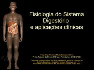
  • 1. Profa. Dra. Cristina Maria Henrique Pinto Profa. Adjunto do Depto. Ciências Fisiológicas-CCB-UFSC Como citar este documento: PINTO, Cristina Maria Henrique. Fisiologia do Sistema Digestório e aplicações clínicas. Disponível em: <http://www.cristina.prof.ufsc.br>. Acesso em: (coloque a data aqui) Fisiologia do Sistema Digestório e aplicações clínicas
  • 2. Esta apresentação foi utilizada em minhas aulas para a graduação em Medicina (6ª fase) até o ano de 2007. Para que este material não se perca, deixo aqui à disposição daqueles que eventualmente tenham interesse. Bons estudos!
  • 3. Bibliografia básica recomendada sobre Fisiologia Humana Livros-textos: “ Berne & Levy: Fisiologia” Koeppen & Stanton, 2009, 6ª Ed. (Ed. Elsevier) “ Tratado de Fisiologia Médica” Guyton & Hall, 2006, 11ª Ed. (Ed. Elsevier) “ Fisiologia” Aires, M. M. 2008, 3ª Ed. (Ed. Guanabara Koogan) “ Fisiologia” Costanzo, 2007, 3ª Ed. (Ed. Elsevier) “ Berne & Levy: Fundamentos de Fisiologia”, Levy et al , 2006, 4ª Ed. (Ed. Elsevier) “ Fundamentos de Fisiologia Médica” Johnson, 2003 (Ed. Guanabara Koogan) “ Fisiologia: texto e atlas” Silbernagl e Despopoulos, 2003 (Ed. Artmed)
  • 4. Profa. Dra. Cristina Maria Henrique Pinto CFS/CCB – 6ª fase - Medicina O Pâncreas como glândula exócrina
  • 5. Figure 3. The pancreas and adjacent anatomy. divisões: cabeça e processo uncinado, colo, corpo e cauda http://hopkins-gi.nts.jhu.edu/pages/latin/templates/index.cfm?pg=disease1&organ=4&disease=22&lang_id=1 PÂNCREAS localização: -órgão retroperitoneal. -anteriores a ele estão o estômago, alças do intestino delgado, colon e omento
  • 6. Duct system of the pancreas. Berne et al., 2004 Ducto pancreático principal formado a partir dos ductos intercalares, intralobares extralobares e ductos coletores principais
  • 7. Ducto pancreático: -desde a cauda até a cabeça do pâncreas -une-se ao ducto colédoco já próximo ao duodeno, formando a ampola hepatopancreática e a papila maior do duodeno ( ou ampola de Vater ). http://hopkins-gi.nts.jhu.edu/pages/latin/templates/index.cfm?pg=disease1&organ=4&disease=22&lang_id=1 É comum a existência de um ducto pancreático secundário , acessório ou dorsal (papila menor)
  • 8. Normally (90% of the time) pancreatic secretions from the entire pancreas and biliary secretions gain access to the duodenum by way of the ventral pancreatic or Wirsung’s duct because of the fusion of the dorsal pancreatic duct during embryological development.  There is only a remnant of the dorsal pancreatic or Santorini’s duct connecting to the duodenum. In 10% of individuals, fusion does not occur. This situation is called pancreas divisum. In pancreas divisum secretions from the dorsal and ventral ducts drain separately into the duodenum. Individuals with pancreas divisum may be at higher risk for pancreatitis (see Acute Pancreatitis for additional information).  Note that the distal common bile duct reaches the duodenum through the head of the pancreas. http://www.gastroslides.org/main/browse_deck.asp?tpc=2&mxpg=390&pg=1844#image DUCTO PANCREÁTICO PRINCIPAL E DUCTO PANCREÁTICO ACESSÓRIO (ocorrência em 90% da população)
  • 9. This figure demonstrates that the stomach, duodenum, liver, biliary system, gallbladder and pancreas are derived from closely related structures in early embryological development. The pancreas, liver, gallbladder and biliary system bud from the duodenum during early embryological development. The pancreas starts as two components, the ventral and dorsal pancreas.  In the process of development, the organs enlarge and the ventral pancreas together with the common bile duct rotates. Then, in most cases, the pancreatic duct from the dorsal pancreas fuses with the pancreatic duct from the ventral pancreas to form the main pancreatic duct. After fusion the pancreatic secretions from the entire pancreas and biliary secretions gain access to the duodenum by way of the ventral pancreatic duct. http://www.gastroslides.org/main/browse_deck.asp?tpc=6&mxpg=390&pg=2233#image EMBRIOLOGIA
  • 10. Normally (90% of the time) pancreatic secretions from the entire pancreas and biliary secretions gain access to the duodenum by way of the ventral pancreatic or Wirsung’s duct because of the fusion of the dorsal pancreatic duct during embryological development.  There is only a remnant of the dorsal pancreatic or Santorini’s duct connecting to the duodenum. In 10% of individuals, fusion does not occur. This situation is called pancreas divisum. In pancreas divisum secretions from the dorsal and ventral ducts drain separately into the duodenum. Individuals with pancreas divisum may be at higher risk for pancreatitis (see Acute Pancreatitis for additional information).  Note that the distal common bile duct reaches the duodenum through the head of the pancreas. http://www.gastroslides.org/main/browse_deck.asp?tpc=2&mxpg=390&pg=1844#image DUCTO PANCREÁTICO PRINCIPAL E DUCTO PANCREÁTICO ACESSÓRIO (ocorrência em 90% da população) PANCREAS DIVISUM (ocorrência em 3 a 7% da pop.)
  • 11. http://hopkins-gi.nts.jhu.edu/pages/latin/templates/index.cfm?pg=disease1&organ=4&disease=22&lang_id=1 -a mais comum anomalia congênita do pâncreas -ocorre em 3 a 7% da população -resulta em fusão incompleta ou inexistente dos brotos dorsal e ventral durante o desenvolvimento embrionário. As regiões da cauda, corpo, colo e pequena porção da cabeça têm suas secreções drenadas para o duodeno através da papila menor via ducto acessório . Obstruções na papila menor podem causar pancreatite aguda (drenagem insuficiente). O ducto pancreático principal que esvazia-se na papila duodenal maior, drena apenas uma pequena porção da secreção exócrina pancreática (porção ventral). CAUSA de pancreatite Pancreas divisum (ou pâncreas dividido )
  • 12. http://hopkins-gi.nts.jhu.edu/pages/latin/templates/index.cfm?pg=disease1&organ=4&disease=22&lang_id=1 Esfíncter de Oddi: células musculares lisas que envolvem a porção terminal dos ductos biliar comum e pancreático principal. É uma estrutura dinâmica que relaxa ou contrai, alterando as dimensões da papila maior duodenal
  • 13. http://hopkins-gi.nts.jhu.edu/pages/latin/templates/index.cfm?pg=disease1&organ=4&disease=22&lang_id=1 Figure 7. Gallstone obstruction. -uma das mais causas comuns de pancreatite (USA e países europeus) -baixa mortalidade -mecanismos causais ainda não totalmente entendidos -obstrução causaria refluxo biliar nas vias pancreáticas o que desencadearia uma complexa cascata de efeitos (p. ex. ativação da tripsina nos ácinos e/ou indução de resposta inflamatória). CAUSA de pancreatite aguda Colelitíase
  • 14. The pancreas is divided into an exocrine portion (acinar and duct tissue) and an endocrine portion (islets of Langerhans). The exocrine portion, comprising 85% of the mass of the pancreas, secretes digestive enzymes, water and NaHCO3 into the duodenum. The endocrine portion, comprising 2% or less of the pancreas, secretes hormones into the blood stream. http://www.gastroslides.org/main/browse_deck.asp?tpc=6&mxpg=390&pg=2240#image O PÂNCREAS: UMA GLÂNDULA ENDÓCRINA E EXÓCRINA
  • 15. ? VOLUME SECRETADO PELO PÂNCREAS NO INTESTINO DELGADO: 1,5 L/DIA
  • 16. secreção hidro-eletrolítica enzimas digestivas (proteases, amilase e lipases) Principais tipos celulares encontrados no pâncreas Ilhotas de Langerhans hormônios: insulina (cél. β ), glucagon (cél. α ), somatostatina (cél. δ ) e polipeptídeo pancreático (cél. θ )
  • 17. ilhota (células alfa: glucagon) ilhota (células beta: insulina) ácinos e ilhota de Langerhans http://www.udel.edu/Biology/Wags/histopage/histopage.htm HISTOLOGIA DO PÂNCREAS PORÇÃO ENDÓCRINA (hormônios: glucagon, insulina, somatostatina e polipeptídeo pancreático) PORÇÃO EXÓCRINA enzimas e secreção hidroeletrolítica http://www.gastroslides.org/main/browse_deck.asp?tpc=6&mxpg=390&pg=2241#image
  • 18. The relationships and major features of the units of the exocrine pancreas. The pancreatic acinar cells of the acinus have prominently stained zymogen granules in the apical area of the cell.  The connecting ductule does not contain zymogen granules. The blue cell in the cartoon depicts the centroacinar cell at the border between the acinus and ductule. The centroacinar cell functions similarly to the duct cell. The major secretory products of the acinus are digestive proenzymes and enzymes with lesser amounts of water and ions. The major secretory products of the duct are water and ions. http://www.gastroslides.org/main/browse_deck.asp?tpc=6&mxpg=390&pg=2243#image AS SECREÇÕES EXÓCRINAS PANCREÁTICAS: água e eletrólitos
  • 19. Neutralization of gastric acid delivered to the duodenum is necessary for optimal digestion and absorption of a meal. Several mechanisms that are not shown are involved in the neutralization process. First, the meal provides buffers from digestion of protein and triglycerides. That is, the peptides and fatty acid products act as pH buffers. Another neutralization process is absorption of hydrogen ion by the duodenal mucosa. Finally, the pancreas, biliary system and duodenal mucosa secrete bicarbonate into the duodenal lumen for neutralization. http://www.gastroslides.org/main/browse_deck.asp?tpc=6&mxpg=390&pg=2287#image Importância da secreção hidroeletrolítica pancreática, rica em HCO 3 - , Na + e água na digestão NaHCO 3 + HCl  NaCl + H 2 O + CO 2 (reabsorção)
  • 20. Secreção hidroeletrolítica pancreática: grande volume e rica em Na + e HCO 3 -
  • 21. Stimulation (i.e. a meal) there is an increase in the flow rate of pancreatic secretions. Furthermore, with increasing flow rates there is a dramatic change in the concentrations of chloride and bicarbonate. The increase in bicarbonate concentration results in a secretion that is alkaline. The bicarbonate ion comes from ductal epithelial cells in the pancreas. In contrast to acinar cells, the ducts secrete a large volume of fluid with a high concentration of bicarbonate. Because the volume of secretion from the acinar cells is thought to be small compared to ductal secretion, with increasing stimulation of the pancreas the concentration of ions approaches that of the ductal secretions. Of note, the alkaline secretion of the pancreas combined with alkaline secretions from the biliary system and the duodenal mucosa neutralize the acid secretion delivered to the duodenum from the stomach. This pH neutral environment is important for optimal digestive enzyme and intestinal mucosal function. http://www.gastroslides.org/main/browse_deck.asp?tpc=6&mxpg=390&pg=2287#image Composição da secreção hidroeletrolítica pancreática em razão da taxa de secreção
  • 22. Cl- secretion drives bicarbonate secretion in the pancreatic duct cell. Cl-  secretion occurs via secretin-stimulated cyclic AMP production. Cyclic AMP, in turn, activates the CFTR Cl-  secretory pathway. In the pancreatic duct cell, the Cl- may exchange with HCO3- resulting in net HCO3- secretion.  Of note, Cl-   delivered from the acinar cells also exchanges with HCO3- in the pancreatic duct. HCO3- may also enter the lumen through CFTR or by other mechanisms (see following slides). Na+ enters the duct lumen via the paracellular space to neutralize the electrical gradient created by the apical HCO3- secretion. Water follows via the paracellular space to maintain iso-osmolality. The combination of these processes determines the composition and volume of pancreatic duct secretions. ( canal regulador da condutância transmembrana da fibrose cística) http://www.gastroslides.org/main/browse_deck.asp?tpc=6&mxpg=390&pg=2332#image Mecanismos celulares de secreção de HCO 3 - , Na + e água pelas células dos ductos pancreáticos
  • 23. Mecanismos celulares de secreção de HCO 3 - , Na + e água pelas células dos ductos pancreáticos Delivery mechanisms to provide a source for HCO3- for the duct cell. There are two delivery systems. In one, membrane diffusible CO2 is catalytically converted to HCO3- and H+ by the action of carbonic anhydrase (CA) which hydrates CO2   thereby forming H2CO3 that then dissociates to HCO3- and H+. The duct cell is rich in CA. The HCO3- is then available for apical secretion. H+ is removed from the cell by a basolateral Na+/H+ antiport to maintain a constant intracellular pH. In the other delivery http://www.gastroslides.org/main/browse_deck.asp?tpc=6&mxpg=390&pg=2322#image
  • 24. The two major classes chloride channels.  CFTR is activated by cAMP dependent protein kinase.  A second family of channels is regulated by calcium.  Both classes of calcium channels may be present in some cells . http://www.gastroslides.org/main/browse_deck.asp?tpc=6&mxpg=390&pg=2332#image CFTR afeta outros canais de cloreto
  • 25. CFTR activation results in activation of conductive pathways for several other molecules as listed. The mechanisms involved in the cooperative effects on transport of these molecules are not established. canal regulador da condutância transmembrana da fibrose cística http://www.gastroslides.org/main/browse_deck.asp?tpc=6&mxpg=390&pg=2332#image
  • 26. Using immunocytochemical staining of CFTR and zymogens, this image shows that CFTR is located on the apical membrane of both pancreatic duct cells and acinar cells. http://www.gastroslides.org/main/browse_deck.asp?tpc=6&mxpg=390&pg=2322#image
  • 27. Chloride is secreted from the apical surface of the acinar cell by two distinct transport processes. In one, agents such as cholecystokinin or muscarinic agents (i.e. acetylcholine) cause an increase in intracellular calcium that, in turn, activates a Ca+2-dependent chloride channel. In the other, agents such as secretin and VIP cause an increase in cyclic AMP that, in turn, activates a cyclic AMP-dependent chloride channel. Of note, both Ca+2 and cyclic AMP activate a basolateral K+ channel. This activation of K+ efflux facilitates apical chloride secretion by make the intracellular environment more electronegative. The Na+ /K+ ATPase is important in facilitating chloride transport because it maintains the electronegative intracellular environment as there are two K+s transported in for three Na+s transported out of the cell. Finally, the Na+/K+/Cl -Cotransport on the basolateral membrane is essential to replenish chloride secreted at the apical surface. CFTR http://www.gastroslides.org/main/browse_deck.asp?tpc=6&mxpg=390&pg=2322#image Mecanismos celulares de secreção de Na + e água pelas células dos ácinos pancreáticos
  • 28. Importância do conhecimento dos mecanismos celulares de secreção de HCO 3 - , Na + e água pelas células centro-acinares, ductos e ácinos Patologias podem ocorrer quando, por diversos fatores, surgem alterações da expressão de canais e/ou transportadores celulares. Exemplo: deficiência da expressão de canais de cloreto na membrana luminal celular na fibrose cística Primary epithelia affected in Cystic Fibrosis http://cellscience.com/reviews2/Cystic_Fibrosis_alternative_Chloride_channel.html acarreta deficiência nutricional especialmente na criança Tissue affected Pancreas Intestine Sweat glands Airways Reproductive organs Pathology Maldigestion Malabsorption/meconium ileus salty sweat Dry airways/infection Sterility Epithelia affected Exocrine cells Crypt epithelium Sweat duct Bronchial epithelium Vas Deferens/various Potential Therapies Enzyme replacement None None UTP + amiloride aerosol/gene therapy None
  • 29. Normal trafficking of CFTR to the plasma membrane where it functions as a cAMP-dependent Cl- channel.  Mutations that lead to protein dysfunction have been classified into five categories: I-no synthesis; II-block in processing, III- block in regulation; IV – altered conductance; V – reduced synthesis. The right panel depicts the two major mechanisms of CFTR dysfunction in cystic fibrosis. The most common mutation that results in cystic fibrosis is delta508 mutation (Type II).  This leads to CFTR misfolding and degradation before it can exit the endoplasmic reticulum.  Type III and IV defects result in CFTR insertion into the membrane, but abnormal regulation.  Type I and V defects are less common causes of cystic fibrosis. Choo-Kang LR, Zeitlin PL. Curr Opin Pulm Med 2000;6:521-529 http://www.gastroslides.org/main/browse_deck.asp?tpc=6&mxpg=390&pg=2330#image
  • 30. The clinical manifestations of cystic fibrosis. Of note, the genetic  defects in chloride transport results in dysfunction in multiple systems including the pulmonary, hepatobiliary, pancreatic, intestinal and reproductive systems http://www.gastroslides.org/main/browse_deck.asp?tpc=6&mxpg=390&pg=2322#image
  • 31. The relationships and major features of the units of the exocrine pancreas. The pancreatic acinar cells of the acinus have prominently stained zymogen granules in the apical area of the cell.  The connecting ductule does not contain zymogen granules. The blue cell in the cartoon depicts the centroacinar cell at the border between the acinus and ductule. The centroacinar cell functions similarly to the duct cell. The major secretory products of the acinus are digestive proenzymes and enzymes with lesser amounts of water and ions. The major secretory products of the duct are water and ions. http://www.gastroslides.org/main/browse_deck.asp?tpc=6&mxpg=390&pg=2243#image AS SECREÇÕES EXÓCRINAS PANCREÁTICAS: ENZIMAS
  • 32. (from duodenal epithelial cells)  -Amylase (no activation needed) secreted by duodenal epithelium (Enterokinase) http://mcb.berkeley.edu/courses/mcb136/topic/Gastrointestinal AS SECREÇÕES EXÓCRINAS PANCREÁTICAS: enzimas lipase
  • 33. Relative amounts (by weight) of the different classes of pancreatic digestive enzymes. Proteases are the most abundant class of enzymes. http://www.gastroslides.org/main/browse_deck.asp?tpc=6&mxpg=390&pg=2273#image
  • 34. (from duodenal epithelial cells)  -Amylase (no activation needed) secreted by duodenal epithelium (Enterokinase) http://mcb.berkeley.edu/courses/mcb136/topic/Gastrointestinal AS SECREÇÕES EXÓCRINAS PANCREÁTICAS: enzimas lipase
  • 35. The non-enzymatic secretory products of the acinar cell. Procolipase when activated facilitates the action of lipase. GP-2 is a glycoprotein linked to the inner zymogen granule membrane and may have a role in protein sorting or membrane recycling.  Some GP-2 is released from the zymogen granule membrane and secreted.  GP-2 is enriched in stones and may have a role in their formation. Lithostathine may prevent stone formation in the pancreas. Pancreatitis-associated protein increases with pancreatitis although the function is not known. Pancreatic secretory trypsin inhibitor has an important role in preventing intrapancreatitic zymogen activation. The ions, especially Na+ and Cl- are important for transport of the secretory products from the acinar lumen into the pancreatic ductal system by pulling water into the luminal space by osmotic forces. Water and ion flow then “wash out” the luminal space http://www.gastroslides.org/main/browse_deck.asp?tpc=6&mxpg=390&pg=2273#image AS SECREÇÕES EXÓCRINAS PANCREÁTICAS: produtos não-enzimáticos
  • 36. Action of major pancreatic lipases. The cleavage of lipids by glycerol ester hydrolase (pancreatic lipase), cholesterol ester hydrolase, and phospholipase A2 is illustrated. P, Phosphate. Berne, 2004 hidrolase de éster de colesterol fosfolipase A 2 A secreção serosa pancreática digestão de lipídeos da dieta do glicerol hidrolase de éster triglicerídeo 2-monoglicerídeo ácido graxo ácido graxo éster do colesterol colesterol ácido graxo ácido graxo lisolecitina lecitina
  • 37.
  • 38. Mucosal Cell Intestinal Lumen TG LIPASE Bile Acids Micelle MG FA A secreção serosa pancreática: lipase e colipase Digestão de lipídeos no ID (meio aquoso) TG=triglyceride; MG=monoglyceride; FA=fatty acid.
  • 39. By demonstrating the relative activity of lipase action with addition of each to a mixture, these experimental results show that the activity of lipase on triglyceride hydrolysis is dependent on both bile salts and colipase. With addition of lipase alone (indicated by the green arrow) there is some triglyceride hydrolysis.  However, the addition of bile salts with colipase to a mixture of triglycerides and lipase results in the greatest activity. The importance of bile salts is that they anchor lipase and colipase together on the oil phase of trigylceride.  This “anchoring” results in the greatest activity. http://www.gastroslides.org/main/browse_deck.asp?tpc=6&mxpg=390&pg=2284#image
  • 40. Mucosal Cell Intestinal Lumen TG LIPASE Bile Acids Micelle MG FA A secreção serosa pancreática: lipases e colipase pancreatite aguda/crônica: deficiência/ausência da lipase pancreática   digestão e absorção da gordura da dieta  perda pelas fezes (esteatorréia) esteatorréia TG=triglyceride; MG=monoglyceride; FA=fatty acid.
  • 41. A digestão e absorção de lipídeos podem ser diminuídas por drogas que ligam-se à lipase e inibem sua ação TG=triglyceride; MG=monoglyceride; FA=fatty acid. Mucosal Cell Intestinal Lumen DROGA TG LIPASE LIPASE LIPASE Bile Acids Micelle MG FA A secreção serosa pancreática: lipases e colipase esteatorréia
  • 42. A secreção serosa pancreática: alfa-amilase digestão de amido/glicogênio Amilopectina (amido) da batata produtos da digestão pela α -amilase pancreática α -amilase pancreática maltose dextrina -limite ligação 1:6 ligação 1:4 maltotriose alfa-amilase Produtos da hidrólise do AMIDO pela alfa-amilase glicose
  • 43. Functions of the major brush border oligosaccharidases. The glucose, galactose, and fructose molecules released by enzymatic hydrolysis are then transported into the epithelial cell by specific transport proteins. The glucose-galactose transporter is also known as SGLT1 and the fructose transporter as GLUT5. G, Glucose; Ga, galactose; F, fructose. Berne et al ., 2004 (SGLT1) Amido glicogênio α -amilase pancreática A secreção serosa pancreática: alfa-amilase digestão de amido/glicogênio para posterior digestão e absorção pelo epitélio do ID produtos da digestão pela α -amilase pancreática α -amilase salivar
  • 44. A secreção serosa pancreática: pró-proteases digestão de proteínas/polipeptídeos ativadas no intestino delgado pela enteropeptidase (ou enteroquinase)
  • 45. proteases pancreáticas PROTEÍNAS Pepsina (gástrica) produtos da digestão pelas proteases pancreáticas A secreção serosa pancreática: pró-proteases digestão de proteínas/polipeptídeos para posterior digestão e absorção pelo epitélio do ID Amino acids Berne et al., 2004
  • 46. A secreção serosa pancreática: pró-proteases ativadas no intestino delgado pela enteropeptidase (ou enteroquinase) Activation pathways of proenzymes by trypsin. Once trypsin is activated, it is capable of activating many other digestive proenzymes. Como é feita a inativação da tripsina enquanto está sendo armazenada e secretada pelo pâncreas? Hirota et al, 2003 JOP http://www.joplink.net/prev/200303/01.html
  • 47. TRIPSINOGÊNIO + Peptídeo inibidor da tripsina http://www.rcsb.org/pdb/static.do?p=education_discussion/molecule_of_the_month/pdb46_2.html O tripsinogênio possui, além da tripsina, o TAP (peptídeo de ativação da tripsina) , uma porção da cadeia que é removida pela enteropeptidase somente no ID TRIPSINOGÊNIO Trypsin is built with an extra piece of protein chain, colored in green in the structure on the left . Actually, only two amino acids of this extra bit are seen in crystal structure, so you have to imagine the rest flopping around away from the protein. This longer form of trypsin, called trypsinogen, is inactive and cannot cut protein chains. Then, when it enters the intestine, the enzyme enteropeptidase makes one cut in the trypsin chain, clipping off the little tail. This allows the new end of the chain, colored here in purple , to tuck into the folded protein and stabilize the active form of the enzyme, as shown on the right. As extra insurance, the pancreas also makes a small protein, trypsin inhibitor (shown in red), that binds to any traces of active trypsin that might be present before it is secreted into the intestine. It binds to the active site of trypsin, blocking its action but not itself being cut into tiny pieces. A inativação da tripsina enquanto está sendo armazenada e secretada pelo pâncreas tripsinogênio armazenado
  • 48. Pancreatic proenzymes (also called zymogens) such as trypsinogen are converted to their active forms with cleavage and removal of an NH2 –terminal portion, the activation peptide. Removal of the activation peptide results in exposure of the catalytic site of the enzyme. In the duodenum, enterokinase activates trypsinogen while trypsin activates other zymogens.  In the case of trypsinogen, the activation peptide is known as TAP (trypsin activation peptide).  In acute pancreatitis, measurements of TAP generation can be used clinically as a predictor of severity. Mecanismo de ativação do tripsinogênio já no ID: hidrólise pela enteropeptidase intestinal http://www.gastroslides.org/main/browse_deck.asp?tpc=6&mxpg=390&pg=2280#image
  • 49. This graphic presents the classes of proteases in the pancreas and their sites of action. Exopeptidases cleave one amino acid at a time from the NH2-terminal or COOH-terminal of a protein. Examples are the carboxypeptidases that cleave one amino acid at a time from the COOH-terminal of a protein. Endopeptidases such as chymotrypsinogen and trypsin each cleave specific bonds irrespective of their location with respect to the NH2-terminal or COOH-terminal of the protein. http://www.gastroslides.org/main/browse_deck.asp?tpc=6&mxpg=390&pg=2280#image Ações das proteases pancreáticas
  • 50. Trypsin is built with an extra piece of protein chain, colored in green in the structure on the left . Actually, only two amino acids of this extra bit are seen in crystal structure, so you have to imagine the rest flopping around away from the protein. This longer form of trypsin, called trypsinogen, is inactive and cannot cut protein chains. Then, when it enters the intestine, the enzyme enteropeptidase makes one cut in the trypsin chain, clipping off the little tail. This allows the new end of the chain, colored here in purple , to tuck into the folded protein and stabilize the active form of the enzyme, as shown on the right. As extra insurance, the pancreas also makes a small protein, trypsin inhibitor ( shown in red ), that binds to any traces of active trypsin that might be present before it is secreted into the intestine. It binds to the active site of trypsin, blocking its action but not itself being cut into tiny pieces. TRIPSINOGÊNIO A inativação da tripsina enquanto está sendo armazenada e secretada pelo pâncreas TAP (peptídeo de ativação da tripsina) porção da cadeia a mais que o tripsinogênio possui, além da tripsina e que será removida no ID pela enteropeptidase tripsinogênio armazenado Pode ocorrer uma ativação “espontânea” do tripsinogênio ainda nos ácinos ou nos ductos pancreáticos. Portanto, deve haver algum “mecanismo de segurança” para evitar que isso aconteça... http://www.rcsb.org/pdb/static.do?p=education_discussion/molecule_of_the_month/pdb46_2.html
  • 51. Trypsin is built with an extra piece of protein chain, colored in green in the structure on the left . Actually, only two amino acids of this extra bit are seen in crystal structure, so you have to imagine the rest flopping around away from the protein. This longer form of trypsin, called trypsinogen, is inactive and cannot cut protein chains. Then, when it enters the intestine, the enzyme enteropeptidase makes one cut in the trypsin chain, clipping off the little tail. This allows the new end of the chain, colored here in purple , to tuck into the folded protein and stabilize the active form of the enzyme, as shown on the right. As extra insurance, the pancreas also makes a small protein, trypsin inhibitor ( shown in red ), that binds to any traces of active trypsin that might be present before it is secreted into the intestine. It binds to the active site of trypsin, blocking its action but not itself being cut into tiny pieces. TRIPSINOGÊNIO + PSTI TRIPSINOGÊNIO Peptídeo inibidor da tripsina (PSTI) A inativação da tripsina enquanto está sendo armazenada e secretada pelo pâncreas TAP (peptídeo de ativação da tripsina) porção da cadeia a mais que o tripsinogênio possui, além da tripsina e que será removida no ID pela enteropeptidase tripsinogênio armazenado tripsinogênio secretado http://www.rcsb.org/pdb/static.do?p=education_discussion/molecule_of_the_month/pdb46_2.html
  • 52. A atividade da tripsina é controlada principalmente pelo peptídeo inibidor da tripsina , PSTI (*) , sintetizado na mesma célula acinar e age como um potente inibidor natural da tripsina. Quando o tripsinogênio é ativado no pâncreas (“espontâneo”), o PSTI liga-se à tripsina e a inativa, prevenindo assim a autodigestão celular. O PSTI também bloqueia futuras ativações que eventualmente venham a ocorrer nos ductos pancreáticos. A inativação da tripsina enquanto está sendo armazenada e secretada pelo pâncreas Activation pathways of proenzymes and PAR-2 by trypsin. Once trypsin is activated, it is capable of activating many other digestive proenzymes. Trypsin also activates pancreatic and inflammatory cells via PAR-2. The trypsin activity in the pancreas is mainly controlled by PSTI. When trypsinogen is activated into trypsin in the pancreas, PSTI binds immediately to trypsin to prevent further activation of pancreatic enzymes. (PAR: protease activated receptors; PSTI: pancreatic secretory trypsin inhibitor). (*) PSTI (pancreatic secretory trypsin inhibitor) ou SPINK1 (serine protease inhibitor Kazal type 1). Hirota et al, 2003 JOP http://www.joplink.net/prev/200303/01.html Mutações no gene PSTI ou da Tripsina podem promover uma predisposição à pancreatite , por diminuir a inibição funcional da tripsina
  • 53. The major organelles involved in protein synthesis and segregation of the newly synthesized proteins into storage compartments. New proteins are synthesized in the rough endoplasmic reticulum of the pancreatic acinar cell. The newly synthesized proteins are then transported to the Golgi apparatus where post-translational modification and targeting occur. http://www.gastroslides.org/main/browse_deck.asp?tpc=6&mxpg=390&pg=2296#image
  • 54. The protective mechanisms present in the pancreas to prevent activation of zymogens in the pancreatic cell and thus prevent that damaging effects of zymogen activation. Zymogens that have been pathologically activated in an acinar cell.. http://www.gastroslides.org/main/browse_deck.asp?tpc=6&mxpg=390&pg=2349#image MECANISMOS DE PROTEÇÃO DAS CÉLULAS ACINARES CONTRA A ATIVAÇÃO INTRACELULAR DAS ENZIMAS
  • 55. Hereditary Pancreatitis: Proposed Mechanism In normal individuals there is presumed to be a continuous low-grade activation and degradation of trypsin in the pancreas. Typsin can be temporarily inactivated by binding of pancreatic secretory trypsin inhibitor (PSTI or SPINK). Both trypsinogen and PSTI are located in the zymogen granules; there is sufficient PSTI to inhibit about 10% of pancreatic trypsin. Thus, as small amounts of active trypsin are formed, PSTI binds.  Proteases, such as trypsin and mesotrypsin, can degrade trypsin.  In the most common form of Hereditary Pancreatitis (HP) the mutation may render trypsin resistant to proteolytic degradation. Thus, trypsin that escapes inhibition by PSTI may remain active and activate the other pancreatic zymogens.  Direct evidence for this mechanism is not available . http://www.gastroslides.org/main/indexcont.asp?tpc=8&mxpg=314&idxpg=73
  • 56. http://www.gastroslides.org/main/indexcont.asp?tpc=8&mxpg=314&idxpg=73 O PSTI ( peptídeo inibidor da tripsina ) LIGA-SE À TRIPSINA E A INATIVA
  • 57. The illustration in this slide shows the effect of a substitution of one amino acid (i.e. histidine for arginine) on the interaction of trypsin with pancreatic trypsin inhibitor and the function of trypsin. The illustrations indicate the proposed mechanism of hereditary pancreatitis as well. In the normal state of folding, active trypsin in the pancreas binds to the trypsin inhibitor at its active site. This interaction leads to degradation of the active trypsin. In contrast, the mutated form of trypsin has altered structure so that the trypsin inhibitor will not interact with its active site and the active trypsin will not degrade. Thus, there is an increase in active trypsin in the pancreas with the potential for causing pancreatitis. http://www.gastroslides.org/main/indexcont.asp?tpc=8&mxpg=314&idxpg=73 FORMA MUTANTE DE TRIPSINA TEM SUA ESTRUTURA ALTERADA, O PSTI ( peptídeo inibidor da tripsina ) NÃO INTERAGE COM SEU SÍTIO ATIVO E A TRIPSINA NÃO É DEGRADADA.
  • 58. http://www.pancreas.org/assets/hp_presentation/index.htm HP: pancreatite hereditária Whitcomb, 2000
  • 59. ALCOOLISMO: CAUSA IMPORTANTE DE PANCREATITE CRÔNICA http://www.gastroslides.org/main/browse_deck.asp?tpc=6&mxpg=390&pg=2357#image
  • 61. Mecanismos que originam a dor na pancreatite aguda The proteinase-activated receptors (PARs) are a family of G-protein coupled receptors that, as their name suggests, are activated in a unique manner by serine proteases such as trypsin and thrombin. Recently PAR-2 has been implicated in the pathogenesis of pain. This may be particularly important in the pancreas where inflammation results in neuronal exposure to activated trypsin (from acinar cells) and tryptase (from mast cells) . Activation of PAR-2 on the peripheral processes of primary spinal afferents may have two effects. First, it may result in release of the neuropeptides CGRP and SP locally further amplifying inflammation and mast cell degranulation.  Secondly, excitation of the neurons also results in central release of neurotransmitters, leading to activation of second order neurons (as indicated by expression of the immediate early gene, c-Fos) in the dorsal horn further along in the nocioceptive pathway.  This pathway may contribute to pain experienced in both acute and chronic pancreatitis. http://www.gastroslides.org/main/browse_deck.asp?tpc=6&mxpg=390&pg=2357#image
  • 62. Exocrine pancreatic secretions are regulated by both endocrine and neurocrine pathways.  In addition, the endocrine and neurocrine mediators regulating secretion differ between the acinus and the duct.  For the acinus, cholecystokinin (CCK), acetylcholine (Ach), gastrin releasing peptide (GRP), vasoactive intestinal polypeptide (VIP) and substance P regulate secretion. In humans, CCK receptors have not been found on acinar cells. However, CCK may stimulate acinar cell secretion indirectly by exciting neural pathways. For the duct, secretin and acetylcholine are the major regulators of secretion.  Serotonin may act through 5-HT receptors to regulate acinar and duct cell secretion, but the importance and mechanism of this pathway in humans has not been defined. REGULAÇÃO DA SECREÇÃO EXÓCRINA PANCREÁTICA
  • 63. PADRÃO DA SECREÇÃO DE ENZIMAS DIGESTIVAS PANCREÁTICAS This graphic shows the output of the digestive enzyme, trypsin, into the duodenum both during the fasting state and after a meal (dog). During fasting there are small periodic increases in trypsin output. These increases correlate with activation of the motility of the intestine during the migrating motor complex (MMC). With a meal, there is a much larger increase in trypsin output due to the inputs of several meal stimuli that mediate secretion through both neural and humoral mechanisms. http://www.gastroslides.org/main/browse_deck.asp?tpc=6&mxpg=390&pg=2256#image
  • 64. Neurohormonal stimulatory and inhibitory mechanisms of pancreatic secretion during cephalic, gastric, and intestinal phases involving long vagovagal and short enteropancreatic reflexes activated by CCK acting on sensory receptors on afferent nerves. Various hormonal and humoral substances stimulate or inhibit the pancreatic secretion acting either through the CNS or directly on the exocrine pancreas. Konturek et al., 2003a
  • 65. REGULAÇÃO DA SECREÇÃO EXÓCRINA PANCREÁTICA estimulação neural pancreática A presença de gordura e de ácido no duodeno estimula o SNE e as terminações aferentes sensoriais que, por sua vez, estimulam (via Vago) a atividade Parassimpática sobre a secreção pancreática
  • 66. Vagal afferent signals from the pancreas are processed in the nodose ganglion and the dorsal vagal complex in the CNS. Afferent signals from the pancreas as well as other afferent signals from the cephalic and gastric phases of a meal are processed in the dorsal vagal complex and efferent signals are directed to the pancreas by the vagal nerves. The vagal efferents synapse with postganglionic neurons in the pancreatic ganglia . These postganglionic neurons then mediate responses in both the islets of Langerhans and pancreatic exocrine tissue through the release of neurotransmitters such as acetylcholine (Ach). http://www.gastroslides.org/main/browse_deck.asp?tpc=2&mxpg=390&pg=1842#image REGULAÇÃO DA SECREÇÃO EXÓCRINA PANCREÁTICA inervação pancreática
  • 67. REGULAÇÃO DA SECREÇÃO EXÓCRINA PANCREÁTICA estimulação hormonal pancreática A presença de gordura e de ácido no duodeno estimula a secreção endócrina intestinal das células endócrinas “S” e “I”. As céls. “S” secretam a SECRETINA que estimula a secreção hidro-eletrolítica dos ductos pancreáticos
  • 68. This image shows the stimulants for release of CCK from I cells and secretin release from S cells. Of note, the I cells and S cells “taste” the specific stimulants in the lumen of the gut to activate release of the hormones. The released CCK can mediate pancreatic secretion by either activating vagal sensory afferents that results in stimulation of pancreatic acinar cell secretion though a vago-vagal reflex involving the CNS or through the circulation (i.e. hormonal action). Secretin also acts through neural (not shown) and hormonal pathways on the pancreas stimulating secretion from both ductal cells and acinar cells. http://www.gastroslides.org/main/browse_deck.asp?tpc=2&mxpg=390&pg=1842#image A REGULAÇÃO DA SECREÇÃO DOS DUCTOS PELA SECRETINA
  • 69. REGULAÇÃO DA SECREÇÃO EXÓCRINA PANCREÁTICA estimulação hormonal pancreática A presença de gordura e de ácido no duodeno estimula a secreção endócrina intestinal das células endócrinas “S” e “I”. As céls. “I” secretam a CCK que estimula a secreção serosa dos ácinos pancreáticos
  • 70. This image shows the stimulants for release of CCK from I cells and secretin release from S cells. Of note, the I cells and S cells “taste” the specific stimulants in the lumen of the gut to activate release of the hormones. The released CCK can mediate pancreatic secretion by either activating vagal sensory afferents that results in stimulation of pancreatic acinar cell secretion though a vago-vagal reflex involving the CNS or through the circulation (i.e. hormonal action). Secretin also acts through neural (not shown) and hormonal pathways on the pancreas stimulating secretion from both ductal cells and acinar cells. http://www.gastroslides.org/main/browse_deck.asp?tpc=2&mxpg=390&pg=1842#image A REGULAÇÃO DA SECREÇÃO DOS DUCTOS PELA CCK
  • 71. The image is an electron photomicrograph of a CCK-secreting I cell. The I cell is one of the enterochromaffin cells. The apical surface of the I cell faces the lumen of the intestine where it “tastes” specific luminal nutrients (i.e. lipid). When it senses the specific nutrients, the I cell is activated and releases CCK from its basal surface. CCK can either act as a hormone to circulate to the pancreas and stimulate a secretory response or interact with sensory afferents containing CCK A (also known as CCK1) receptors. The image demonstrates the interaction of CCK with sensory receptors that are part of the enteric nervous system (ENS) of the gut. These neural receptors are located in proximity to the I cells. Vagal nerves to the CNS carry the afferent sensory signals. The afferent information going to the dorsal motor complex is processed and leads to stimulation of responses in the pancreas via vagal efferents. http://www.gastroslides.org/main/browse_deck.asp?tpc=6&mxpg=390&pg=2263#image A CCK atuaria também de maneira parácrina, estimulando as terminações sensitivas do PS (e do SNE)
  • 72. This image shows the several pathways mediating meal-stimulated pancreatic secretion that involve CCK. First, meal nutrients such as fatty acids, amino acids and peptides delivered from the duodenum stimulate the release of CCK from the CCK-containing I cell to the area around the basolateral surface of the I cell. The CCK released can activate vagal afferent neurons that carry the signal to the dorsal vagal complex where the sensory information is integrated and vagal efferents are activated. Vagal efferents synapse with neurons in the pancreatic ganglia. In turn, via the neurotransmitters, acetylcholine (Ach), gastrin releasing peptide (GRP; also known as bombesin) and vasoactive intestinal polypeptide (VIP), effector neurons in the pancreatic ganglia activate secretion by pancreatic parenchymal cells. In addition to activating the neural pathway, CCK released by the I cell enters the general circulation and may act as a hormone on the pancreatic acinar cells to cause secretion. Since CCK receptors have not been demonstrated on human acinar cells, the importance of direct hormonal stimulation is questionable. http://www.gastroslides.org/main/browse_deck.asp?tpc=6&mxpg=390&pg=2263#image
  • 73. The graphic in this image depicts the neurotransmission systems and neurotransmitters involved in regulating acinar and ductal secretion. The blue cell is called a centroacinar cell. These cells have characteristics more similar to ductal cells although their exact function is not known. Vagal efferents activate neurons in the intrapancreatic ganglia. Neurons from the ganglia then have processes that are in proximity to the parenchymal cells. These processes release the neurotransmitters, acetylcholine (Ach), gastrin releasing peptide (GRP) and vasoactive intestinal polypeptide (VIP) when their neurons are activated. Ach causes secretion from both acinar cells and ducts while all three neurotransmitters can cause secretion from acinar cells. http://www.gastroslides.org/main/browse_deck.asp?tpc=2&mxpg=390&pg=1842#image
  • 74. http://www.gastroslides.org/main/browse_deck.asp?tpc=2&mxpg=390&pg=1842#image Pancreatic acinar cell agonists that stimulate digestive enzyme secretion act through two separate pathways. In one pathway, agonists such as gastrin-releasing peptide, cholecytokinin and acetylcholine mediate secretion through increases in cellular calcium. In the other pathway, agonists such as vasoactive intestinal polypeptide and secretin mediate secretion through increases in cyclic AMP. Of note, a simultaneous increase in both calcium and cyclic AMP following stimulation with a combination of agonists results in a synergistic effect on secretion. That is, the observed response is greater than would be expected from the additive responses of the individual agonists acting alone. The double arrows indicate that the cellular mechanisms that cause secretion are largely unknown. http://www.gastroslides.org/main/browse_deck.asp?tpc=2&mxpg=390&pg=1842#image
  • 75. toxinas de escorpiões inseticidas organofosforados Exposição a inseticidas organofosforados (inibidores da acetilcolinesterase) ou toxinas específicas de escorpiões causam um aumento nos níveis de liberação de acetilcolina nas terminações nervosas, podendo causar pancreatite. A picada do escorpião Tityus serrulatus também tem sido causa de pancreatite. (G. Robles-Diaz, Mexico) . RELEVÂNCIA CLÍNICA DE TAIS CONHECIMENTOS http://www.gastroslides.org/main/browse_deck.asp?tpc=6&mxpg=390&pg=2317#image
  • 76. Optimal digestion and absorption occur near neutral pH. Sensors in the duodenal   lumen activate pathways leading to bicarbonate secretion. The result is neutralization of acidic contents from the stomach delivered to the duodenum. The major sensor in the duodenum that mediates pancreatic bicarbonate secretion is the secretin or S cell. The apical microvilli of the S cell face the luminal contents. The S cells respond to a low pH in the duodenal lumen by releasing secretin from the basolateral region of the cell. Secretin, in turn, enters the circulation and acts as a hormone to stimulate sodium bicarbonate and water secretion from the pancreatic ducts as well as the biliary system. The threshold for secretin release and water and bicarbonate secretion is pH 4.5. Below this pH pancreatic water and bicarbonate secretion are related to the total amount of titratable acid delivered to the duodenum.  In addition to acid, other luminal factors including lipids may contribute to secretin release. http://www.gastroslides.org/main/browse_deck.asp?tpc=2&mxpg=390&pg=1842#image
  • 77. Profa. Dra. Cristina Maria Henrique Pinto Profa. Adjunto do Depto. Ciências Fisiológicas-CCB-UFSC Como citar este documento: PINTO, Cristina Maria Henrique. Fisiologia do Sistema Digestório e aplicações clínicas. Disponível em: <http://www.cristina.prof.ufsc.br>. Acesso em: (coloque a data aqui) Fisiologia do Sistema Digestório e aplicações clínicas V eja mais aqui

Editor's Notes

  1. Orlistat prevents fat digestion and absorption by binding to gastrointestinal lipases Orlistat is a chemically synthesized hydrogenated derivative of lipstatin (a natural product of Streptomyces toxyricini) . Orlistat binds to gastric, pancreatic, and carboxylester lipases in the gut lumen and blocks the digestion of dietary fat by preventing lipase from interacting with its lipid target [1]. Inhibition of fat digestion decreases mixed micelle formation and absorption of long-chain fatty acids, cholesterol, and certain fat-soluble vitamins. Orlistat does not affect the action of systemic lipases or lipases located in other organ systems because it is minimally (&lt;1%) absorbed from the gastrointestinal tract. Drent ML, van der Veen EA. Lipase inhibition: A novel concept in the treatment of obesity. Int J Obes Relat Metab Disord 1993;17:241-244.
  2. Orlistat prevents fat digestion and absorption by binding to gastrointestinal lipases Orlistat is a chemically synthesized hydrogenated derivative of lipstatin (a natural product of Streptomyces toxyricini) . Orlistat binds to gastric, pancreatic, and carboxylester lipases in the gut lumen and blocks the digestion of dietary fat by preventing lipase from interacting with its lipid target [1]. Inhibition of fat digestion decreases mixed micelle formation and absorption of long-chain fatty acids, cholesterol, and certain fat-soluble vitamins. Orlistat does not affect the action of systemic lipases or lipases located in other organ systems because it is minimally (&lt;1%) absorbed from the gastrointestinal tract. Drent ML, van der Veen EA. Lipase inhibition: A novel concept in the treatment of obesity. Int J Obes Relat Metab Disord 1993;17:241-244.
  3. Orlistat prevents fat digestion and absorption by binding to gastrointestinal lipases Orlistat is a chemically synthesized hydrogenated derivative of lipstatin (a natural product of Streptomyces toxyricini) . Orlistat binds to gastric, pancreatic, and carboxylester lipases in the gut lumen and blocks the digestion of dietary fat by preventing lipase from interacting with its lipid target [1]. Inhibition of fat digestion decreases mixed micelle formation and absorption of long-chain fatty acids, cholesterol, and certain fat-soluble vitamins. Orlistat does not affect the action of systemic lipases or lipases located in other organ systems because it is minimally (&lt;1%) absorbed from the gastrointestinal tract. Drent ML, van der Veen EA. Lipase inhibition: A novel concept in the treatment of obesity. Int J Obes Relat Metab Disord 1993;17:241-244.