What is the
cardiovascular
system?
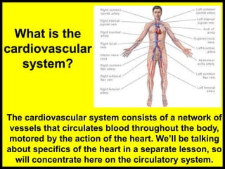
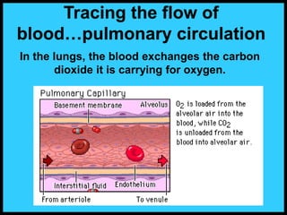
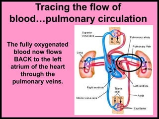
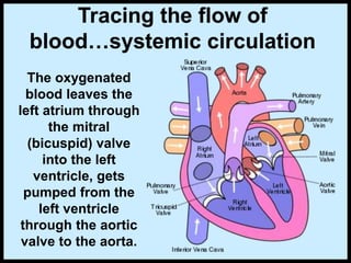
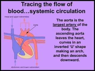
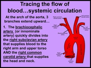
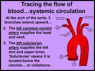
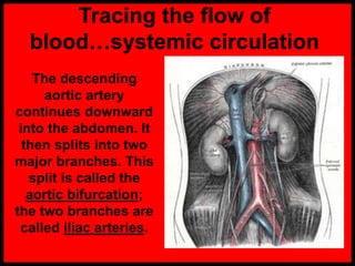
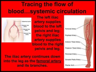
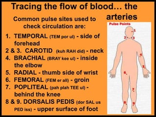
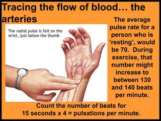
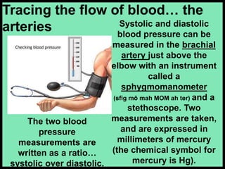
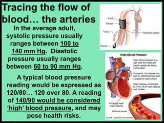
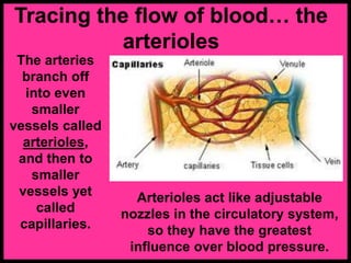
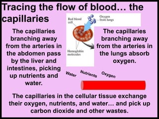
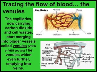
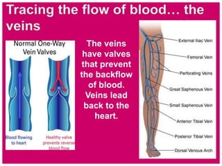
The cardiovascular system consists of a network of
vessels that circulates blood throughout the body,
motored by the action of the heart. We’ll be talking
about specifics of the heart in a separate lesson, so
will concentrate here on the circulatory system.
Tracing the flow of
blood…pulmonary circulation
The inferior vena cava is
the largest vein of the body.
It carries de-oxygenated
blood back from the lower
part of the body to the right
atrium of the heart. This
blood is carrying carbon
dioxide.
The superior vena cava is above the heart and
carries de-oxygenated blood from the head and
arms to the right atrium of the heart.
Tracing the flow of
blood…pulmonary circulation
From the right
atrium, the blood
flows through the
tricuspid valve to
the right ventricle
and then onto the
lungs through the
pulmonary valve
and pulmonary
artery.
In the lungs, the blood exchanges the carbon
dioxide it is carrying for oxygen.
Tracing the flow of
blood…pulmonary circulation
Tracing the flow of
blood…pulmonary circulation
The fully oxygenated
blood now flows
BACK to the left
atrium of the heart
through the
pulmonary veins.
Tracing the flow of
blood…systemic circulation
The oxygenated
blood leaves the
left atrium through
the mitral
(bicuspid) valve
into the left
ventricle, gets
pumped from the
left ventricle
through the aortic
valve to the aorta.
Tracing the flow of
blood…systemic circulation
The aorta is the
largest artery of the
body. The
ascending aorta
leaves the heart,
curves in an
inverted ‘U’ shape
making an arch,
and then descends
downward.
Tracing the flow of
blood…systemic circulation
At the arch of the aorta, 3
branches extend upward…
1. The brachiocephalic
artery (or innominate
artery) quickly divides into
the right subclavian artery
that supplies blood to the
right arm and upper torso
AND the right common
carotid artery that supplies
the head and neck.
Tracing the flow of
blood…systemic circulation
At the arch of the aorta, 3
branches extend upward…
2. The left common carotid
artery supplies the head
and neck.
3. The left subclavian
artery supplies the left
arm and upper torso.
‘Subclavian’ means it is
located below the
clavicle… or collarbone.
Tracing the flow of blood…systemic
circulation
The descending aortic
artery leads downward
through the diaphragm
and chest…and into the
abdomen. About 1/5 to 1/3
of the blood passes
through the renal artery
into the kidney. The
kidney is a filter, and takes
some water and waste
products out of the blood.
The kidneys excrete the waste products and
water out of the body as urine.
Tracing the flow of
blood…systemic circulation
The descending
aortic artery
continues downward
into the abdomen. It
then splits into two
major branches. This
split is called the
aortic bifurcation;
the two branches are
called iliac arteries.
Tracing the flow of
blood…systemic circulation
The left iliac
artery supplies
blood to the left
pelvis and leg;
the right iliac
artery supplies
blood to the right
pelvis and leg.
The iliac artery continues down
into the leg as the femoral artery
and its branches.
Tracing the flow of
blood… the arteries
Arteries are elastic tubes that carry
blood in pulsating waves. The blood
exerts pressure against the walls of
the arteries as it passes through. The
peak pressure occurs during the
heart’s contraction, and is called
systolic pressure. The minimum
pressure occurs between
contractions when the heart expands
and refills, and is called diastolic
pressure. This pressure variation
within the artery produces a pulse.
All arteries have a pulse.
Tracing the flow of blood… the
arteries
Common pulse sites used to
check circulation are:
1. TEMPORAL (TEM por ul) - side of
forehead
2 & 3. CAROTID (kuh RAH did) - neck
4. BRACHIAL (BRAY kee ul) - inside
the elbow
5. RADIAL - thumb side of wrist
6. FEMORAL (FEM er all) - groin
7. POPLITEAL (pah plah TEE ul) -
behind the knee
8 & 9. DORSALIS PEDIS (dor SAL us
PED iss) - upper surface of foot
Tracing the flow of blood… the
arteries
Count the number of beats for
15 seconds x 4 = pulsations per minute.
The average
pulse rate for a
person who is
‘resting’, would
be 70. During
exercise, that
number might
increase to
between 130
and 140 beats
per minute.
Systolic and diastolic
blood pressure can be
measured in the brachial
artery just above the
elbow with an instrument
called a
sphygmomanometer
(sfig mō mah MOM ah ter) and a
stethoscope. Two
measurements are taken,
and are expressed in
millimeters of mercury
(the chemical symbol for
mercury is Hg).
Tracing the flow of blood… the
arteries
The two blood
pressure
measurements are
written as a ratio…
systolic over diastolic.
In the average adult,
systolic pressure usually
ranges between 100 to
140 mm Hg. Diastolic
pressure usually ranges
between 60 to 90 mm Hg.
Tracing the flow of
blood… the arteries
A typical blood pressure
reading would be expressed as
120/80… 120 over 80. A reading
of 140/90 would be considered
‘high’ blood pressure, and may
pose health risks.
Arterioles act like adjustable
nozzles in the circulatory system,
so they have the greatest
influence over blood pressure.
Tracing the flow of blood… the
arterioles
The arteries
branch off
into even
smaller
vessels called
arterioles,
and then to
smaller
vessels yet
called
capillaries.
Tracing the flow of blood… the
capillaries
The capillaries are the
smallest of the blood
vessels, and the walls
are so thin that
molecules can pass
through them. They
branch out from the
arterioles, passing
next to the organs,
intestines, and
through all the
cellular tissue.
In the cellular tissue, the
capillaries provide the
means of exchange,
through the process of
absorption.
Tracing the flow of blood… the
capillaries
The capillaries
branching away
from the arteries in
the abdomen pass
by the liver and
intestines, picking
up nutrients and
water.
The capillaries in the cellular tissue exchange
their oxygen, nutrients, and water… and pick up
carbon dioxide and other wastes.
The capillaries
branching away
from the arteries in
the lungs absorb
oxygen.
Tracing the flow of blood… the
venules
The capillaries,
now carrying
carbon dioxide
and cell wastes,
start merging
into bigger vessels
called venules (VEEN
or VEN yoo als) The
venules widen
even further,
emptying into
veins.
The veins
have valves
that prevent
the backflow
of blood.
Veins lead
back to the
heart.
Tracing the flow of blood… the
veins
Veins are the vessels
that are used to
remove blood from
the body for analysis.
Tracing the flow of blood… the
veins
This procedure is called a
venipuncture (VEEN ah punk chur)
and the medical personnel
that specializes in this
procedure is called a
phlebotomist (flah BOTT ah mist).
The veins carry the
blood BACK toward the
heart. The blood still
carries a small amount
of oxygen along with
cellular waste, but has
fairly low pressure
compared to blood in
arteries. It finally travels
through the superior
and inferior vena cava,
and back into the right
atrium of the heart.
Tracing the flow of blood… the
veins
Circulation is complete, and starts over again.
170-Anatomy-Circulatory-System.ppt

170-Anatomy-Circulatory-System.ppt

  • 2.
    What is the cardiovascular system? Thecardiovascular system consists of a network of vessels that circulates blood throughout the body, motored by the action of the heart. We’ll be talking about specifics of the heart in a separate lesson, so will concentrate here on the circulatory system.
  • 3.
    Tracing the flowof blood…pulmonary circulation The inferior vena cava is the largest vein of the body. It carries de-oxygenated blood back from the lower part of the body to the right atrium of the heart. This blood is carrying carbon dioxide. The superior vena cava is above the heart and carries de-oxygenated blood from the head and arms to the right atrium of the heart.
  • 4.
    Tracing the flowof blood…pulmonary circulation From the right atrium, the blood flows through the tricuspid valve to the right ventricle and then onto the lungs through the pulmonary valve and pulmonary artery.
  • 5.
    In the lungs,the blood exchanges the carbon dioxide it is carrying for oxygen. Tracing the flow of blood…pulmonary circulation
  • 6.
    Tracing the flowof blood…pulmonary circulation The fully oxygenated blood now flows BACK to the left atrium of the heart through the pulmonary veins.
  • 7.
    Tracing the flowof blood…systemic circulation The oxygenated blood leaves the left atrium through the mitral (bicuspid) valve into the left ventricle, gets pumped from the left ventricle through the aortic valve to the aorta.
  • 8.
    Tracing the flowof blood…systemic circulation The aorta is the largest artery of the body. The ascending aorta leaves the heart, curves in an inverted ‘U’ shape making an arch, and then descends downward.
  • 9.
    Tracing the flowof blood…systemic circulation At the arch of the aorta, 3 branches extend upward… 1. The brachiocephalic artery (or innominate artery) quickly divides into the right subclavian artery that supplies blood to the right arm and upper torso AND the right common carotid artery that supplies the head and neck.
  • 10.
    Tracing the flowof blood…systemic circulation At the arch of the aorta, 3 branches extend upward… 2. The left common carotid artery supplies the head and neck. 3. The left subclavian artery supplies the left arm and upper torso. ‘Subclavian’ means it is located below the clavicle… or collarbone.
  • 11.
    Tracing the flowof blood…systemic circulation The descending aortic artery leads downward through the diaphragm and chest…and into the abdomen. About 1/5 to 1/3 of the blood passes through the renal artery into the kidney. The kidney is a filter, and takes some water and waste products out of the blood. The kidneys excrete the waste products and water out of the body as urine.
  • 12.
    Tracing the flowof blood…systemic circulation The descending aortic artery continues downward into the abdomen. It then splits into two major branches. This split is called the aortic bifurcation; the two branches are called iliac arteries.
  • 13.
    Tracing the flowof blood…systemic circulation The left iliac artery supplies blood to the left pelvis and leg; the right iliac artery supplies blood to the right pelvis and leg. The iliac artery continues down into the leg as the femoral artery and its branches.
  • 14.
    Tracing the flowof blood… the arteries Arteries are elastic tubes that carry blood in pulsating waves. The blood exerts pressure against the walls of the arteries as it passes through. The peak pressure occurs during the heart’s contraction, and is called systolic pressure. The minimum pressure occurs between contractions when the heart expands and refills, and is called diastolic pressure. This pressure variation within the artery produces a pulse. All arteries have a pulse.
  • 15.
    Tracing the flowof blood… the arteries Common pulse sites used to check circulation are: 1. TEMPORAL (TEM por ul) - side of forehead 2 & 3. CAROTID (kuh RAH did) - neck 4. BRACHIAL (BRAY kee ul) - inside the elbow 5. RADIAL - thumb side of wrist 6. FEMORAL (FEM er all) - groin 7. POPLITEAL (pah plah TEE ul) - behind the knee 8 & 9. DORSALIS PEDIS (dor SAL us PED iss) - upper surface of foot
  • 16.
    Tracing the flowof blood… the arteries Count the number of beats for 15 seconds x 4 = pulsations per minute. The average pulse rate for a person who is ‘resting’, would be 70. During exercise, that number might increase to between 130 and 140 beats per minute.
  • 17.
    Systolic and diastolic bloodpressure can be measured in the brachial artery just above the elbow with an instrument called a sphygmomanometer (sfig mō mah MOM ah ter) and a stethoscope. Two measurements are taken, and are expressed in millimeters of mercury (the chemical symbol for mercury is Hg). Tracing the flow of blood… the arteries The two blood pressure measurements are written as a ratio… systolic over diastolic.
  • 18.
    In the averageadult, systolic pressure usually ranges between 100 to 140 mm Hg. Diastolic pressure usually ranges between 60 to 90 mm Hg. Tracing the flow of blood… the arteries A typical blood pressure reading would be expressed as 120/80… 120 over 80. A reading of 140/90 would be considered ‘high’ blood pressure, and may pose health risks.
  • 19.
    Arterioles act likeadjustable nozzles in the circulatory system, so they have the greatest influence over blood pressure. Tracing the flow of blood… the arterioles The arteries branch off into even smaller vessels called arterioles, and then to smaller vessels yet called capillaries.
  • 20.
    Tracing the flowof blood… the capillaries The capillaries are the smallest of the blood vessels, and the walls are so thin that molecules can pass through them. They branch out from the arterioles, passing next to the organs, intestines, and through all the cellular tissue. In the cellular tissue, the capillaries provide the means of exchange, through the process of absorption.
  • 21.
    Tracing the flowof blood… the capillaries The capillaries branching away from the arteries in the abdomen pass by the liver and intestines, picking up nutrients and water. The capillaries in the cellular tissue exchange their oxygen, nutrients, and water… and pick up carbon dioxide and other wastes. The capillaries branching away from the arteries in the lungs absorb oxygen.
  • 22.
    Tracing the flowof blood… the venules The capillaries, now carrying carbon dioxide and cell wastes, start merging into bigger vessels called venules (VEEN or VEN yoo als) The venules widen even further, emptying into veins.
  • 23.
    The veins have valves thatprevent the backflow of blood. Veins lead back to the heart. Tracing the flow of blood… the veins
  • 24.
    Veins are thevessels that are used to remove blood from the body for analysis. Tracing the flow of blood… the veins This procedure is called a venipuncture (VEEN ah punk chur) and the medical personnel that specializes in this procedure is called a phlebotomist (flah BOTT ah mist).
  • 25.
    The veins carrythe blood BACK toward the heart. The blood still carries a small amount of oxygen along with cellular waste, but has fairly low pressure compared to blood in arteries. It finally travels through the superior and inferior vena cava, and back into the right atrium of the heart. Tracing the flow of blood… the veins Circulation is complete, and starts over again.