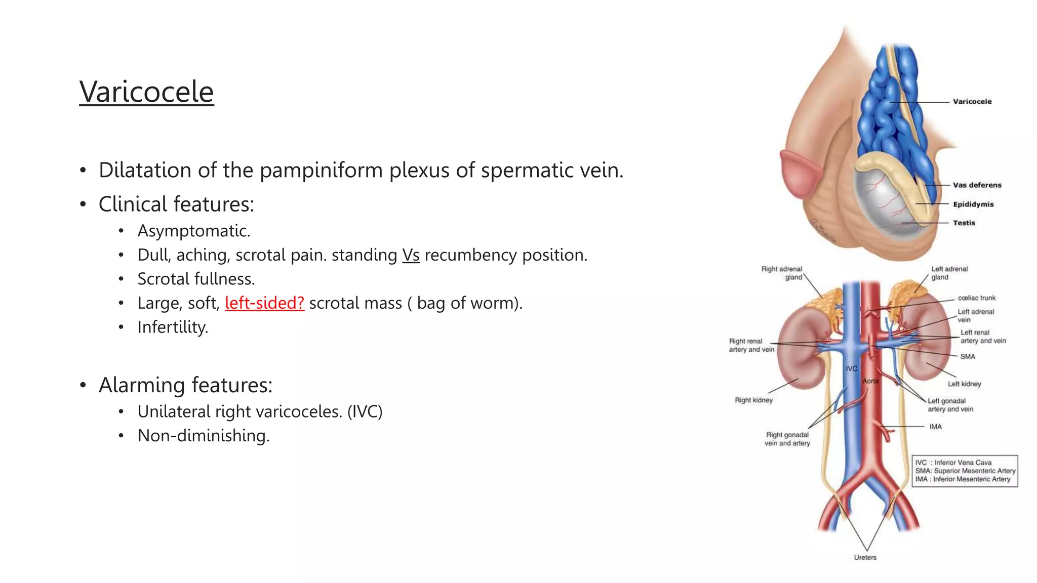
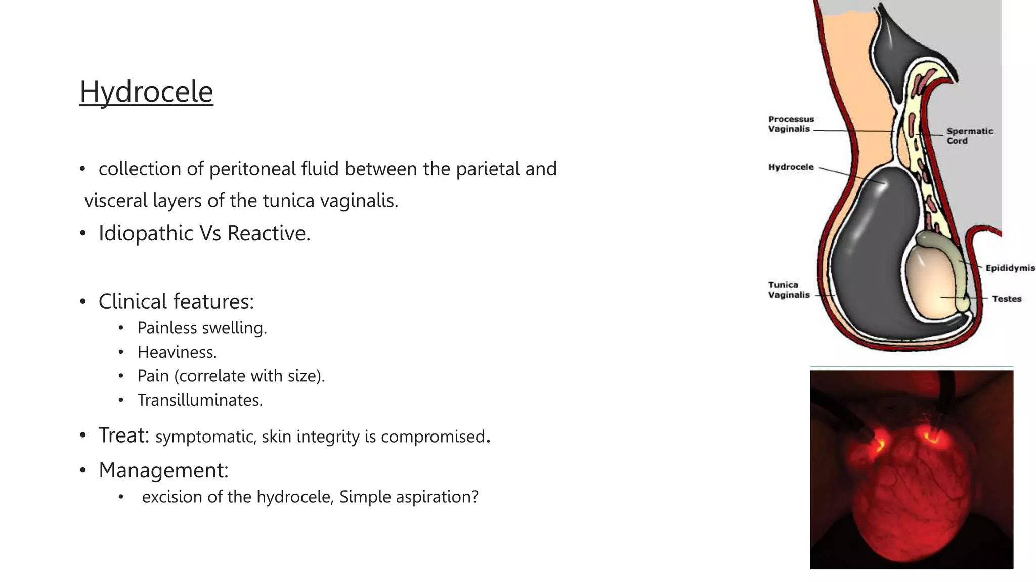
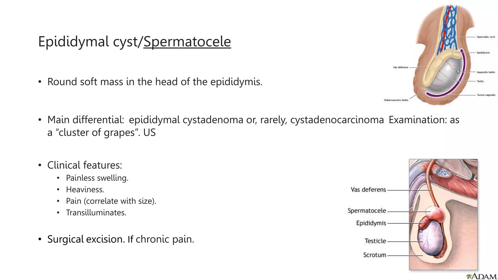
Overview of
Chronic Scrotal Pain
Hassan Almarzooq
Why This Topic?
• Limitations to daily activities.
• Limited ability to work and sexual dysfunction.
• Psychological disorders, depression, somatization disorder.
• Social isolation.
• Decreased quality of life.
Definition
• intermittent or constant, unilateral or bilateral
• localized to the scrotal structures
• 3 months or longer in duration
• significantly interferes with daily activities
• and prompts the patient to seek medical attention.
ETIOLOGY
• 50% of patients will not have an identifiable etiology.
• “self-palpation” orchitis.
• Psychological.
Varicocele
• Dilatation of the pampiniform plexus of spermatic vein.
• Clinical features:
• Asymptomatic.
• Dull, aching, scrotal pain. standing Vs recumbency position.
• Scrotal fullness.
• Large, soft, left-sided? scrotal mass ( bag of worm).
• Infertility.
• Alarming features:
• Unilateral right varicoceles. (IVC)
• Non-diminishing.
Management
Up to date
Hydrocele
• collection of peritoneal fluid between the parietal and
visceral layers of the tunica vaginalis.
• Idiopathic Vs Reactive.
• Clinical features:
• Painless swelling.
• Heaviness.
• Pain (correlate with size).
• Transilluminates.
• Treat: symptomatic, skin integrity is compromised.
• Management:
• excision of the hydrocele, Simple aspiration?
Epididymal cyst/Spermatocele
• Round soft mass in the head of the epididymis.
• Main differential: epididymal cystadenoma or, rarely, cystadenocarcinoma Examination: as
a “cluster of grapes”. US
• Clinical features:
• Painless swelling.
• Heaviness.
• Pain (correlate with size).
• Transilluminates.
• Surgical excision. If chronic pain.
Testicular cancer
• Painless, firm, nontender nodule or mass that does not transilluminate.
• +- gynecomastia, elevated Beta- hCG
• Scrotal ultrasound is the diagnostic test of choice to evaluate a testicular nodule or mass.
• MRI if US inconclusive.
• AFP, beta-hCG. normal serum values do not exclude testicular cancer.
DIAGNOSIS - CSP
• History
- Pain: SOCRATE
- Urinary, Sexual and Bowel functions.
- Hx of: scrotal, inguinal, abdominal or pelvic surgeries. PVPS
- history of psychological, physical or sexual abuse.
• PE:
- scrotal structures: concentrating on the testis, epididymides and vasa for any
anatomic abnormalities and to localization.
- scars, hernias or areas of tenderness.
- PR; Tone, tenderness, enlargement.
- Neurological examination of the lower limbs for sensory deficits and radiculopathy.
Diagnosis
• Lab: Hx and PE directed
• lower urinary tract symptoms or hematuria; Urine analysis.
• Urethral discharge, or penile pain; gonorrhea and chlamydia.
• Palpable abnormality/ mass; Scrotal ultrasound
• routine scrotal ultrasound is debated.
• Spermatic cord blockade is used as a diagnostic and therapeutic measure.
TREATMENT
• Conservative therapies.
• watchful waiting.
• scrotal support.
• heat or cold therapy.
• avoidance of aggravating activities.
• Psychological therapy & CBT.
• catastrophic thinking.
• irrational fear of injury.
• pain distorted thinking.
• Medical management
• NSAIDs.
• Antibiotics.
• Tricyclic antidepressants.
• Neuropathic Medications.
Reference
• Diagnosis and Management of Chronic Scrotal Pain, (AUA)
https://drive.google.com/file/d/1-q44O1SFjMK2OoefMK3rSf96L9-55-
xW/view
• Nonacute scrotal conditions in adults, (UTD)
Thank you

chronic scrotal pain.pptx

  • 1.
    Overview of Chronic ScrotalPain Hassan Almarzooq
  • 2.
    Why This Topic? •Limitations to daily activities. • Limited ability to work and sexual dysfunction. • Psychological disorders, depression, somatization disorder. • Social isolation. • Decreased quality of life.
  • 3.
    Definition • intermittent orconstant, unilateral or bilateral • localized to the scrotal structures • 3 months or longer in duration • significantly interferes with daily activities • and prompts the patient to seek medical attention.
  • 4.
    ETIOLOGY • 50% ofpatients will not have an identifiable etiology. • “self-palpation” orchitis. • Psychological.
  • 5.
    Varicocele • Dilatation ofthe pampiniform plexus of spermatic vein. • Clinical features: • Asymptomatic. • Dull, aching, scrotal pain. standing Vs recumbency position. • Scrotal fullness. • Large, soft, left-sided? scrotal mass ( bag of worm). • Infertility. • Alarming features: • Unilateral right varicoceles. (IVC) • Non-diminishing.
  • 6.
  • 7.
    Hydrocele • collection ofperitoneal fluid between the parietal and visceral layers of the tunica vaginalis. • Idiopathic Vs Reactive. • Clinical features: • Painless swelling. • Heaviness. • Pain (correlate with size). • Transilluminates. • Treat: symptomatic, skin integrity is compromised. • Management: • excision of the hydrocele, Simple aspiration?
  • 8.
    Epididymal cyst/Spermatocele • Roundsoft mass in the head of the epididymis. • Main differential: epididymal cystadenoma or, rarely, cystadenocarcinoma Examination: as a “cluster of grapes”. US • Clinical features: • Painless swelling. • Heaviness. • Pain (correlate with size). • Transilluminates. • Surgical excision. If chronic pain.
  • 9.
    Testicular cancer • Painless,firm, nontender nodule or mass that does not transilluminate. • +- gynecomastia, elevated Beta- hCG • Scrotal ultrasound is the diagnostic test of choice to evaluate a testicular nodule or mass. • MRI if US inconclusive. • AFP, beta-hCG. normal serum values do not exclude testicular cancer.
  • 10.
    DIAGNOSIS - CSP •History - Pain: SOCRATE - Urinary, Sexual and Bowel functions. - Hx of: scrotal, inguinal, abdominal or pelvic surgeries. PVPS - history of psychological, physical or sexual abuse. • PE: - scrotal structures: concentrating on the testis, epididymides and vasa for any anatomic abnormalities and to localization. - scars, hernias or areas of tenderness. - PR; Tone, tenderness, enlargement. - Neurological examination of the lower limbs for sensory deficits and radiculopathy.
  • 11.
    Diagnosis • Lab: Hxand PE directed • lower urinary tract symptoms or hematuria; Urine analysis. • Urethral discharge, or penile pain; gonorrhea and chlamydia. • Palpable abnormality/ mass; Scrotal ultrasound • routine scrotal ultrasound is debated. • Spermatic cord blockade is used as a diagnostic and therapeutic measure.
  • 12.
    TREATMENT • Conservative therapies. •watchful waiting. • scrotal support. • heat or cold therapy. • avoidance of aggravating activities. • Psychological therapy & CBT. • catastrophic thinking. • irrational fear of injury. • pain distorted thinking. • Medical management • NSAIDs. • Antibiotics. • Tricyclic antidepressants. • Neuropathic Medications.
  • 16.
    Reference • Diagnosis andManagement of Chronic Scrotal Pain, (AUA) https://drive.google.com/file/d/1-q44O1SFjMK2OoefMK3rSf96L9-55- xW/view • Nonacute scrotal conditions in adults, (UTD)
  • 17.

Editor's Notes

  • #5 a variety of psychiatric conditions, including depression, anxiety and schizophrenia may manifest as psychogenic pain.13, Its hard to direct this relation, which cause which
  • #8 epididymitis, torsion, appendiceal torsion. cancer
  • #11 Men with a history of abuse are at an increased risk for CSP
  • #12 easy, safe and inexpensive imaging modality, and a normal ultrasound may help reassure Ultrasound has been shown to be highly sensitive and specific for detecting intrascrotal anatomic abnormalities, such as testicular tumors.33