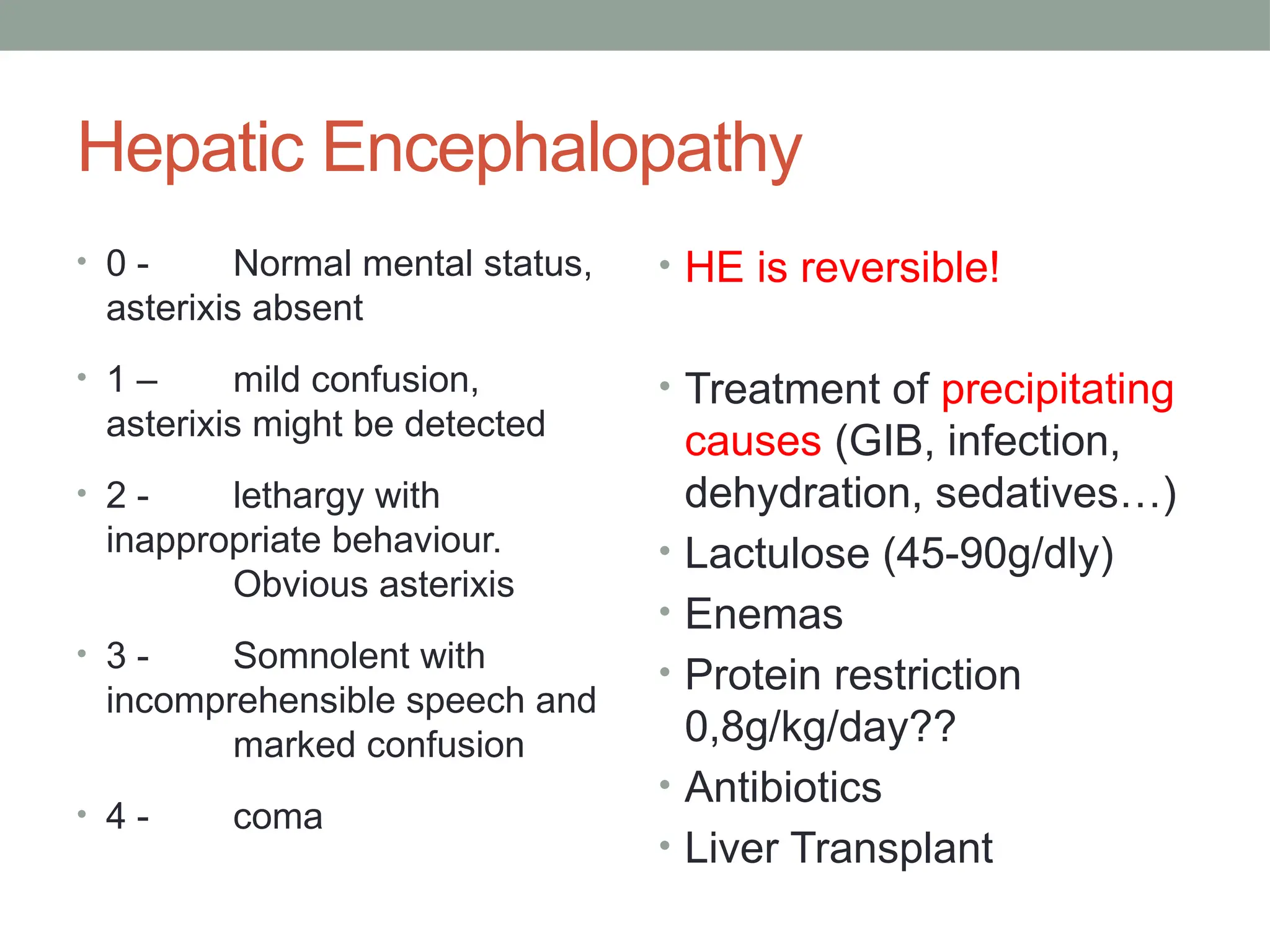
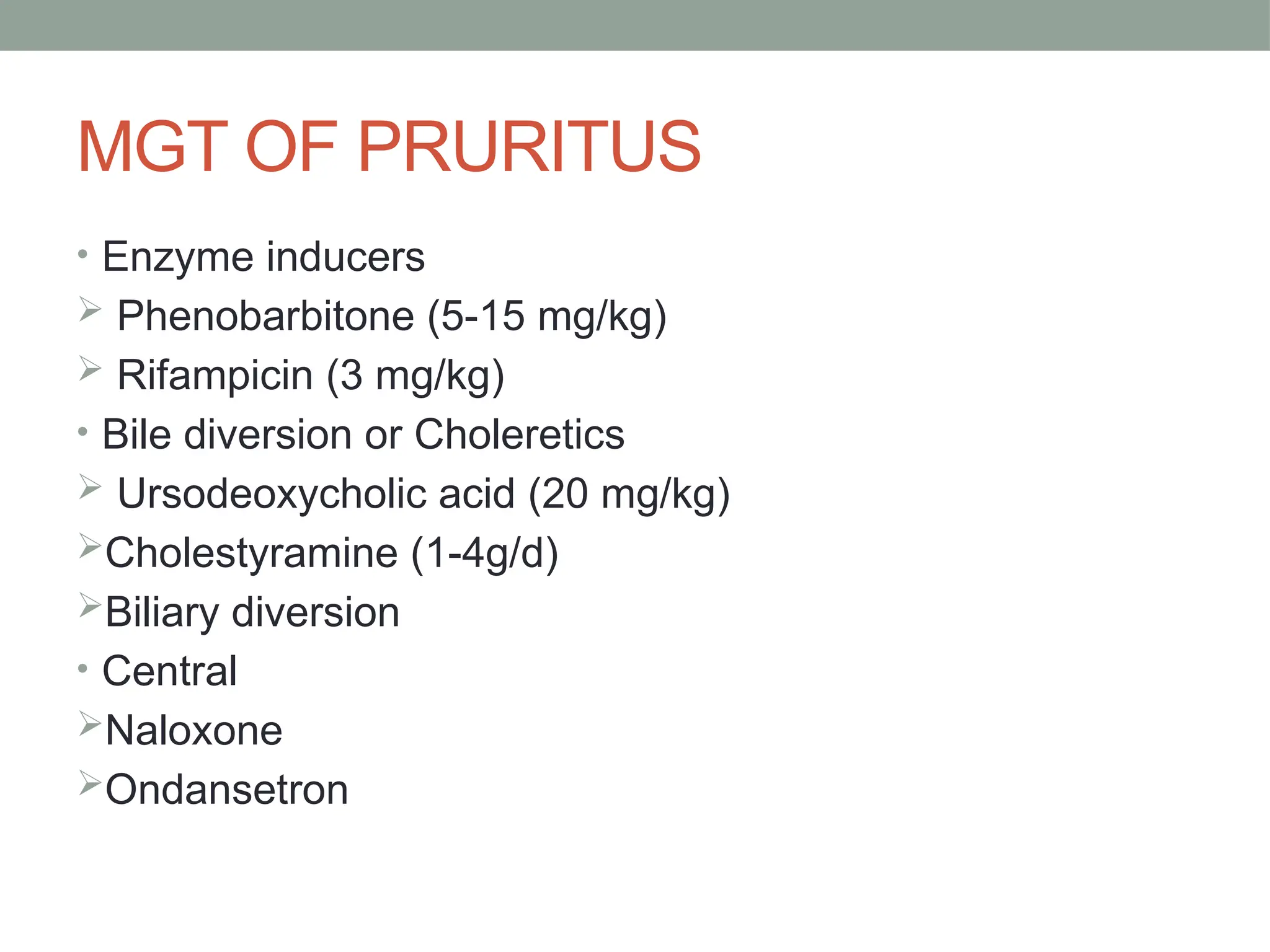
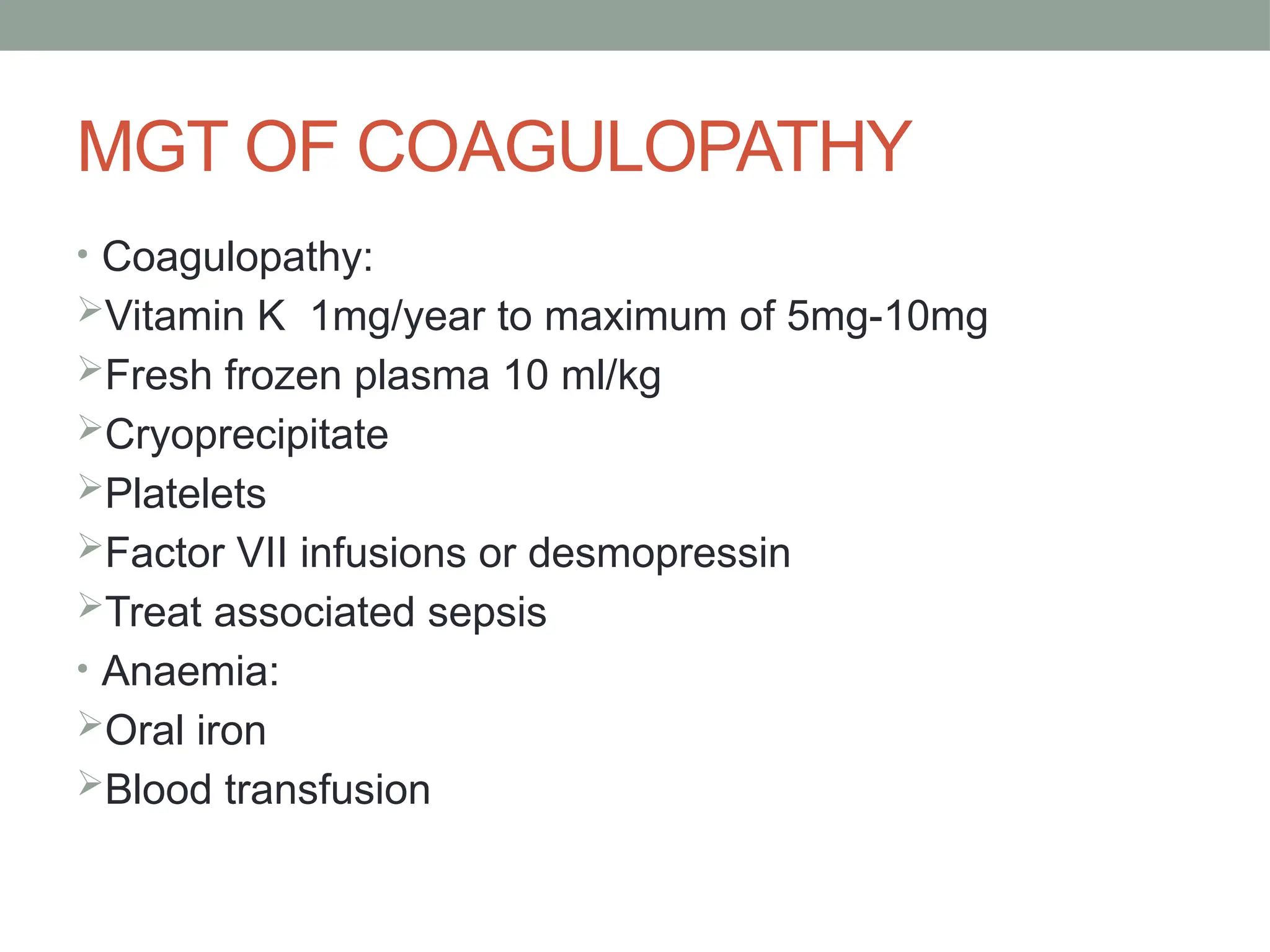
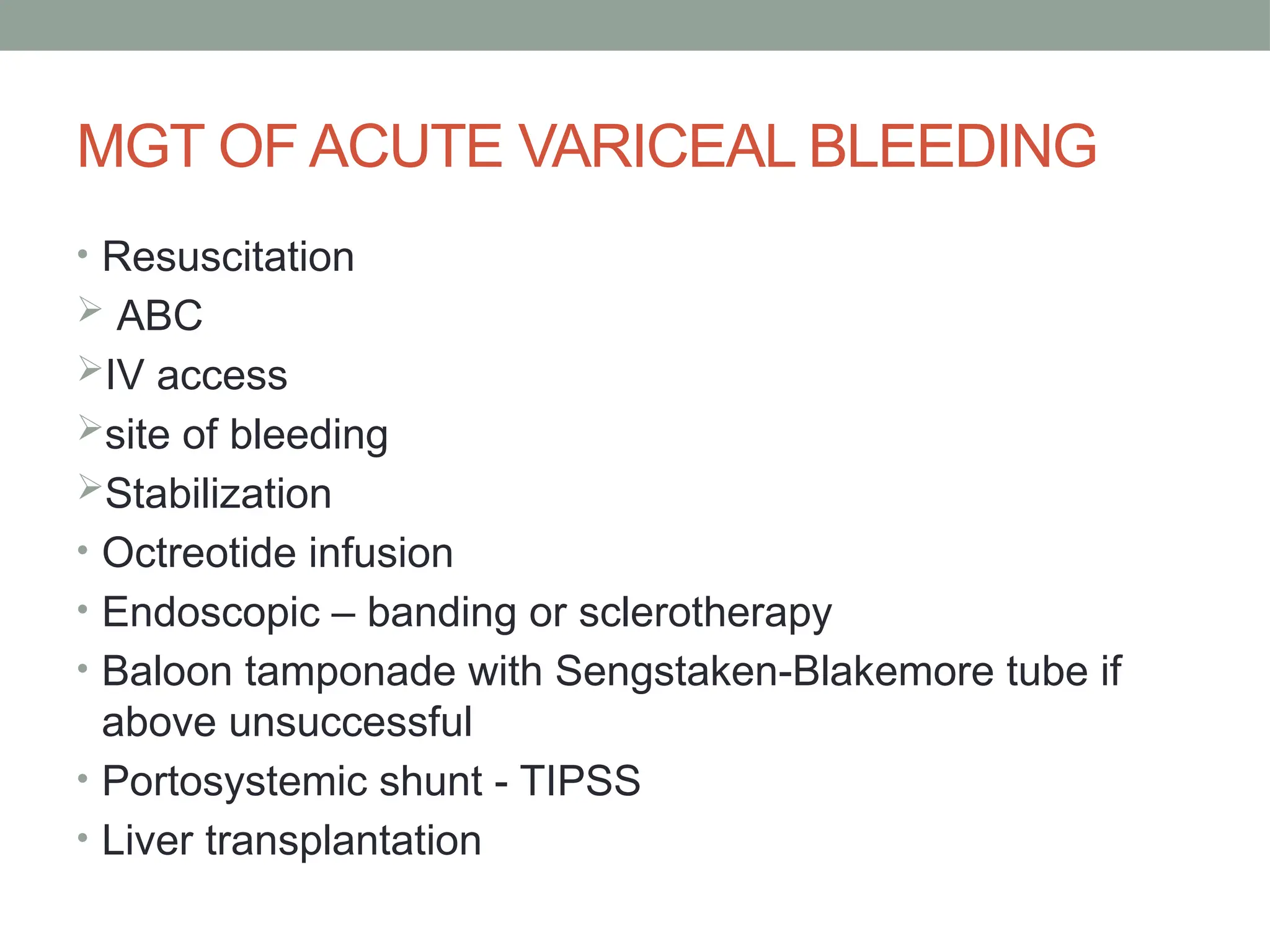
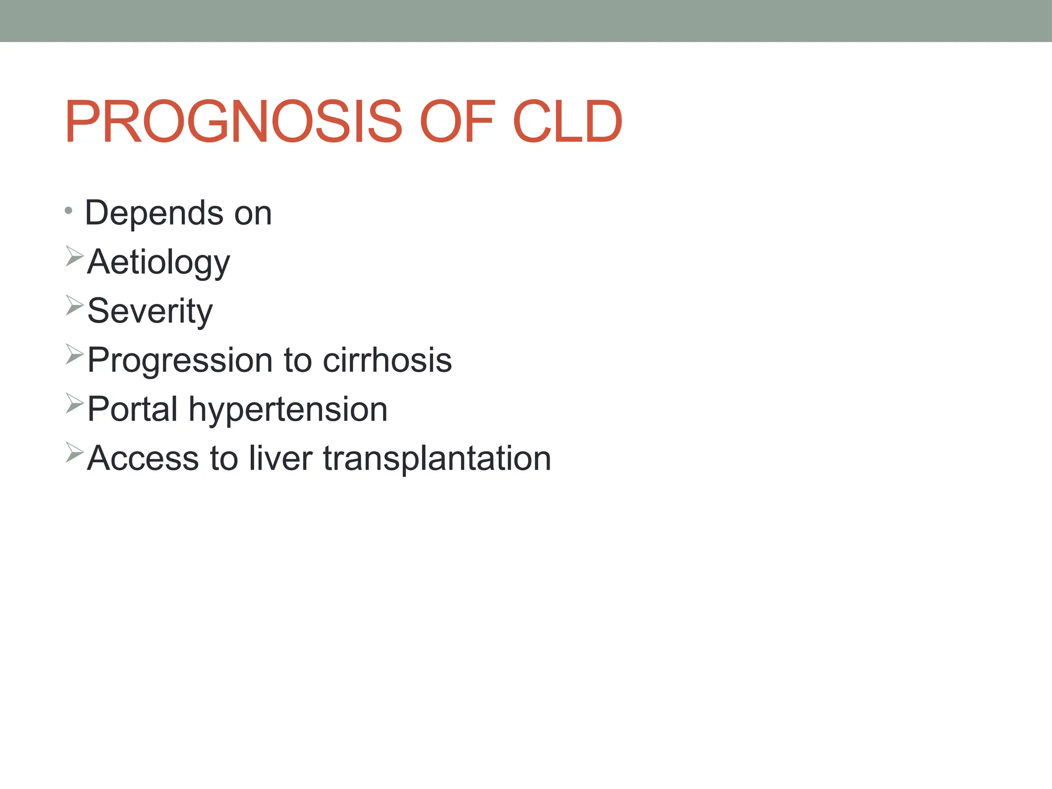
Azong Abdul-Wahid Akanlug-Ba
Chronic Liver Disease
OUTLINE
• DEFINITION
• BRIEF ANATOMY AND PHYSIOLOGY OF THE LIVER
• AETIOLOGY
• CLINICAL BREAKDOWN
• INVESTIGATIONS
• APPROACH
• COMPLICATIONS
• MANAGEMENT
• PROGNOSIS
• CASE SCENARIO
DEFINITION
• Definition: persistent liver disease for more than 8 weeks
• Persistent hepatic injury, with or without clinical
symptoms, resulting in progressive histological alterations
• Chronic liver disease in the clinical context is a disease
process of the liver that involves a process of progressive
destruction and regeneration of the liver parenchyma
leading to fibrosis and cirrhosis.
CIRRHOSIS
• Definition: histological presence of bands of fibrous tissue
that link central and portal areas and form parenchymal
nodules, is a potential end stage of any acute or chronic
liver dx
• Aetiological type
Posthepatitic – after acute or chronic hepatitis
Postnecrotic – after toxic injury
May follow chronic biliary obstruction – biliary cirrhosis
CIRRHOSIS
• Morphological type
Macronodular – nodules of various sizes up to 5cm
separated by broad septa
Micronodular – with nodules of uniform size <1cm
separated by fine septa
BREIF ANATOMY AND PHYSIOLOGY OF THE LIVER
AETIOLOGY
 Major congenital disorders ;
Biliary atresia, Idiopathic neonatal hepatitis,Tyrosinaemia ,Untreated
galactosaemia, ETC
Toxic :Alcohol ,Glucocorticoids,Amiodarone ,Tamoxifen,
Antiretroviral agents,Tetracyline, Herbal preparations
Autoimmune Hepatitis (AIH) ,Clinically
indistinguishable .
Infectious : Hepatitis A-E , EBV, CMV,Yellow Fever, Lues,
Typhoid, Leptosp
Others:Wilson disease, Cystic fibrosis, Non-alcoholic fatty liver
disease
CLINICAL PRESENTATION
• Common signs and symptoms may stem from decreased hepatic synthetic
function eg, coagulopathy, portal hypertension, hepatic encephalopathy.
• Jaundice
• Clubbing
• Itchiness
• Fatigue
• confusion
• Oedema
• Bleeding disorders such as epistaxis
• Palmer erythema
• Leuconychia
• Spider naevae
• Growth failure
• Infections
• Gynecomastia
CLINICAL PRESENTATION
• Portal hypertension causes may be
Prehepatic eg. Portal vein thrombosis, splenic vein
thrombosis
Intrahepatic eg. Schistosomiasis, Cirrhosis, Veno-
occlusive disease
Posthepatic eg. Budd-Chiari syndrome (hepatomegaly,
abdominal pain, ascites)
CLINICAL PRESENTATION
• Ascites is suggested by the following findings on physical
examination:
Abdominal distention
Bulging flanks
Shifting dullness
Fluid thrill
INVESTIGATION
• These will depend to a considerable extent upon clinical
suspicion of the aetiology.
• FBC: occult bleeding may produce anaemia;
hypersplenism may cause thrombocytopenia
• LFTs: Aspartate transaminase (AST), alanine
transaminase (ALT), alkaline phosphatase (ALP), bilirubin,
gamma-glutamyltransferase (gamma-GT), bilirubin,
albumin, Coagulation profile
INVESTIGATION
• Renal function tests and electrolytes
• Ferritin: low ferritin may indicate iron deficiency from diet
or blood loss; ferritin is raised in haemochromatosis.
• Viral antibody screen: to look for evidence of hepatitis B or
C infection.
• Fasting glucose/insulin/triglycerides and uric acid levels:
these should be measured if non-alcoholic steatohepatitis
(NASH) is suspected.
INVESTIGATION
• Autoantibody screen: ANA, SMA, anti-LK microsomal,
soluble liver
• Alpha-1-antitrypsin level: to assess for alpha-1-antitrypsin
deficiency
• Ceruloplasmin and urinary copper: to look for Wilson's
disease.
• Fasting transferrin saturation and HFE
(haemochromatosis C282Y) mutation: along with a raised
ferritin, these tests can screen for haemochromatosis.
• Urine for reducing sugars to look for Galactosaemia. If
galactose positive check blood for GALT deficiency
• Fat soluble Vitamin levels in the blood
INVESTIGATION
• USG with doppler
• CT Scan
• MRI
• Liver biopsy
Histology is usually needed for the definitive diagnosis of
cirrhosis and liver biopsy is the gold standard.
It may also give a clue to the underlying cause.
If there are clear signs of cirrhosis, such as ascites,
coagulopathy, and a shrunken nodular-appearing liver,
then confirmation of diagnosis by biopsy may not be
necessary.
Approach to the Patient
• Stepwise
• Assess for alcohol use
• Identify medications and supplements that can cause
elevation of the LFT
• Test for viral hepatitis B and C
• Test for fatty liver
Prognosis in Cirrhosis
• Fibrosis
• Reversible in initial stages
• Cirrhosis
• Irreversible
• High susceptibility for complications
• Life expectancy markedly reduced
• Quality of life impaired
Child-Pugh Score
A=5-6 (2 yr survival 85%)
B=7-9 (2 yr survival 57%)
C=10-15 (2 yr survival 35%)
1 2 3
Bilirubin <2 2-3 >3
Albumin >3.5 3.5-2.8 <2.8
INR <1.7 1.7-2.3 >2.3
Ascites Absent Mild-Moderate Severe /
Refractory
Encephalopathy Absent Mild (I-II) Severe (III-IV)
COMPLICATIONS OF CLD
• Definition: complications of CLD and cirrhosis are a
consequence of the impaired metabolic and synthetic
function and structural alterations of the parenchyma
leading to elevated portal pressure.
COMPLICATIONS
• Growth faltering and malnutrition
• Hepatic encephalopathy
• Coagulopathy
• Hepatopulmonary syndrome
• Portal hypertension and variceal bleeding
• Ascites
• Spontaneous bacterial peritonitis
• Hepatorenal syndrome
• Hepatocellular carcinoma
• Endocrine dysfunction
MANAGEMENT OF CLD
• SUPPORTIVE
 Preventive therapy
Malnutrition
Fat soluble vitamin deficiency
Variceal bleeding and coagulopathy
Ascites
Sepsis
 Nutritional support
 Psychosocial – both child and parents
NUTRITIONAL SUPPORT
• Modular feed
High protein/CHO
Balanced MCT/LCT
Low salt
150% RDA
Fat soluble vitamins A, D, E, K
• Route of feeding
 Oral
 Nasogastric
 Parenteral
Hepatic Encephalopathy
• 0 - Normal mental status,
asterixis absent
• 1 – mild confusion,
asterixis might be detected
• 2 - lethargy with
inappropriate behaviour.
Obvious asterixis
• 3 - Somnolent with
incomprehensible speech and
marked confusion
• 4 - coma
• HE is reversible!
• Treatment of precipitating
causes (GIB, infection,
dehydration, sedatives…)
• Lactulose (45-90g/dly)
• Enemas
• Protein restriction
0,8g/kg/day??
• Antibiotics
• Liver Transplant
MANAGEMENT OF CLF – FLUID MGT
• Management of ascites
Diagnosis: fluid thrill/shifting dullness, USG and
abdominal paracentesis
 Fluid restriction
Nutritional support
ASCITES AND FLUID RETENTION
• Fluid and salt restriction
(2/3 maintenance)
Diuretics: Spironolactone 2-3mg/kg/d up to 7mg/kg/d
Furosemide 1-2 mg/kg or hydrochlorothiazide
If resistant consider 5ml/kg 20% albumin infusion over 4-
6h with furosemide
• Therapeutic paracentesis if ascites is resistant esp if
compromising respiratory function
• Shunt – peritoneal-jugular or TIPSS
• Haemofiltration/Dialysis
MGT OF PRURITUS
• Enzyme inducers
 Phenobarbitone (5-15 mg/kg)
 Rifampicin (3 mg/kg)
• Bile diversion or Choleretics
 Ursodeoxycholic acid (20 mg/kg)
Cholestyramine (1-4g/d)
Biliary diversion
• Central
Naloxone
Ondansetron
MGT OF COAGULOPATHY
• Coagulopathy:
Vitamin K 1mg/year to maximum of 5mg-10mg
Fresh frozen plasma 10 ml/kg
Cryoprecipitate
Platelets
Factor VII infusions or desmopressin
Treat associated sepsis
• Anaemia:
Oral iron
Blood transfusion
SPECIFIC MGT
• Immunosuppression for Autoimmune hepatitis
• Penicillamine , Zinc, trientine for Wilsons
• Ursodeoxycholic acid for Cystic Fibrosis
• Anti-viral therapy for Hep B and C
• Diet
• Liver Transplantation
Treatment of alcoholic liver disease
• Abstinence
• Single most predictive factor for progression
• Can achieve improvement in fibrosis, reduction or
normalisation of portal pressure, resolution of ascites.
SCHISTOSOMIASIS
• Caused by the deposition of Schistosoma oocytes in
presinusoidal portal venules
• Development of granulomata and portal fibrosis.
Schistosomiasis is the most common noncirrhotic cause
of variceal bleeding worldwide
• Schistosoma mansoni infection is described in Puerto
Rico, Central and South America, the Middle East, and
Africa.
• S japonicum is described in the Far East.
• S hematobium, observed in the Middle East and Africa,
can produce portal fibrosis but more commonly is
associated with urinary tract deposition of eggs.
PORTAL HYPERTENSION
• Results in
Varices – oesophageal, gastric, rectal
Portal gastropathy
Hypersplenism
• Implications for a normal life
 School
Contact sports
MGT OF PORTAL HYPERTENSION
• Medical Therapy
UGI endoscopy - diagnostic and therapeutic
Drug therapy – somatostatin analogues, Vasopressin
propranolol
• Transjugular Intrahepatic Porto-systemic Shunt
stent shunt (TIPS)
• Prophylaxis - propanolol
MGT OF ACUTE VARICEAL BLEEDING
• Resuscitation
 ABC
IV access
site of bleeding
Stabilization
• Octreotide infusion
• Endoscopic – banding or sclerotherapy
• Baloon tamponade with Sengstaken-Blakemore tube if
above unsuccessful
• Portosystemic shunt - TIPSS
• Liver transplantation
HEPATORENAL SYNDROME
• Definition: Functional renal failure in patients with severe
liver disease
• Two types: I and II
• Pathophysiology –intense renal vasoconstriction with co-
existent systemic vasodilation thereby reducing renal
blood flow
• Diagnosis: by exclusion, rule out
Hypovolaemic shock, nephrotoxic drugs or kidney dx
Hereditary tyrosinaemia, Alagilles and polycystic kidney dx
GFR is markedly reduced
• Treatment – heamofiltration, liver transplantation
ENDOCRINE DYSFUNCTION
• Regulation and function of multiple endocrine systems is
affected in CLD
• Increased growth hormone resistance due to reduced
synthesis IGF-1 and IGF-3
• Feminization and hypogonadism in males – reduced
testosterone and relative increase in oesterone and
oestradiol
• Hypothyroidism – reduced T3 and T4
SPONTANEOUS BACTERIAL PERITONITIS
• Bacterial infection of ascitic fluid in the absence of
secondary causes such as bowel perforation or intra-
abdominal abscess
• Presentation – subtle with fever and irritability
• Diagnosis
high index of suspicion
Non-specific deterioration in condition
Ascitic fluid for microscopy, culture (monomicrobial)
PMN >250/mm cube is diagnostic
• Treat with antibiotics for 5-7days
PROGNOSIS OF CLD
• Depends on
Aetiology
Severity
Progression to cirrhosis
Portal hypertension
Access to liver transplantation
CLIP Score
Prospective validation of the CLIP score: A new prognostic system for patients with cirrhosis and hepatocellular carcinoma. Hepatology 2000; 31:840
Child-Pugh
A 0
B 1
C 2
Tumor morphology
Uninodular and extension 50% 0
Multinodular and extension 50% 1
Massive or extension >50% 2
AFP
<400 0
>400 1
Portal Vein Thrombosis
No 0
Yes 1
Survival curves based on the CLIP score
Farinati, F, Rinaldi, M, Gianni, S, Naccarato. Cancer 2000; 89:2266.
INDICATIONS FOR TRANSPLANT
• Laboratory Indices
Serum cholesterol <2.6 mmol/l (100 mg/dl)
Indirect bilirubin >100 mmol/l (6mg/dl)
Serum albumin <35 g/l (3.5 mg/dl)
Prothrombin ratio (INR) >1.4
Portal vein reverse flow on ultrasonography
INDICATIONS FOR TRANSPLANT
• Clinical
Severe portal hypertension
Recurrent variceal bleeding
Refractory ascites
Intractable pruritus
Growth retardation
Unacceptable quality of life
REFERRAL OF CLD
• Early referral and recognition of liver disease
• Referral to Specialist Centre
Early diagnosis
Consideration of Treatment
Improve outcomes for children
Case Senario
• 75 year old woman who
• HBsAg (+)
• HBeAg (+)
• ALT of 50 IU/L
• HBV DNA is 8000 copies/mL.
• She has ascites and known esophageal varices..
• Is she a Candidate for Liver Transplant??
THANK YOU

CHRONIC LIVER DISEASE ksdh a presentation

  • 1.
  • 2.
    OUTLINE • DEFINITION • BRIEFANATOMY AND PHYSIOLOGY OF THE LIVER • AETIOLOGY • CLINICAL BREAKDOWN • INVESTIGATIONS • APPROACH • COMPLICATIONS • MANAGEMENT • PROGNOSIS • CASE SCENARIO
  • 3.
    DEFINITION • Definition: persistentliver disease for more than 8 weeks • Persistent hepatic injury, with or without clinical symptoms, resulting in progressive histological alterations • Chronic liver disease in the clinical context is a disease process of the liver that involves a process of progressive destruction and regeneration of the liver parenchyma leading to fibrosis and cirrhosis.
  • 4.
    CIRRHOSIS • Definition: histologicalpresence of bands of fibrous tissue that link central and portal areas and form parenchymal nodules, is a potential end stage of any acute or chronic liver dx • Aetiological type Posthepatitic – after acute or chronic hepatitis Postnecrotic – after toxic injury May follow chronic biliary obstruction – biliary cirrhosis
  • 5.
    CIRRHOSIS • Morphological type Macronodular– nodules of various sizes up to 5cm separated by broad septa Micronodular – with nodules of uniform size <1cm separated by fine septa
  • 6.
    BREIF ANATOMY ANDPHYSIOLOGY OF THE LIVER
  • 8.
    AETIOLOGY  Major congenitaldisorders ; Biliary atresia, Idiopathic neonatal hepatitis,Tyrosinaemia ,Untreated galactosaemia, ETC Toxic :Alcohol ,Glucocorticoids,Amiodarone ,Tamoxifen, Antiretroviral agents,Tetracyline, Herbal preparations Autoimmune Hepatitis (AIH) ,Clinically indistinguishable . Infectious : Hepatitis A-E , EBV, CMV,Yellow Fever, Lues, Typhoid, Leptosp Others:Wilson disease, Cystic fibrosis, Non-alcoholic fatty liver disease
  • 9.
    CLINICAL PRESENTATION • Commonsigns and symptoms may stem from decreased hepatic synthetic function eg, coagulopathy, portal hypertension, hepatic encephalopathy. • Jaundice • Clubbing • Itchiness • Fatigue • confusion • Oedema • Bleeding disorders such as epistaxis • Palmer erythema • Leuconychia • Spider naevae • Growth failure • Infections • Gynecomastia
  • 10.
    CLINICAL PRESENTATION • Portalhypertension causes may be Prehepatic eg. Portal vein thrombosis, splenic vein thrombosis Intrahepatic eg. Schistosomiasis, Cirrhosis, Veno- occlusive disease Posthepatic eg. Budd-Chiari syndrome (hepatomegaly, abdominal pain, ascites)
  • 11.
    CLINICAL PRESENTATION • Ascitesis suggested by the following findings on physical examination: Abdominal distention Bulging flanks Shifting dullness Fluid thrill
  • 12.
    INVESTIGATION • These willdepend to a considerable extent upon clinical suspicion of the aetiology. • FBC: occult bleeding may produce anaemia; hypersplenism may cause thrombocytopenia • LFTs: Aspartate transaminase (AST), alanine transaminase (ALT), alkaline phosphatase (ALP), bilirubin, gamma-glutamyltransferase (gamma-GT), bilirubin, albumin, Coagulation profile
  • 13.
    INVESTIGATION • Renal functiontests and electrolytes • Ferritin: low ferritin may indicate iron deficiency from diet or blood loss; ferritin is raised in haemochromatosis. • Viral antibody screen: to look for evidence of hepatitis B or C infection. • Fasting glucose/insulin/triglycerides and uric acid levels: these should be measured if non-alcoholic steatohepatitis (NASH) is suspected.
  • 14.
    INVESTIGATION • Autoantibody screen:ANA, SMA, anti-LK microsomal, soluble liver • Alpha-1-antitrypsin level: to assess for alpha-1-antitrypsin deficiency • Ceruloplasmin and urinary copper: to look for Wilson's disease. • Fasting transferrin saturation and HFE (haemochromatosis C282Y) mutation: along with a raised ferritin, these tests can screen for haemochromatosis. • Urine for reducing sugars to look for Galactosaemia. If galactose positive check blood for GALT deficiency • Fat soluble Vitamin levels in the blood
  • 15.
    INVESTIGATION • USG withdoppler • CT Scan • MRI • Liver biopsy Histology is usually needed for the definitive diagnosis of cirrhosis and liver biopsy is the gold standard. It may also give a clue to the underlying cause. If there are clear signs of cirrhosis, such as ascites, coagulopathy, and a shrunken nodular-appearing liver, then confirmation of diagnosis by biopsy may not be necessary.
  • 16.
    Approach to thePatient • Stepwise • Assess for alcohol use • Identify medications and supplements that can cause elevation of the LFT • Test for viral hepatitis B and C • Test for fatty liver
  • 17.
    Prognosis in Cirrhosis •Fibrosis • Reversible in initial stages • Cirrhosis • Irreversible • High susceptibility for complications • Life expectancy markedly reduced • Quality of life impaired
  • 18.
    Child-Pugh Score A=5-6 (2yr survival 85%) B=7-9 (2 yr survival 57%) C=10-15 (2 yr survival 35%) 1 2 3 Bilirubin <2 2-3 >3 Albumin >3.5 3.5-2.8 <2.8 INR <1.7 1.7-2.3 >2.3 Ascites Absent Mild-Moderate Severe / Refractory Encephalopathy Absent Mild (I-II) Severe (III-IV)
  • 19.
    COMPLICATIONS OF CLD •Definition: complications of CLD and cirrhosis are a consequence of the impaired metabolic and synthetic function and structural alterations of the parenchyma leading to elevated portal pressure.
  • 20.
    COMPLICATIONS • Growth falteringand malnutrition • Hepatic encephalopathy • Coagulopathy • Hepatopulmonary syndrome • Portal hypertension and variceal bleeding • Ascites • Spontaneous bacterial peritonitis • Hepatorenal syndrome • Hepatocellular carcinoma • Endocrine dysfunction
  • 21.
    MANAGEMENT OF CLD •SUPPORTIVE  Preventive therapy Malnutrition Fat soluble vitamin deficiency Variceal bleeding and coagulopathy Ascites Sepsis  Nutritional support  Psychosocial – both child and parents
  • 22.
    NUTRITIONAL SUPPORT • Modularfeed High protein/CHO Balanced MCT/LCT Low salt 150% RDA Fat soluble vitamins A, D, E, K • Route of feeding  Oral  Nasogastric  Parenteral
  • 23.
    Hepatic Encephalopathy • 0- Normal mental status, asterixis absent • 1 – mild confusion, asterixis might be detected • 2 - lethargy with inappropriate behaviour. Obvious asterixis • 3 - Somnolent with incomprehensible speech and marked confusion • 4 - coma • HE is reversible! • Treatment of precipitating causes (GIB, infection, dehydration, sedatives…) • Lactulose (45-90g/dly) • Enemas • Protein restriction 0,8g/kg/day?? • Antibiotics • Liver Transplant
  • 24.
    MANAGEMENT OF CLF– FLUID MGT • Management of ascites Diagnosis: fluid thrill/shifting dullness, USG and abdominal paracentesis  Fluid restriction Nutritional support
  • 25.
    ASCITES AND FLUIDRETENTION • Fluid and salt restriction (2/3 maintenance) Diuretics: Spironolactone 2-3mg/kg/d up to 7mg/kg/d Furosemide 1-2 mg/kg or hydrochlorothiazide If resistant consider 5ml/kg 20% albumin infusion over 4- 6h with furosemide • Therapeutic paracentesis if ascites is resistant esp if compromising respiratory function • Shunt – peritoneal-jugular or TIPSS • Haemofiltration/Dialysis
  • 26.
    MGT OF PRURITUS •Enzyme inducers  Phenobarbitone (5-15 mg/kg)  Rifampicin (3 mg/kg) • Bile diversion or Choleretics  Ursodeoxycholic acid (20 mg/kg) Cholestyramine (1-4g/d) Biliary diversion • Central Naloxone Ondansetron
  • 27.
    MGT OF COAGULOPATHY •Coagulopathy: Vitamin K 1mg/year to maximum of 5mg-10mg Fresh frozen plasma 10 ml/kg Cryoprecipitate Platelets Factor VII infusions or desmopressin Treat associated sepsis • Anaemia: Oral iron Blood transfusion
  • 28.
    SPECIFIC MGT • Immunosuppressionfor Autoimmune hepatitis • Penicillamine , Zinc, trientine for Wilsons • Ursodeoxycholic acid for Cystic Fibrosis • Anti-viral therapy for Hep B and C • Diet • Liver Transplantation
  • 29.
    Treatment of alcoholicliver disease • Abstinence • Single most predictive factor for progression • Can achieve improvement in fibrosis, reduction or normalisation of portal pressure, resolution of ascites.
  • 30.
    SCHISTOSOMIASIS • Caused bythe deposition of Schistosoma oocytes in presinusoidal portal venules • Development of granulomata and portal fibrosis. Schistosomiasis is the most common noncirrhotic cause of variceal bleeding worldwide • Schistosoma mansoni infection is described in Puerto Rico, Central and South America, the Middle East, and Africa. • S japonicum is described in the Far East. • S hematobium, observed in the Middle East and Africa, can produce portal fibrosis but more commonly is associated with urinary tract deposition of eggs.
  • 31.
    PORTAL HYPERTENSION • Resultsin Varices – oesophageal, gastric, rectal Portal gastropathy Hypersplenism • Implications for a normal life  School Contact sports
  • 32.
    MGT OF PORTALHYPERTENSION • Medical Therapy UGI endoscopy - diagnostic and therapeutic Drug therapy – somatostatin analogues, Vasopressin propranolol • Transjugular Intrahepatic Porto-systemic Shunt stent shunt (TIPS) • Prophylaxis - propanolol
  • 33.
    MGT OF ACUTEVARICEAL BLEEDING • Resuscitation  ABC IV access site of bleeding Stabilization • Octreotide infusion • Endoscopic – banding or sclerotherapy • Baloon tamponade with Sengstaken-Blakemore tube if above unsuccessful • Portosystemic shunt - TIPSS • Liver transplantation
  • 34.
    HEPATORENAL SYNDROME • Definition:Functional renal failure in patients with severe liver disease • Two types: I and II • Pathophysiology –intense renal vasoconstriction with co- existent systemic vasodilation thereby reducing renal blood flow • Diagnosis: by exclusion, rule out Hypovolaemic shock, nephrotoxic drugs or kidney dx Hereditary tyrosinaemia, Alagilles and polycystic kidney dx GFR is markedly reduced • Treatment – heamofiltration, liver transplantation
  • 35.
    ENDOCRINE DYSFUNCTION • Regulationand function of multiple endocrine systems is affected in CLD • Increased growth hormone resistance due to reduced synthesis IGF-1 and IGF-3 • Feminization and hypogonadism in males – reduced testosterone and relative increase in oesterone and oestradiol • Hypothyroidism – reduced T3 and T4
  • 36.
    SPONTANEOUS BACTERIAL PERITONITIS •Bacterial infection of ascitic fluid in the absence of secondary causes such as bowel perforation or intra- abdominal abscess • Presentation – subtle with fever and irritability • Diagnosis high index of suspicion Non-specific deterioration in condition Ascitic fluid for microscopy, culture (monomicrobial) PMN >250/mm cube is diagnostic • Treat with antibiotics for 5-7days
  • 37.
    PROGNOSIS OF CLD •Depends on Aetiology Severity Progression to cirrhosis Portal hypertension Access to liver transplantation
  • 38.
    CLIP Score Prospective validationof the CLIP score: A new prognostic system for patients with cirrhosis and hepatocellular carcinoma. Hepatology 2000; 31:840 Child-Pugh A 0 B 1 C 2 Tumor morphology Uninodular and extension 50% 0 Multinodular and extension 50% 1 Massive or extension >50% 2 AFP <400 0 >400 1 Portal Vein Thrombosis No 0 Yes 1
  • 39.
    Survival curves basedon the CLIP score Farinati, F, Rinaldi, M, Gianni, S, Naccarato. Cancer 2000; 89:2266.
  • 40.
    INDICATIONS FOR TRANSPLANT •Laboratory Indices Serum cholesterol <2.6 mmol/l (100 mg/dl) Indirect bilirubin >100 mmol/l (6mg/dl) Serum albumin <35 g/l (3.5 mg/dl) Prothrombin ratio (INR) >1.4 Portal vein reverse flow on ultrasonography
  • 41.
    INDICATIONS FOR TRANSPLANT •Clinical Severe portal hypertension Recurrent variceal bleeding Refractory ascites Intractable pruritus Growth retardation Unacceptable quality of life
  • 42.
    REFERRAL OF CLD •Early referral and recognition of liver disease • Referral to Specialist Centre Early diagnosis Consideration of Treatment Improve outcomes for children
  • 43.
    Case Senario • 75year old woman who • HBsAg (+) • HBeAg (+) • ALT of 50 IU/L • HBV DNA is 8000 copies/mL. • She has ascites and known esophageal varices.. • Is she a Candidate for Liver Transplant??
  • 44.