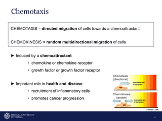
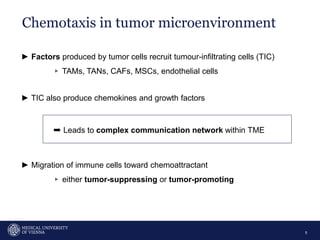
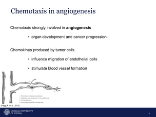
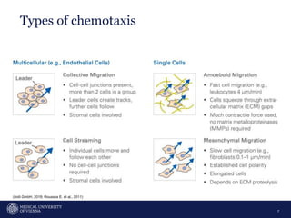
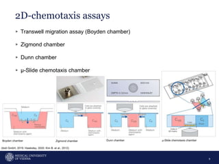
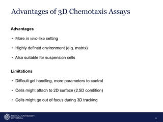
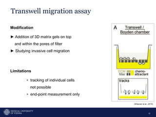
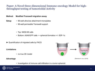
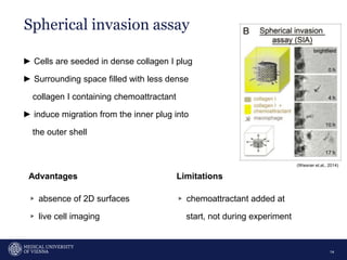
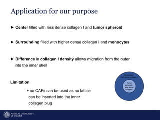
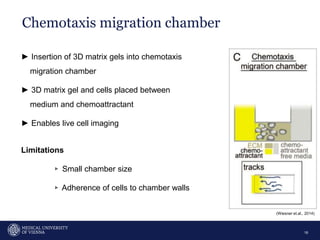
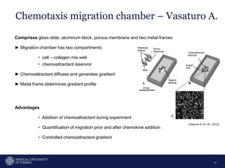
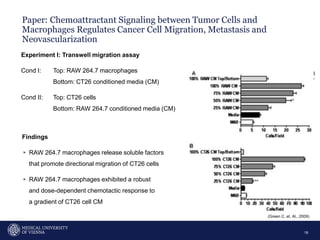
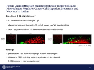
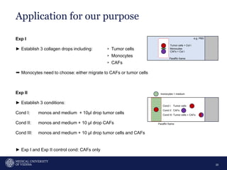
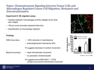
Organisational unit
Presentation title / topic OR Presenter's name
1
Case presentation
Rainer Marlene
2
•
32-jähriger Patient mit Kopfschmerzen und verschwommenem
Sehen
3
Ein 32-jähriger Mann kommt in die Ambulanz wegen Kopfschmerzen
und klagt über verschwommenes Sehen. Die Beschwerden dauern
bereits seit 3 Monaten an. Er bemerkt ebenso eine verminderte
Libido. In der Anamnese fällt auf dass der Patient an einem
peptischen Ulcus gelitten hat, weswegen er nun Omeprazol
einnimmt. Die Familienanamnese zeigt wiederkehrende peptische
Ulcera mütterlicherseits und einen Onkel welcher an einem
Pankreastumor verstarb. Der Status ist unauffällig. Ein Schädel-MRT
wird veranlasst und zeigt eine Raumforderung in der Hypophyse.
Welche der folgenden Untersuchungen sollte bei diesem Patienten
vorgenommen werden? Wählen Sie eine Antwort:
Chemotaxis
4
CHEMOTAXIS = directed migration of cells towards a chemoattractant
CHEMOKINESIS = random multidirectional migration of cells
► Induced by a chemoattractant
▹ chemokine or chemokine receptor
▹ growth factor or growth factor receptor
► Important role in health and disease
▹ recruitment of inflammatory cells
▹ promotes cancer progression
Chemotaxis in tumor microenvironment
5
► Factors produced by tumor cells recruit tumour-infiltrating cells (TIC)
▹ TAMs, TANs, CAFs, MSCs, endothelial cells
► TIC also produce chemokines and growth factors
➨ Leads to complex communication network within TME
► Migration of immune cells toward chemoattractant
▹ either tumor-suppressing or tumor-promoting
Chemotaxis in angiogenesis
6
Chemotaxis strongly involved in angiogenesis
▹ organ development and cancer progression
Chemokines produced by tumor cells
▹ influence migration of endothelial cells
▹ stimulate blood vessel formation
(Flegg A. et al., 2015)
Types of chemotaxis
7
(ibidi GmbH, 2019; Roussos E. et al., 2011)
Why conduct chemotaxis assays?
8
Analyzation whether a certain cell type directly migrates towards a defined
chemoattractant, or not.
Allow investigation of:
▹ Chemotactic behavior of cells after KO, KD, overexpression of GOI
▹ Differentiation between chemotaxis and chemokinesis
▹ Testing effect of inhibitors or enhancers of cell migration
▹ Determination of chemotactic potential of substances
2D-chemotaxis assays
9
▹ Transwell migration assay (Boyden chamber)
▹ Zigmond chamber
▹ Dunn chamber
▹ µ-Slide chemotaxis chamber
Boyden chamber Zigmond chamber Dunn chamber µ-Slide chemotaxis chamber
(ibidi GmbH, 2019; Hawksley, 2020; Kim B. et al., 2012)
3D-chemotaxis assays
10
▹ Modified Transwell migration assay
▹ Chemotaxis chamber (µ-Slide ibidi; Vasaturo A.)
▹ Spherical invasion assay
▹ Migration in 3D Environments
Advantages of 3D Chemotaxis Assays
11
Advantages
▹ More in vivo-like setting
▹ Highly defined environment (e.g. matrix)
▹ Also suitable for suspension cells
Limitations
▹ Difficult gel handling, more parameters to control
▹ Cells might attach to 2D surface (2.5D condition)
▹ Cells might go out of focus during 3D tracking
Transwell migration assay
12
Modification
► Addition of 3D matrix gels on top
and within the pores of filter
► Studying invasive cell migration
Limitations
▹ tracking of individual cells
not possible
▹ end-point measurement only
(Wiesner et.al., 2014)
Paper: A Novel three-dimensional Immune oncology Model for high-
throughput testing of tumoricidal Activity
13
Method: Modified Transwell migration assay
Setup ▹ 96-well ultra-low attachment microplates
▹ 96-well permeable Transwell support
▹ Top: NK92-MI cells
▹ Bottom: A549/GFP cells ➙ spheroid formation +/- SDF-1ɑ
► Quantification of migrated cells by FACS
Limitations
▹ no true 3D model
Advantage
▹ Investigation of immune cell infiltration in a tumor spheroid
(Sherman H. et. al, 2018)
Spherical invasion assay
14
► Cells are seeded in dense collagen I plug
► Surrounding space filled with less dense
collagen I containing chemoattractant
► induce migration from the inner plug into
the outer shell
Limitations
▹ chemoattractant added at
start, not during experiment
Advantages
▹ absence of 2D surfaces
▹ live cell imaging
(Wiesner et.al., 2014)
Tumor
spheroid +
less dense
collagen I
Application for our purpose
15
► Center filled with less dense collagen I and tumor spheroid
► Surrounding filled with higher dense collagen I and monocytes
► Difference in collagen I density allows migration from the outer
into the inner shell
Limitation
▹no CAFs can be used as no lattice
can be inserted into the inner
collagen plug
Monocytes +
high dense collagen I
Chemotaxis migration chamber
16
► Insertion of 3D matrix gels into chemotaxis
migration chamber
► 3D matrix gel and cells placed between
medium and chemoattractant
► Enables live cell imaging
Limitations
▹ Small chamber size
▹ Adherence of cells to chamber walls
(Wiesner et.al., 2014)
Chemotaxis migration chamber – Vasaturo A.
17
Comprises glass slide, aluminum block, porous membrane and two metal frames
► Migration chamber has two compartments
▹ cell – collagen mix well
▹ chemoattractant reservoir
► Chemoattractant diffuses and generates gradient
► Metal frame determines gradient profile
Advantages
▹ Addition of chemoattractant during experiment
▹ Quantification of migration prior and after chemokine addition
▹ Controlled chemoattractant gradient
(Vasturo A. et. Al., 2012)
Paper: Chemoattractant Signaling between Tumor Cells and
Macrophages Regulates Cancer Cell Migration, Metastasis and
Neovascularization
18
Experiment I: Transwell migration assay
Cond I: Top: RAW 264.7 macrophages
Bottom: CT26 conditioned media (CM)
Cond II: Top: CT26 cells
Bottom: RAW 264.7 conditioned media (CM)
Findings
▹ RAW 264.7 macrophages release soluble factors
that promote directional migration of CT26 cells
▹ RAW 264.7 macrophages exhibited a robust
and dose-dependent chemotactic response to
a gradient of CT26 cell CM
(Green C. et. Al., 2009)
Paper: Chemoattractant Signaling between Tumor Cells and
Macrophages Regulates Cancer Cell Migration, Metastasis and
Neovascularization
19
Experiment II: 3D migration assay
▹ CT26 cells embedded in collagen I gel
▹ place drop-wise on a fibronectin (10 mg/ml) coated Lab-Tek chamber slides
▹ after 7 days of incubation: 15–30 randomly selected fields evaluated
Findings
▹ presence of CT26: active macrophage invasion into collagen I
▹ absence of CT26: only little macrophage invasion into collagen I
▹ 8-fold increase in macrophage invasion
(Green C. et. Al., 2009)
Exp I
► Establish 3 collagen drops including: ▹ Tumor cells
▹ Monocytes
▹ CAFs
➨ Monocytes need to choose: either migrate to CAFs or tumor cells
Exp II
► Establish 3 conditions:
Cond I: monos and medium + 10µl drop tumor cells
Cond II: monos and medium + 10 µl drop CAFs
Cond III: monos and medium + 10 µl drop tumor cells and CAFs
► Exp I and Exp II control cond: CAFs only
Application for our purpose
20
Tumor cells + Col I
Monocytes
CAFs + Col I
Cond I: Tumor cells
Cond II: CAFs
Cond III: Tumor cells + CAFs
monocytes + medium
Paraffin frame
e.g. PBS
Paraffin frame
Paper: Chemoattractant Signaling between Tumor Cells and
Macrophages Regulates Cancer Cell Migration, Metastasis and
Neovascularization
21
Experiment II: 3D migration assay
▹ Interface between macrophages and the collagen tumor drop
was imaged
▹ 100 µm tumor boundary (dashed white line)
▹ Quantification of macrophage migration
Findings
Within boundary: ▹ ~40% reduction in total distance
▹ total displacement reduced by 50%
➨ suggests decrease in random movement
Beyond boundary: ▹ rapid chemokinetic movement
▹ no persistent, directional path
➨ suggests direct RAW 264.7 – CT26
contact may prevent chemotactic movement
(Green C. et. Al., 2009)
In Vitro Analysis of Chemotactic Leukocyte Migration in 3D
Environments
22
Comprises glass slide, paraffin mix and coverslip
► Migration chamber filled with
▹ collagen–cell mixture (2/3)
▹ chemoattractant (1/3)
► Exp e.g. Dendritic cell migration toward the chemokine CCL19
Advantages
▹ quickly established and cheap
▹ allows visualization of chemotactic cells
Limitations
▹ closed system, gas exchange with surrounding is limited
▹for short-term experiments only!
(Sixt M. et. al, 2011)
thin collagen
I
layer
Application for our purpose
23
Migration chamber comprises four layers:
▹ L1: tumor cells + CAFs + collagen I
▹ L2: collagen I
▹ L3: monocytes + collagen I
▹ L4: medium
Frame = silicon mold
height: ~3mm
► Control cond: CAFs only (w/o tumor cells)
vmonos = 0.3-0.8 µm/min
Migration through collagen I layer
▹ 1mm collagen I in 55h at 0.3 µm/min
Limitation: for short-term experiments only!
tumor cells + CAFs
monocytes
medium
L1
L2
L3
L4
tumor cells + CAFs
monocytes
medium
L1
L2
L3
~3 mm
top view
side view
Paper: The Role of Phosphoinositide 3-Kinases in Neutrophil
Migration in 3D Collagen Gels
24
Aim: study involvement of class I PI3Ks in CXCL8- and GMCSF-induced neutrophil migration
► Used 3D collagen gel to mimic asthmatic lung
► Used chemoattractants:
▹ CXCL8: known chemoattractant
▹ GMCSF: known priming and survival agent
► gradient and non-gradient 3D gel
Findings
▹ Strong migration behavior in response to CXCL8 or GM-CSF
▹ Response to CXCL8: chemokinesis and chemotaxis
▹ Response to GM-CSF: chemokinesis
▹ Inhibitor PIK-294 significantly reduced migration in response to CXCL8 stimulation
(Martin K. et. al, 2015)
Paper: Neutrophil swarms require LTB4 and integrins at sites of cell
death in vivo
25
► Induced dermis-restricted tissue damage in mice via two-photon laser pulse (80mW)
► Two-photon intravital microscopy (2P-IVM) - recording time up to 2h
► Lipid leukotriene B4 amplifies
▹ interstitial death signals
▹ leads to highly directed neutrophil recruitment
► Neutrophils rearrange collagen fibers and form
collagen-free zone at wound center
► Late recruitment of monocytes and macrophages at
edge of displaced collagen fibers
(Lämmermann T. et. al, 2013)
Chemotactic index, radial velocity and
accumulation index
26
(Lämmermann T. et. al, 2013)
Chemotactic index
27
► The smaller the angle, the more direct migration (red) cos ɑ → 1
► The larger the angle, the less direct migration (blue) cos ɑ → -1
(Lämmermann T. et. al, 2013)
Radial velocity – time plot
28
vtrack migrating towards target endpoint = pos. values
vtrack migrating opposing to target endpoint = neg. values
0°
90°
180°
(Lämmermann T. et. al, 2013)
target
Accumulation index
29
Accumulation index = measure of cell entry into the collagen-free zone
>1 higher neutrophil entry in collagen-free
wound in gene-deficient mice
<1 lower neutrophil entry in collagen-free
wound in gene-deficient mice
► Quantification of fluorescent signals via ImageJ
fluorescent signal from gene−deficient cells in collagen−free zone
total signal at wound site
Accumulation index =
fluorescent signal from control cells in collagen−free zone
total signal at wound site
(Lämmermann T. et. al, 2013)
References I
30
Biswenger, V., Baumann, N., Jürschick, J., Häckl, M., Battle, C., Schwarz, J., Horn, E., & Zantl, R. (2018). Characterization of EGF-guided
MDA-MB-231 cell chemotaxis in vitro using a physiological and highly sensitive assay system. PloS one, 13(9), e0203040.
https://doi.org/10.1371/journal.pone.0203040
Flegg, J. A., Menon, S. N., Maini, P. K., & McElwain, D. L. (2015). On the mathematical modeling of wound healing angiogenesis in skin as
a reaction-transport process. Frontiers in physiology, 6, 262. https://doi.org/10.3389/fphys.2015.00262
Green, C. E., Liu, T., Montel, V., Hsiao, G., Lester, R. D., Subramaniam, S., … Klemke, R. L. (2009). Chemoattractant signaling between
tumor cells and macrophages regulates cancer cell migration, metastasis and neovascularization. PLoS ONE, 4(8).
https://doi.org/10.1371/journal.pone.0006713
Hawksley (2020). Dunn Chamber. Retrieved from: https://hawksley.co.uk/products/dunn-chamber (Accessed on: 24.03.2020)
ibidi GmbH. (2019). Chemotaxis Assays. Ibidi Application Guide, v1.1. Retrieved from:
https://ibidi.com/img/cms/resources/AG/FL_AG_035_Chemotaxis_150dpi.pdf
Irimia, D. (2010). Microfluidic Technologies for Temporal Perturbations of Chemotaxis. Annual Review of Biomedical Engineering, 12(1),
259–284. doi:10.1146/annurev-bioeng-070909-105241
Kim, B.J., Wu, M. (2012). Microfluidics for Mammalian Cell Chemotaxis. Annals of Biomedical Engineering, 40, 1316–1327.
https://doi.org/10.1007/s10439-011-0489-9
Martin, K. J. S., Muessel, M. J., Pullar, C. E., Willars, G. B., & Wardlaw, A. J. (2015). The role of phosphoinositide 3-kinases in neutrophil
migration in 3D collagen gels. PLoS ONE, 10(2), 1–18. doi:10.1371/journal.pone.0116250
References II
31
Ratajczak, M. Z., Suszynska, M., & Kucia, M. (2016). Does it make sense to target one tumor cell chemotactic factor or its receptor when
several chemotactic axes are involved in metastasis of the same cancer?. Clinical and translational medicine, 5(1), 28.
https://doi.org/10.1186/s40169-016-0113-6
Roussos, E. T., Condeelis, J. S., & Patsialou, A. (2011). Chemotaxis in cancer. Nature reviews. Cancer, 11(8), 573–587.
https://doi.org/10.1038/nrc3078
Trepat, X., Chen, Z., & Jacobson, K. (2012). Cell migration. Comprehensive Physiology, 2(4), 2369–2392.
https://doi.org/10.1002/cphy.c110012
Sherman, H., Gitschier, H. J., & Rossi, A. E. (2018). A Novel Three-Dimensional Immune Oncology Model for High-Throughput Testing of
Tumoricidal Activity. Frontiers in immunology, 9, 857. https://doi.org/10.3389/fimmu.2018.00857
Sixt, M., Lämmermann T. (2011). In Vitro Analysis of Chemotactic Leukocyte Migration in 3D Environments. Methods in Molecular Biology,
769, 149-165. doi: 10.1007/978-1-61779-207-6_11
Wiesner, C., Le-Cabec, V., El Azzouzi, K., Maridonneau-Parini, I., & Linder, S. (2014). Podosomes in space: macrophage migration and
matrix degradation in 2D and 3D settings. Cell adhesion & migration, 8(3), 179–191. https://doi.org/10.4161/cam.28116
Vasaturo, A., Caserta, S., Russo, I., Preziosi, V., Ciacci, C., & Guido, S. (2012). A novel chemotaxis assay in 3-D collagen gels by time-
lapse microscopy. PloS one, 7(12), e52251. https://doi.org/10.1371/journal.pone.0052251

Chemotaxis.pptx

  • 1.
    Organisational unit Presentation title/ topic OR Presenter's name 1 Case presentation Rainer Marlene
  • 2.
    2 • 32-jähriger Patient mitKopfschmerzen und verschwommenem Sehen
  • 3.
    3 Ein 32-jähriger Mannkommt in die Ambulanz wegen Kopfschmerzen und klagt über verschwommenes Sehen. Die Beschwerden dauern bereits seit 3 Monaten an. Er bemerkt ebenso eine verminderte Libido. In der Anamnese fällt auf dass der Patient an einem peptischen Ulcus gelitten hat, weswegen er nun Omeprazol einnimmt. Die Familienanamnese zeigt wiederkehrende peptische Ulcera mütterlicherseits und einen Onkel welcher an einem Pankreastumor verstarb. Der Status ist unauffällig. Ein Schädel-MRT wird veranlasst und zeigt eine Raumforderung in der Hypophyse. Welche der folgenden Untersuchungen sollte bei diesem Patienten vorgenommen werden? Wählen Sie eine Antwort:
  • 4.
    Chemotaxis 4 CHEMOTAXIS = directedmigration of cells towards a chemoattractant CHEMOKINESIS = random multidirectional migration of cells ► Induced by a chemoattractant ▹ chemokine or chemokine receptor ▹ growth factor or growth factor receptor ► Important role in health and disease ▹ recruitment of inflammatory cells ▹ promotes cancer progression
  • 5.
    Chemotaxis in tumormicroenvironment 5 ► Factors produced by tumor cells recruit tumour-infiltrating cells (TIC) ▹ TAMs, TANs, CAFs, MSCs, endothelial cells ► TIC also produce chemokines and growth factors ➨ Leads to complex communication network within TME ► Migration of immune cells toward chemoattractant ▹ either tumor-suppressing or tumor-promoting
  • 6.
    Chemotaxis in angiogenesis 6 Chemotaxisstrongly involved in angiogenesis ▹ organ development and cancer progression Chemokines produced by tumor cells ▹ influence migration of endothelial cells ▹ stimulate blood vessel formation (Flegg A. et al., 2015)
  • 7.
    Types of chemotaxis 7 (ibidiGmbH, 2019; Roussos E. et al., 2011)
  • 8.
    Why conduct chemotaxisassays? 8 Analyzation whether a certain cell type directly migrates towards a defined chemoattractant, or not. Allow investigation of: ▹ Chemotactic behavior of cells after KO, KD, overexpression of GOI ▹ Differentiation between chemotaxis and chemokinesis ▹ Testing effect of inhibitors or enhancers of cell migration ▹ Determination of chemotactic potential of substances
  • 9.
    2D-chemotaxis assays 9 ▹ Transwellmigration assay (Boyden chamber) ▹ Zigmond chamber ▹ Dunn chamber ▹ µ-Slide chemotaxis chamber Boyden chamber Zigmond chamber Dunn chamber µ-Slide chemotaxis chamber (ibidi GmbH, 2019; Hawksley, 2020; Kim B. et al., 2012)
  • 10.
    3D-chemotaxis assays 10 ▹ ModifiedTranswell migration assay ▹ Chemotaxis chamber (µ-Slide ibidi; Vasaturo A.) ▹ Spherical invasion assay ▹ Migration in 3D Environments
  • 11.
    Advantages of 3DChemotaxis Assays 11 Advantages ▹ More in vivo-like setting ▹ Highly defined environment (e.g. matrix) ▹ Also suitable for suspension cells Limitations ▹ Difficult gel handling, more parameters to control ▹ Cells might attach to 2D surface (2.5D condition) ▹ Cells might go out of focus during 3D tracking
  • 12.
    Transwell migration assay 12 Modification ►Addition of 3D matrix gels on top and within the pores of filter ► Studying invasive cell migration Limitations ▹ tracking of individual cells not possible ▹ end-point measurement only (Wiesner et.al., 2014)
  • 13.
    Paper: A Novelthree-dimensional Immune oncology Model for high- throughput testing of tumoricidal Activity 13 Method: Modified Transwell migration assay Setup ▹ 96-well ultra-low attachment microplates ▹ 96-well permeable Transwell support ▹ Top: NK92-MI cells ▹ Bottom: A549/GFP cells ➙ spheroid formation +/- SDF-1ɑ ► Quantification of migrated cells by FACS Limitations ▹ no true 3D model Advantage ▹ Investigation of immune cell infiltration in a tumor spheroid (Sherman H. et. al, 2018)
  • 14.
    Spherical invasion assay 14 ►Cells are seeded in dense collagen I plug ► Surrounding space filled with less dense collagen I containing chemoattractant ► induce migration from the inner plug into the outer shell Limitations ▹ chemoattractant added at start, not during experiment Advantages ▹ absence of 2D surfaces ▹ live cell imaging (Wiesner et.al., 2014)
  • 15.
    Tumor spheroid + less dense collagenI Application for our purpose 15 ► Center filled with less dense collagen I and tumor spheroid ► Surrounding filled with higher dense collagen I and monocytes ► Difference in collagen I density allows migration from the outer into the inner shell Limitation ▹no CAFs can be used as no lattice can be inserted into the inner collagen plug Monocytes + high dense collagen I
  • 16.
    Chemotaxis migration chamber 16 ►Insertion of 3D matrix gels into chemotaxis migration chamber ► 3D matrix gel and cells placed between medium and chemoattractant ► Enables live cell imaging Limitations ▹ Small chamber size ▹ Adherence of cells to chamber walls (Wiesner et.al., 2014)
  • 17.
    Chemotaxis migration chamber– Vasaturo A. 17 Comprises glass slide, aluminum block, porous membrane and two metal frames ► Migration chamber has two compartments ▹ cell – collagen mix well ▹ chemoattractant reservoir ► Chemoattractant diffuses and generates gradient ► Metal frame determines gradient profile Advantages ▹ Addition of chemoattractant during experiment ▹ Quantification of migration prior and after chemokine addition ▹ Controlled chemoattractant gradient (Vasturo A. et. Al., 2012)
  • 18.
    Paper: Chemoattractant Signalingbetween Tumor Cells and Macrophages Regulates Cancer Cell Migration, Metastasis and Neovascularization 18 Experiment I: Transwell migration assay Cond I: Top: RAW 264.7 macrophages Bottom: CT26 conditioned media (CM) Cond II: Top: CT26 cells Bottom: RAW 264.7 conditioned media (CM) Findings ▹ RAW 264.7 macrophages release soluble factors that promote directional migration of CT26 cells ▹ RAW 264.7 macrophages exhibited a robust and dose-dependent chemotactic response to a gradient of CT26 cell CM (Green C. et. Al., 2009)
  • 19.
    Paper: Chemoattractant Signalingbetween Tumor Cells and Macrophages Regulates Cancer Cell Migration, Metastasis and Neovascularization 19 Experiment II: 3D migration assay ▹ CT26 cells embedded in collagen I gel ▹ place drop-wise on a fibronectin (10 mg/ml) coated Lab-Tek chamber slides ▹ after 7 days of incubation: 15–30 randomly selected fields evaluated Findings ▹ presence of CT26: active macrophage invasion into collagen I ▹ absence of CT26: only little macrophage invasion into collagen I ▹ 8-fold increase in macrophage invasion (Green C. et. Al., 2009)
  • 20.
    Exp I ► Establish3 collagen drops including: ▹ Tumor cells ▹ Monocytes ▹ CAFs ➨ Monocytes need to choose: either migrate to CAFs or tumor cells Exp II ► Establish 3 conditions: Cond I: monos and medium + 10µl drop tumor cells Cond II: monos and medium + 10 µl drop CAFs Cond III: monos and medium + 10 µl drop tumor cells and CAFs ► Exp I and Exp II control cond: CAFs only Application for our purpose 20 Tumor cells + Col I Monocytes CAFs + Col I Cond I: Tumor cells Cond II: CAFs Cond III: Tumor cells + CAFs monocytes + medium Paraffin frame e.g. PBS Paraffin frame
  • 21.
    Paper: Chemoattractant Signalingbetween Tumor Cells and Macrophages Regulates Cancer Cell Migration, Metastasis and Neovascularization 21 Experiment II: 3D migration assay ▹ Interface between macrophages and the collagen tumor drop was imaged ▹ 100 µm tumor boundary (dashed white line) ▹ Quantification of macrophage migration Findings Within boundary: ▹ ~40% reduction in total distance ▹ total displacement reduced by 50% ➨ suggests decrease in random movement Beyond boundary: ▹ rapid chemokinetic movement ▹ no persistent, directional path ➨ suggests direct RAW 264.7 – CT26 contact may prevent chemotactic movement (Green C. et. Al., 2009)
  • 22.
    In Vitro Analysisof Chemotactic Leukocyte Migration in 3D Environments 22 Comprises glass slide, paraffin mix and coverslip ► Migration chamber filled with ▹ collagen–cell mixture (2/3) ▹ chemoattractant (1/3) ► Exp e.g. Dendritic cell migration toward the chemokine CCL19 Advantages ▹ quickly established and cheap ▹ allows visualization of chemotactic cells Limitations ▹ closed system, gas exchange with surrounding is limited ▹for short-term experiments only! (Sixt M. et. al, 2011)
  • 23.
    thin collagen I layer Application forour purpose 23 Migration chamber comprises four layers: ▹ L1: tumor cells + CAFs + collagen I ▹ L2: collagen I ▹ L3: monocytes + collagen I ▹ L4: medium Frame = silicon mold height: ~3mm ► Control cond: CAFs only (w/o tumor cells) vmonos = 0.3-0.8 µm/min Migration through collagen I layer ▹ 1mm collagen I in 55h at 0.3 µm/min Limitation: for short-term experiments only! tumor cells + CAFs monocytes medium L1 L2 L3 L4 tumor cells + CAFs monocytes medium L1 L2 L3 ~3 mm top view side view
  • 24.
    Paper: The Roleof Phosphoinositide 3-Kinases in Neutrophil Migration in 3D Collagen Gels 24 Aim: study involvement of class I PI3Ks in CXCL8- and GMCSF-induced neutrophil migration ► Used 3D collagen gel to mimic asthmatic lung ► Used chemoattractants: ▹ CXCL8: known chemoattractant ▹ GMCSF: known priming and survival agent ► gradient and non-gradient 3D gel Findings ▹ Strong migration behavior in response to CXCL8 or GM-CSF ▹ Response to CXCL8: chemokinesis and chemotaxis ▹ Response to GM-CSF: chemokinesis ▹ Inhibitor PIK-294 significantly reduced migration in response to CXCL8 stimulation (Martin K. et. al, 2015)
  • 25.
    Paper: Neutrophil swarmsrequire LTB4 and integrins at sites of cell death in vivo 25 ► Induced dermis-restricted tissue damage in mice via two-photon laser pulse (80mW) ► Two-photon intravital microscopy (2P-IVM) - recording time up to 2h ► Lipid leukotriene B4 amplifies ▹ interstitial death signals ▹ leads to highly directed neutrophil recruitment ► Neutrophils rearrange collagen fibers and form collagen-free zone at wound center ► Late recruitment of monocytes and macrophages at edge of displaced collagen fibers (Lämmermann T. et. al, 2013)
  • 26.
    Chemotactic index, radialvelocity and accumulation index 26 (Lämmermann T. et. al, 2013)
  • 27.
    Chemotactic index 27 ► Thesmaller the angle, the more direct migration (red) cos ɑ → 1 ► The larger the angle, the less direct migration (blue) cos ɑ → -1 (Lämmermann T. et. al, 2013)
  • 28.
    Radial velocity –time plot 28 vtrack migrating towards target endpoint = pos. values vtrack migrating opposing to target endpoint = neg. values 0° 90° 180° (Lämmermann T. et. al, 2013) target
  • 29.
    Accumulation index 29 Accumulation index= measure of cell entry into the collagen-free zone >1 higher neutrophil entry in collagen-free wound in gene-deficient mice <1 lower neutrophil entry in collagen-free wound in gene-deficient mice ► Quantification of fluorescent signals via ImageJ fluorescent signal from gene−deficient cells in collagen−free zone total signal at wound site Accumulation index = fluorescent signal from control cells in collagen−free zone total signal at wound site (Lämmermann T. et. al, 2013)
  • 30.
    References I 30 Biswenger, V.,Baumann, N., Jürschick, J., Häckl, M., Battle, C., Schwarz, J., Horn, E., & Zantl, R. (2018). Characterization of EGF-guided MDA-MB-231 cell chemotaxis in vitro using a physiological and highly sensitive assay system. PloS one, 13(9), e0203040. https://doi.org/10.1371/journal.pone.0203040 Flegg, J. A., Menon, S. N., Maini, P. K., & McElwain, D. L. (2015). On the mathematical modeling of wound healing angiogenesis in skin as a reaction-transport process. Frontiers in physiology, 6, 262. https://doi.org/10.3389/fphys.2015.00262 Green, C. E., Liu, T., Montel, V., Hsiao, G., Lester, R. D., Subramaniam, S., … Klemke, R. L. (2009). Chemoattractant signaling between tumor cells and macrophages regulates cancer cell migration, metastasis and neovascularization. PLoS ONE, 4(8). https://doi.org/10.1371/journal.pone.0006713 Hawksley (2020). Dunn Chamber. Retrieved from: https://hawksley.co.uk/products/dunn-chamber (Accessed on: 24.03.2020) ibidi GmbH. (2019). Chemotaxis Assays. Ibidi Application Guide, v1.1. Retrieved from: https://ibidi.com/img/cms/resources/AG/FL_AG_035_Chemotaxis_150dpi.pdf Irimia, D. (2010). Microfluidic Technologies for Temporal Perturbations of Chemotaxis. Annual Review of Biomedical Engineering, 12(1), 259–284. doi:10.1146/annurev-bioeng-070909-105241 Kim, B.J., Wu, M. (2012). Microfluidics for Mammalian Cell Chemotaxis. Annals of Biomedical Engineering, 40, 1316–1327. https://doi.org/10.1007/s10439-011-0489-9 Martin, K. J. S., Muessel, M. J., Pullar, C. E., Willars, G. B., & Wardlaw, A. J. (2015). The role of phosphoinositide 3-kinases in neutrophil migration in 3D collagen gels. PLoS ONE, 10(2), 1–18. doi:10.1371/journal.pone.0116250
  • 31.
    References II 31 Ratajczak, M.Z., Suszynska, M., & Kucia, M. (2016). Does it make sense to target one tumor cell chemotactic factor or its receptor when several chemotactic axes are involved in metastasis of the same cancer?. Clinical and translational medicine, 5(1), 28. https://doi.org/10.1186/s40169-016-0113-6 Roussos, E. T., Condeelis, J. S., & Patsialou, A. (2011). Chemotaxis in cancer. Nature reviews. Cancer, 11(8), 573–587. https://doi.org/10.1038/nrc3078 Trepat, X., Chen, Z., & Jacobson, K. (2012). Cell migration. Comprehensive Physiology, 2(4), 2369–2392. https://doi.org/10.1002/cphy.c110012 Sherman, H., Gitschier, H. J., & Rossi, A. E. (2018). A Novel Three-Dimensional Immune Oncology Model for High-Throughput Testing of Tumoricidal Activity. Frontiers in immunology, 9, 857. https://doi.org/10.3389/fimmu.2018.00857 Sixt, M., Lämmermann T. (2011). In Vitro Analysis of Chemotactic Leukocyte Migration in 3D Environments. Methods in Molecular Biology, 769, 149-165. doi: 10.1007/978-1-61779-207-6_11 Wiesner, C., Le-Cabec, V., El Azzouzi, K., Maridonneau-Parini, I., & Linder, S. (2014). Podosomes in space: macrophage migration and matrix degradation in 2D and 3D settings. Cell adhesion & migration, 8(3), 179–191. https://doi.org/10.4161/cam.28116 Vasaturo, A., Caserta, S., Russo, I., Preziosi, V., Ciacci, C., & Guido, S. (2012). A novel chemotaxis assay in 3-D collagen gels by time- lapse microscopy. PloS one, 7(12), e52251. https://doi.org/10.1371/journal.pone.0052251