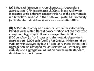
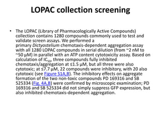
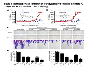
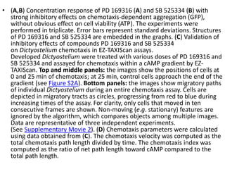
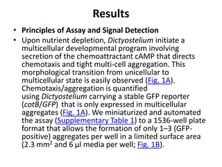
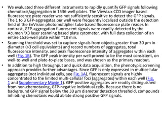
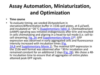
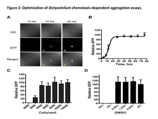
The document describes the development of a high-throughput screening assay using Dictyostelium discoideum for identifying novel chemotaxis inhibitors, which are essential in various biological processes and disease pathogenesis. The assay utilizes a GFP marker to quantify cell aggregation in response to chemoattractants and has successfully identified two inhibitors from a screen of 1,280 compounds, confirming their effects on both Dictyostelium and human cells. The approach enables efficient screening of small molecule libraries, potentially leading to new therapeutic agents targeting chemotactic pathways.

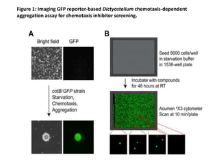

![• Since the GFP-expressing multicellular aggregates formed within each well have
different sizes and numbers, the total fluorescence intensity signals can vary
[coefficient of variation (CV) = 27%)] from well-to-well and plate-to-plate, with a Z’
factor of 0.47. Importantly, the parameters of the laser scanning plate cytometer
are set to only quantify GFP fluorescence presented in multicellular aggregates.
The design of the assay results in a “zero” signal when aggregation is completely
inhibited (see Fig. 1) and, consequently, in non-conventional signal-to-basal ratio
calculations. This high throughput assay is extremely robust, sensitive, and reliable
for the identification of novel chemotaxis inhibitors within small molecule libraries.
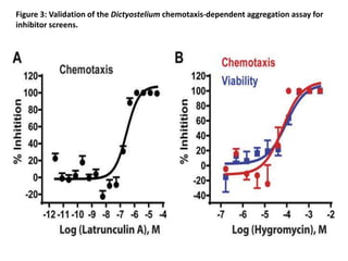
• Latrunculin A is a natural product, which binds actin monomers at 1:1 with a Kd of
200 nM in vitro. Inhibition of actin polymerization by latrunculin A disrupts actin
filament formation, cytoskeletal organization, cell migration, and chemotaxis. We
evaluated inhibitory activity of latrunculin A on chemotaxis-dependent aggregation
in the 1536-well format. Latrunculin A inhibited chemotaxis-dependent aggregation
with an IC50 of 282 nM (95% confidence interval, 193–412 nM; Fig. 3A), similar to
that of mammalian cells. Results demonstrated that the Dictyostelium assay is an
effective alternate approach for compound screening to identify chemotaxis
inhibitors. While latrunculin A may cause lethality of oncogenic mammalian cells, it
is not toxic to Dictyostelium in our assay. When Dictyostelium are seeded in
starvation buffer, growth is arrested and non-dividing cells survive without actin-
dependent processes for external nutrient capture, cell division, or motility; cellular
energy, etc. derive from autophagy. When we simultaneously monitored dose
response effects of latrunculin A on aggregation and cell viability, complete
inhibition of aggregation is confirmed at 1 μM, whereas cells remained viable to
93+/−2.3%, as assayed by vital dye staining. Latrunculin A is more cytotoxic at
10 μM (~30% viability).](https://image.slidesharecdn.com/cellbiologyandgeneticsda-2-170421171942/85/Cell-biology-and-genetics-da-2-18-320.jpg)