More Related Content

PPTX

PPT

PPTX
Mitochondrial disorders overview 
PPTX
mitochondrialdiseases-140122103237-phpapp02.pptx 
PPTX
Mitochondrial-Diseases after all ninu wine shop store tappu.pptx 
PPTX

PPTX
MITOCHONDRIAL DISEASES PPT SANCHI.pptxycuvu 
PPTX
Mitochondrial genes and neurology Similar to Chapter05 mitochondria and deiseases.ppt

PPT
Lecture 3- Mitochondrial disease.ppt,,,,,,,,,, 
PPTX

PDF
Mitochondrial Function and Dysfunction 1st Edition A. Shapira (Eds.) 
PPTX

PPTX
Mitochondrial dysfunction in medical.pptx 
PDF

PPTX
GENETIC HETEROGENEITY OF MITOCHONDRIAL DISORDERS - Agnès Rötig 
PPT
Genetics of Mitochondrial disorders 
PDF
What is mitochondrial disease 
PPT

PPTX

PPTX

PPTX
Mitochondrial dna and dysfunctions 
PPTX
mitochondrial. disease.pptx 
PPTX
Mitochondria , its importance in neurosciences 
PPTX
everything about mitochondria, discovery and transfer 
PPTX
Mitochondrial Medicine - Mosul project 
PPTX

PPTX
Mitochondrial inheritance 
PPT
More from Thuyamani M

PPTX
Tolperisone-A-Comprehensive-Overview for painful(1).pptx 
PPTX
SWOT_Infographic_Light slides for business infographics found in business ana... 
PPTX
Stroke presentation-170712123573032.pptx 
PPT
Management_stroke in stroke patients.ppt 
PPTX
ICU Nursing Intervention Clinical Case by Slidesgo.pptx 
PPTX
Stroke Program Orientation for Medical Staff.pptx 
PPTX
notes cell respiration mitochondria.pptx 
PPT
Natural Trees for the future oil part 3.ppt 
PPT
Natural Trees for the future oil part 1.ppt 
PPT
CS All About Sunscreens UV Radiation Slides.ppt 
PDF
Sun Safety and Skin Cancer Prevention.pdf 
PPTX
Wed journaldsfgojggfd;msdfgmdfs;lfgsd;g.pptx 
PPTX
Stroke for nurses shadkfahfjflkfdslkxm.pptx 
PPTX
brivaracetam drug information for patients.pptx 
PPTX
NATIONAL EPILEPSY DAY POSTER CONTENT.pptx 
PPTX
20 Best sales objections handling techniques.pptx 
PDF
Use of New Antiepileptic Agents, Esteem Pharma 
PPTX
Allergic Conjunctivitis, Global Market Analysis, Insights, and Forecast, 2021... 
PPTX
Sales by Month Infographics by Slidesgo.pptx 
PPTX
Sleep deprivation and memory in Insomnia.pptx Recently uploaded

PPTX
Biotransformation and Excretion 
PDF
Top 9 Websites for Purchasing Google Voice Accounts Online.pdf 
PPTX
From Planning to Delivery: Precision Workflows That Reduce Errors, Toxicity, ... 
PPTX
EBOLA VIRUS DISEASE ppt -Ayurveda Detailing 
PPTX
Chemistry of Hemoglobin (Types, Derivatives and Disorders) 
PPTX
Anaesthesia in Hypertension (anaesthesia in intercurrent diseases) 
PDF
Scholar.postdoc - Anaesthesia: Dalamagka, Maria 
PDF
4_5857478915135641976.pdf physiology of menstruation 
PPTX
Back Pain Evaluation: Red Flags, Neurological Examination & Special Tests | S... 
PPTX
Neonatal intestinal obstruction - MIRACLE.pptx 
PPTX
Streptococcus pathogenic species....pptx 
PPTX
BILIARY TREE RADIOLOGY ADVANCE IMAGING (USG, CT SCAN, MRI AND ERCP) OF ITS AN... 
PPTX
Diabetic retinopathy- Investigation and Management 
PDF
From Classification to Collaboration: Applying the International Classificati... ![Hypothalamus short ppt by Dr. Neha [PT].pptx](https://cdn.slidesharecdn.com/ss_thumbnails/hypothalamusbydr-260124145759-b9f94a93-thumbnail.jpg?width=640&height=640&fit=bounds)
PPTX
Hypothalamus short ppt by Dr. Neha [PT].pptx 
PPTX
Infective Endocarditis: Epidemiology, Diagnosis, Imaging, and Management 
PDF
Abdominal Muscles (Ant. & Post).pdf anatomy 
PPTX
COLORECTAL CARCINOMA GDMC for UG Class Presentation 
PPTX
Hormonal IUS (Mirena®) Common Myths.pptx 
PDF
Autoimmune diseases in children and anesthesia. Dalamagka, Maria Chapter05 mitochondria and deiseases.ppt
- 1.
© 2020 ElsevierInc. All rights reserved.
Chapter 5
Mitochondria and Diseases
- 2.
© 2020 ElsevierInc. All rights reserved. 2
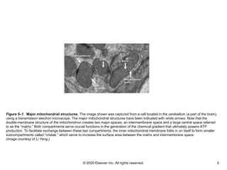
Figure 5–1. Major mitochondrial structures. The image shown was captured from a cell located in the cerebellum (a part of the brain),
using a transmission electron microscope. The major mitochondrial structures have been indicated with white arrows. Note that the
double-membrane structure of the mitochondrion creates two major spaces: an intermembrane space and a large central space referred
to as the “matrix.” Both compartments serve crucial functions in the generation of the chemical gradient that ultimately powers ATP
production. To facilitate exchange between these two compartments, the inner mitochondrial membrane folds in on itself to form smaller
subcompartments called “cristae,” which serve to increase the surface area between the matrix and intermembrane space.
(Image courtesy of Li Yang.)
- 3.
© 2020 ElsevierInc. All rights reserved. 3
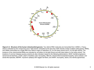
Figure 5–2. Structure of the human mitochondrial genome. Two distinct RNA molecules are transcribed from mtDNA: a “heavy
strand” (indicated by the orange line) and a “light strand” (indicated by the blue line). Both strands contain multiple coding genes, tRNAs,
and rRNAs transcribed as a single RNA from distinct origins of replication (OH for the heavy strand and OL for the light strand). The
locations of the mitochondrial tRNAs are indicated by red letters on the light strand and with black letters on the heavy strand. The
locations of common disease-causing variants are also indicated, based on the following abbreviations: DEAF, familial progressive
sensorineural deafness; LHON, Leber’s hereditary optic neuropathy; MELAS, mitochondrial encephalomyopathy, lactic acidosis, and
stroke-like episodes; MERRF, myoclonic epilepsy with ragged red fibers; and NARP, neuropathy, ataxia, and retinitis pigmentosa.
- 4.
© 2020 ElsevierInc. All rights reserved. 4
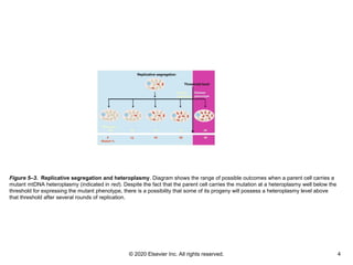
Figure 5–3. Replicative segregation and heteroplasmy. Diagram shows the range of possible outcomes when a parent cell carries a
mutant mtDNA heteroplasmy (indicated in red). Despite the fact that the parent cell carries the mutation at a heteroplasmy well below the
threshold for expressing the mutant phenotype, there is a possibility that some of its progeny will possess a heteroplasmy level above
that threshold after several rounds of replication.
- 5.
© 2020 ElsevierInc. All rights reserved. 5
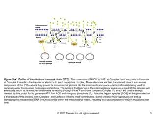
Figure 5–4. Outline of the electron transport chain (ETC). The conversion of NADH to NAD+
at Complex I and succinate to fumarate
at Complex II results in the transfer of electrons to each respective complex. These electrons are then transferred to each successive
component of the ETC—where they power the movement of protons into the intermembrane space—before ultimately being used to
generate water from oxygen molecules and protons. The protons that build up in the intermembrane space as a result of this process will
eventually return to the mitochondrial matrix by moving through the ATP synthase complex (Complex V), which will use the energy
created by this proton flux to generate ATP from ADP and inorganic phosphate (Pi). Reactive oxygen species (ROS) will be generated as
a byproduct of this process, with Complex I and Complex III being major contributors. Some of these ROS byproducts will end up
damaging the mitochondrial DNA (mtDNA) carried within the mitochondrial matrix, resulting in an accumulation of mtDNA mutations over
time.
- 6.
© 2020 ElsevierInc. All rights reserved. 6
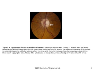
Figure 5–5. Optic atrophy induced by mitochondrial disease. The image shown is of the fundus (i.e., the back of the eye) from a
patient carrying a mutation associated with both mitochondrial dysfunction and optic atrophy. The white spot in the center of the image is
the optic disk (the point of exit for the optic nerve from the retina), while the rest of the image shows the retina proper, along with the
blood vessels supplying the retina. Atrophy of the optic disk can be clearly observed along the edge of the optic disk (white arrow).
- 7.
© 2020 ElsevierInc. All rights reserved. 7
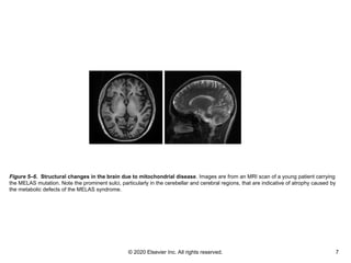
Figure 5–6. Structural changes in the brain due to mitochondrial disease. Images are from an MRI scan of a young patient carrying
the MELAS mutation. Note the prominent sulci, particularly in the cerebellar and cerebral regions, that are indicative of atrophy caused by
the metabolic defects of the MELAS syndrome.
- 8.
© 2020 ElsevierInc. All rights reserved. 8
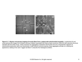
Figure 5–7. Electron microscopy imaging of muscle fibers from a patient with mitochondrial myopathy. Longitudinal (A) and
cross-sectional (B) images from a patient carrying a deletion associated with Kearns-Sayre syndrome. Note the dark, granular structures
indicated with the white arrows. These inclusions are aggregates of diseased mitochondria and are a characteristic signature of
mitochondrial dysfunction within the mitochondria. Under Gömöri trichrome staining, these aggregates will take on a striking red
appearance, leading to the name “ragged red fibers” to describe this histological condition.
- 9.
© 2020 ElsevierInc. All rights reserved. 9
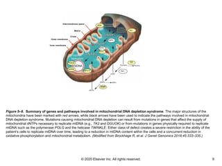
Figure 5–8. Summary of genes and pathways involved in mitochondrial DNA depletion syndrome. The major structures of the
mitochondria have been marked with red arrows, while black arrows have been used to indicate the pathways involved in mitochondrial
DNA depletion syndrome. Mutations causing mitochondrial DNA depletion can result from mutations in genes that affect the supply of
mitochondrial dNTPs necessary to replicate mtDNA (e.g., TK2 and DGUOK) or from mutations in genes physically required to replicate
mtDNA such as the polymerase POLG and the helicase TWINKLE. Either class of defect creates a severe restriction in the ability of the
patient’s cells to replicate mtDNA over time, leading to a reduction in mtDNA content within the cells and a concurrent reduction in
oxidative phosphorylation and mitochondrial metabolism. (Modified from Brockhage R, et al. J Genet Genomics 2018;45:333–335.)
- 10.
© 2020 ElsevierInc. All rights reserved. 10
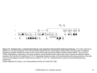
Figure 5–9. Pedigree from a mitochondrial disease case treated by mitochondrial replacement therapy. The mother referred for
treatment in this case (labeled as “III-9”) had previously experienced four miscarriages and two children who died early in childhood.
Sequencing analysis revealed the mother to be a carrier of the Leigh syndrome mutation (mtDNA mutation 8993T > G) at 23.27%–
33.65% heteroplasmy, depending on the tissue analyzed. Oocyte spindle transfer method was used to replace the defective mtDNA,
resulting in the live birth of a health boy (IV-12). To separately indicate the status of the mitochondrial and nuclear genomes in this child,
his box on the pedigree has been divided into two sections: “N” and “mt,” representing the nuclear and mitochondrial genomes,
respectively.
(Image originally from Zhang J, et al. Reprod Biomed Online 2017;34(4):361–368.)