Introduction to ArtificiaI Intelligence in Higher Education
Chapter 2 Hematopoiesis.ppt
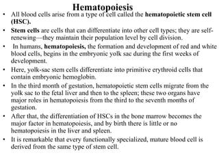
1. Hematopoiesis
• All blood cells arise from a type of cell called the hematopoietic stem cell
(HSC).
• Stem cells are cells that can differentiate into other cell types; they are self-
renewing—they maintain their population level by cell division.
• In humans, hematopoiesis, the formation and development of red and white
blood cells, begins in the embryonic yolk sac during the first weeks of
development.
• Here, yolk-sac stem cells differentiate into primitive erythroid cells that
contain embryonic hemoglobin.
• In the third month of gestation, hematopoietic stem cells migrate from the
yolk sac to the fetal liver and then to the spleen; these two organs have
major roles in hematopoiesis from the third to the seventh months of
gestation.
• After that, the differentiation of HSCs in the bone marrow becomes the
major factor in hematopoiesis, and by birth there is little or no
hematopoiesis in the liver and spleen.
• It is remarkable that every functionally specialized, mature blood cell is
derived from the same type of stem cell.
2. Hematopoiesis Cont----
• Incontrast to a unipotent cell, which differentiates into a single cell
type.
• A hematopoietic stem cell is multipotent, or pluripotent, able to
differentiate in various ways.
• And thereby generate erythrocytes, granulocytes, monocytes, mast
cells, lymphocytes, and megakaryocytes.
HSCs display an enormous proliferative capacity.
• This can be demonstrated in mice whose hematopoietic systems have
been completely destroyed by a lethal dose of x-rays.
• Such irradiated mice will die within 10 days unless they are infused
with normal bone-marrow cells from a syngeneic (genetically
identical) mouse.
• Although a normal mouse has 3x 10⁸ bone-marrow cells, infusion of
only 104–105 bone-marrow cells (i.e., 0.01%–0.1% of the normal
amount) from a donor is sufficient to completely restore the
hematopoietic system which demonstrates the enormous proliferative
and differentiative capacity of the stem cells.
3. Hematopoiesis Cont----
• Early in hematopoiesis, a multipotent stem cell differentiates along one
of two pathways, giving rise to either a common lymphoid progenitor
cell or a common myeloid progenitor cell.
4. Development of the immune system
NK cell
Stem cell
Macrophage
Lymphoid
progenitor
Myeloid
progenitor
T cell
B cell
Plasma Cell
Granulocyte
Monocyte
Mast cell
Dendritic cell
5. Cells of the immune system: innate
• Phagocytes
– Monocytes/macrophages
– PMNs/neutrophils
• NK cells
• Basophils and mast cells
• Eosinophils
• Platelets
Cells of the immune system: adaptive
• Lymphocytes
– B cells
• Plasma cells (Ab producing)
– T cells
• Cytotoxic (Cytotoxic T Lymphocyte)
• Helper (T helper)
• Th1 & Th2
6. Phagocytes – neutrophils (PMNs)
Neutrophil
Geimsa stain
• Characteristic nucleus, cytoplasm &
Granules
• CD66 membrane marker protein
• Represents approximately 50 to 70
percent of the total peripheral white blood
cells.
• They are released into the peripheral
blood and circulate for 7–10 h before
migrating into the tissues, where they
have a life span of only a 5 days.
• These are around 10 to 15 μm in diameter,
with a nucleus that has between two and
five lobes.
• The resulting transient increase in the
number of circulating neutrophils, called
leukocytosis, indication of infection.
7. Characteristics of neutrophil granules
Primary granules Secondary granules
Azurophilic; young neutrophils Specific for mature neutrophils
Contain:
cationic proteins, lysozyme, defensins,
elastase and
Contain:
Lysozyme,
NADPH oxidase components and
myeloperoxidase Lactoferrin
8. Eosinophils
• Characteristic bi-lobed nucleus
• Cytoplasmic granules, stain with
acidic dyes (eosin)
• Kill parasitic worms
• like neutrophils, are motile
phagocytic cells that can migrate
from the blood into the tissue
spaces.
• Eosinophils are approximately 12
to 15 μm in diameter.
• They normally make up between 1
and 3 percent of the circulating
white blood cells.
• Their most important role is
neutralizing basophil and mast cell
products and killing certain
parasites.
9. Characteristics of Eosinophil granules
Primary granules Secondary granules
Contain Specific for mature Eosinophil
Contain
acid phosphatase and arylsulfatase major basic protein
eosinophil cationic protein
eosinophil peroxidase
and eosinophil-derived neurotoxin
10. Basophils
• Basophils are found in very small
numbers, representing less than 1
percent of all circulating white
blood cells.
• Stained with Basic Dye (Methylene
Blue)
• 10 to 15 μm in diameter.
• Constituents of the granules are
histamine, a small amount of
heparin, and eosinophil chemotactic
factor-A.
• All have an important function in
inducing and maintaining
immediate hypersensitivity
reactions.
Basophil
11. Mast Cells
Mast Cell
• The precursors of these cells, are
formed in the bone marrow by
hematopoiesis, are released into the
blood as undifferentiated cells.
• They differentiate when they leave
the blood and enter the tissues.
• These can be found in a wide variety
of tissues, skin, connective tissues of
various organs, mucosal epithelial
tissue of the respiratory,genitourinary,
and digestive tracts.
• Have large numbers of cytoplasmic
granules that contain histamine and
pharmacologically active substances.
• Play an important role in the
development of allergies with
basophils.