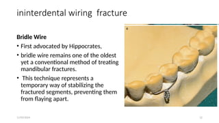
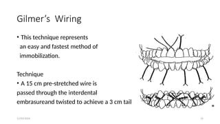
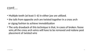
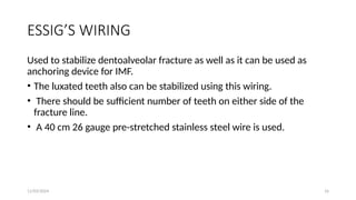
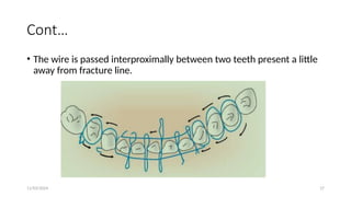
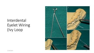
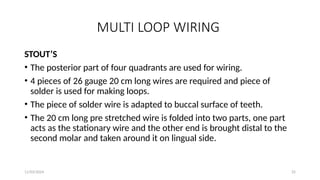
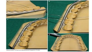
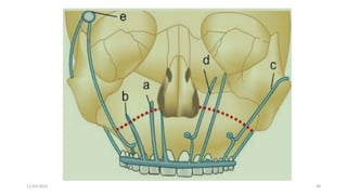
11/03/2024 1
Wire and Wiring Techniques
Prepared by Dr.Alex Asmamaw( DMD, OCMFS )
11/03/2024 2
outline
• Objective
• Introduction
• Closed reduction
• Armamentarium and Principles
• Direct ,indirect, multiloop wiring and Arch bare
• Internal skeletal suspension wiring
• Intraosseous wiring
• Summary
• Reference
11/03/2024 3
Objective
in this seminar we will cover
• different types of wire like ss & k wires
• Different types of wiring technique
• Wiring in edentulous patients
• Use of suspension wire in mid face fractures
11/03/2024 4
INTRODUCTION
•wiring is one of the oldiest techniques truma mangment
• The wire can only provide stability by means of its ability
to prevent gap
• the wire cannot neutralize torsion or shear forces
• wireing technique can be applied for MMF , suspensions or
interossious fixation
11/03/2024 5
closed reduction
indication of closed reduction
• Nondisplaced favorable fractures
• Grossly comminuted fractures
• Fractures in children involving the developing dentition
• Coronoid fractures
• treatment of condylar fractures
Contraindicated in asthmatic, in parkinsonism, mentally challenged
patients
11/03/2024 6
cont...
advantage of closed reduction
1. Day-care procedure, not require hospitalization
2. Cost-effective and safe
3. Avoid damage to the vital structure
disadvantage
1. Restriction of mouth opening for minimum of 4 weeks
2. Patients can take only liquid diet
11/03/2024 7
cont...
3. Diffcult to maintain oral hygiene
4. Challenging in partially edentulous patients
5. Weight loss
6. Partial trismus for few weeks
11/03/2024 8
Armamentarium and Principles
Basic principles to be followed are :
1. Continuous tension on the wire while twisting and tightening.
2. Force should be apically directed while tightening.
3. Utilizing a clock wise direction for all tightening/twisting.
4. Only half a turn to be given at a time at the end.
11/03/2024 9
CONT...
5. Once the tail is cut, it is turned and placed into the inter-proximal
embrasures.
6. Once all the wires are in place, move the fnger around the arch
bar/wire to check for any sharp edge/margin/ wire ending.
11/03/2024 10
Armamentarium
1. Presterilized 26 gauge stainless steel wires cut into
lengths of 20 cm each.
• Wires are always cut on a bevel,.
• The wires should be prestretched about 10 % to prevent
loosening after fixing it to the teeth.
2. Two needle holders or wire holders.
3. Wire cutters
4. Kelsey-Fry bone-awl introducer
11/03/2024 11
Direct ,indirect,multiloop wiring and Arch bare
Direct Indirect
• BRIDLE WIRE IVY LOOPING
• GILMER’S ERNST
• ESSIG’S WIRING
• RISDON’S WIRING
Continuous or Multiloop
STOUT’S
OBWEGESER
11/03/2024 12
ininterdental wiring fracture
Bridle Wire
• First advocated by Hippocrates,
• bridle wire remains one of the oldest
yet a conventional method of treating
mandibular fractures.
• This technique represents a
temporary way of stabilizing the
fractured segments, preventing them
from flaying apart.
11/03/2024 13
cont...
Advantages
• include stabilization of the two fractured
• segments preventing further damage to the adjacent soft tissues,
• maintenance of the airway patency (especially advantageous for the
fractures of the anterior mandible),
• decrease pain, and reducing damage to the neurovascular bundle
from immobilizing the fractured fragments,
11/03/2024 14
Gilmer’s Wiring
• This technique represents
an easy and fastest method of
immobilization.
Technique
• A 15 cm pre-stretched wire is
passed through the interdental
embrasureand twisted to achieve a 3 cm tail
11/03/2024 15
cont...
• Multiple teeth (at least 5–6) in either jaw are utilized.
• the tails from opposite arch are twisted together in a cross arch
or zigzag fashion to achieve immobilization
• The only drawback of this technique is that, in cases of broken /loose
wire,all the cross arch wires will have to be removed and redone post
placement of twisted wire
11/03/2024 16
ESSIG’S WIRING
Used to stabilize dentoalveolar fracture as well as it can be used as
anchoring device for IMF.
• The luxated teeth also can be stabilized using this wiring.
• There should be sufficient number of teeth on either side of the
fracture line.
• A 40 cm 26 gauge pre-stretched stainless steel wire is used.
11/03/2024 17
Cont…
• The wire is passed interproximally between two teeth present a little
away from fracture line.
11/03/2024 18
cont...
• The wires are passed around the teeth in a figure of 8 manner until
they reach 2 to 3 teeth away from the fracture line.
• Now the wires are passed without looping to the other side of the
fracture line and 2 to 3 teeth away from the fracture line on the
opposite side.
• Again the wires are taken around 2 to 3 teeth in a figure of 8 manner.
• Now this acts as an arch bar on which the other smaller wires are
tightened to stabilize the fracture.
11/03/2024 19
RISDON’S WIRING
• It is commonly used method of horizontal wire fixation.
• This can be a substitute technique for arch bar.
• In this method second molars are usually chosen for anchorage on
either side.
• A 25 cm long 26 guage wire is passed around the neck of second
molar on each side and both the ends are brought in buccal side
11/03/2024 20
11/03/2024 21
Cont…
• The ends are twisted for entire length thus forming a strong base wire
that comes towards the midline from each second molars.
• Two base wires are grasped and twisted at mid line and adapted to
the necks of the teeth on the buccal side . 
• This base wire is secured to individual teeth by using additional
interdental wires. 
• This type of horizontal wiring offers strong fixation.
11/03/2024 22
Indirect wireing
Eyelet Wiring (Ivy Loop Method
• eyelet wiring is a simple and effective method of reduction and
immobilization of jaw fracture
• Used when fracture lines are favorable and displacement minimal
• It is advisable in cases where many teeth are present in pairs in both
arches.
• May also be used in combination with gunning type splints in an
apposing edentulous jaw, and arch bars or partially dentate jaw.
11/03/2024 23
Interdental
Eyelet Wiring
(Ivy Loop
11/03/2024 24
Hallam modification Button wiring clove hitch
technique
11/03/2024 25
MULTI LOOP WIRING
STOUT’S
• The posterior part of four quadrants are used for wiring.
• 4 pieces of 26 gauge 20 cm long wires are required and piece of
solder is used for making loops.
• The piece of solder wire is adapted to buccal surface of teeth.
• The 20 cm long pre stretched wire is folded into two parts, one part
acts as the stationary wire and the other end is brought distal to the
second molar and taken around it on lingual side.
11/03/2024 26
11/03/2024 27
cont...
• This working end is threaded through the mesial side of second molar
to the buccal side under the solder wire.
• It is then looped around the stationary wire and solder wire and back
into the interdental space from buccal to lingual.
• The same procedure is repeated for each tooth up to midline.
11/03/2024 28
Cont…
• The solder wire is removed after the loops are formed and the loops
are twisted to form eyelets.
• Finally the stationary and working ends of the wires are twisted
together.
11/03/2024 29
Multiple loop wiring (Obwegeser method)
11/03/2024 30
Cont…
• This technique utilizes a 30 cm-long wire that is bent into a
continuous “W” form
• The elevated portion of the arcade acts to form the loop, whereas the
depressed portion adapts to the contour of the teeth palatally or
lingually.
• The distal end of the wire arcade is kept long enough so that it will
follow the buccal contour of the arch, when it is passed through the
interdental space of the most posterior tooth from lingual/palatal to
buccal side.
•
11/03/2024 31
Cont…
• The elevated ends of the wire are held together and twisted
• The loops can be used for both elastic traction and wires for securing
MMF.
• The disadvantage of this technique is that it is very cumbersome and
adequate expertise is needed to carry out this technique.
11/03/2024 32
ARCH BAR FIXATION
• The arch bar is a flat, sturdy stainless steel bar on which fleats or
hooks are attached.
• It is an effective, quick and inexpensive method of fixation.
• Commercially available arch bars are usually customized or
prefabricated.
• In most clinical scenarios, prefabricated arch bars like Erich’s, Jelenko
are the need for placement
11/03/2024 33
Jelenko arch Erich’s arch bar
11/03/2024 34
Cont…
Indications
1. Partially edentulous patients where placement of eyelets is not an
option due to insufficient number of teeth.
2. The teeth present are far apart making MMF impossible.
3. For alignment of avulsed teeth/displaced dentoalveolar fracture into
proper arch form.
4. Reduction and immobilization of dentoalveolar component/ fracture.
11/03/2024 35
Cont…
• 5. As a part of integrated treatment for suspension for treatment of
maxillofacial fractures.
• The arch bar is measured to fit from first molar to first molar.
• The arch bar is placed in such a way that the fleats or hooks face
towards the gingival margin.
11/03/2024 36
Cont…
• Now 15 cm of 26 guage wire is taken and starting from the distal
tooth, the wire is passed from buccal to lingual side below the arch
bar and from lingual to buccal above the arch bar and twisted
together.
• This is continued for all the teeth and the arch bar is secured.
• When placing an arch bar across a displaced fracture segment,it is cut
at the fracture site and placed separately.
11/03/2024 37
acrylic splint
Lateral compression splint/ open cap acrylic splint
• for the stabilization of mandibular arch.
• Mainly used in cases of children, where there is
mixed dentition and presence of developing teeth
buds
• It can be also used in adult mandibular body
fracture,
11/03/2024 38
GUNNING –TYPE SPLINTS
11/03/2024 39
Cont…
Indications:
• For the unilateral and bilateral fractures of the edentulous
mandible, where the fractures lying proximal to these areas can
be controlled by inter maxillary fixation.
Contraindications:
• Unfavourably displaced fracture lying outside the denture
bearing areas.
11/03/2024 40
Cont…
• Severe posterior displacement of fractures of the anterior part of the
mandible which will probably be inadequately controlled by this
method alone and requires additional fixation.
• Projectile injuries, involving grossly comminuted soft tissue and bone
loss.
• Extreme atrophy of the maxilla or mandible.
11/03/2024 41
PER ALVEOLAR WIRING
• Two perialveolar wires are placed in the canine region on either side
for fitting patient’s own denture to alveolar ridge.
• The splint is firmly placed in the position in the upper jaw.
• A kelsey-Fry bone awl introducer is pushed from buccal to palatal
aspect.
• A 26 gauge wire is threaded through the eye and the wire is
withdrawn with it the wire on the buccal surface.
11/03/2024 42
• Same procedure is
repeated on
opposite side and
then the splint is
placed in the mouth
and wires adjusted
over it and twisted
over the grooves
and the ends tucked
inwards
11/03/2024 43
CIRCUMMANDIBULAR WIRING
OBWEGESER’S PROCEDURE
• It is used for fixation of lateral compression splint to the mandibular
bone.
• Lower border of mandible is palpated in the canine region and the
skin is pierced beneath the lower border of the mandible by Kelsey-
Fry bone awl and it emerges through the floor of mouth.
11/03/2024 44
Cont…
• A 26 or 28 guage wire is inserted through the eye of the awl and
the awl is withdrawn till the lower border and directed upward
along the buccal surface of mandible to pierce through the buccal
sulcus.
• The two ends of the wire are adjusted and the splint is adjusted
and the lingual and buccal wires are held together and twisted in
the region of canine grooves, cut and finished inward
11/03/2024 45
11/03/2024 46
Prolonged use of MMF
• IMF are placed in the for 4- 6 weeks
• prolonged placement of will cause
- ankylosis of TMJ
- wight loss
- Poor oral hygiene
- Malnutrition
11/03/2024 47
Internal Suspension wiring
• The principle of internal wire suspension was first described by
Adams in 1943
• the wires are passed subcutaneously from stable skeletal
points into the oral cavity and are fixed on both sides to the
arch bars under traction (craniofacial suspension).
• Depending on the type of fracture, the point of fixation may vary.
• The wires are always passed from a stable point above the fracture
line.
11/03/2024 48
11/03/2024 49
Lateral frontal suspension
• le Fort III and II (Mandible stable)
• An incision is made in the lateral 3rd of the eyebrow to expose the
zygomatic process of the frontal bone
• A bur hole is drilled 5mm above the suture line and angulated to
emerge on the post of infra-temporal fossa.
• A 40cm length of soft stainless steel wire is passed through this
and bent backwards and then threaded through the eye of a Rowe
zygomatic awl.
11/03/2024 50
• The awl is then passed
downwards and forward
behind the frontal
process of the
zygomatic bone to
pierce the oral mucosa
adjacent to the molar
tooth and is secured to
a splint or arch bar.
11/03/2024 51
Central frontal wire
• leFort III and II (Mandible unstable)
• 2cm horizontal incision is done on central forehead
• Roger “Anderson pin” inserted about 2cm subcutaneous tissue
tunnel.
• An awl is then passed upwards through the soft tissues, entering via
the vestibule of the mouth in the upper canine region
• passing posterolateral to the piriform margin of the nose and in front
of the lacrimal
11/03/2024 52
a silicone tube below the pin to allow them to diverge at a lower point over
the glabella
11/03/2024 53
Circum zygomatic
• Le Fort II and I
• The instrument pierces the temporal fascia and keeping the point
close to the deep or temporal surface
• enter the upper buccal sulcus in the first molar region.
• The wire is attached, and the tip of the introducer withdrawn to lie
just above the arch without emerging from the skin and movelateral
to and down to the arch
• emerge through the original point of entry of the wire in the buccal
sulcus and fix on arch bar
11/03/2024 54
Obwegeser technique
11/03/2024 55
Zygomatic suspension
• This method was advocated by Guerman (1957).
• Used in le fort I
• A 3 cm incision is made in the upper buccal sulcus above the
premolar and molar teeth and the soft tissues are dissected
subperiosteally to expose the base of the zygomatic buttress
• A bur hole is drilled posteriorly and slightly laterally and a
soft stainless-steel wire is passed.
11/03/2024 56
Cont…
• The two ends are then attached to a suitable loop on either
the arch bar or silver cap splint.
• The deep aspect of the periosteum should not be
penetrated so as to avoid herniation of the buccal pad of
fat.
Infra-orbital suspension
• A 3 cm incision is made in the upper buccal sulcus above the
canine tooth,
• the soft tissues are dissected subperiosteally to expose the
inferior orbital margin lateral to the infra-orbital foramen.
• A bur hole is drilled upwards and posteriorly and a soft
stainless-steel wire is passed.
• During the drilling of the bur hole and the threading of the wire
the globe of the eye should be protected with the rounded end
of a Howarth’s periosteal elevator.
11/03/2024 58
Cont…
• The end of the wire is then withdrawn
into the mouth and the two ends are
attached to a suitable loop or cleat on
the arch bar or silver cap splint
Piriform aperture suspension
• A transverse 2 cm incision is made in the upper labial sulcus
directly above the lateral incisor tooth
• the periosteum is reflected to expose the bony piriform aperture of
the nose,
• The nasal mucosa is then elevated from the medial aspect to a
depth of 2 cm
11/03/2024 60
• the periosteal elevator is left in position to act as a guard for this
vascular tissue.
Cont…
• A bur hole is drilled threading the wires as described above and
subsequent fsoft stainless-steel wire is passed.
• The two ends are withdrawn into the mouth and attached to a
suitable loop or cleat depending on the method of intermaxillary
fixation used.
• In children, particularly when united to a circum mandibular wire
(also repeated on the contralateral side), a very rigid and stable
fixation can be achieved in this way
11/03/2024 62
Transosseous or intraosseous wiring
Direct bone wiring in the mandible
1. the posterior fragment fractures,
2. the edentulous mandibular fracture,
3. Compound mandibular fracture, and
4. the lower border of the mandible in the presence of multiple
fractures where the upper border of the jaw has been secured by one
of the conventional methods of fixation,
11/03/2024 63
Cont…
• The direct wiring of mid-face fractures
which occur at normal suture lines
1.Fronto-nasal;
2. Fronto-maxillary;
3. Fronto-zygomatic
4.Maxillary-zygomatic.
11/03/2024 64
Cont…
When the fracture does not occur at a normal suture line
the commoner sites for direct wiring are:
1. Inferior and
lateral orbital margins;
2. Superior orbital margin;
3. Zygomatic arch;
4. Palatal processes of the maxillae
11/03/2024 65
Transfixation with Kirschner wire
• comparison with other techniques the appliances are extremely
simple and unobtrusive and
• nursing care is, in many respects, less of a problem.
• the transfixation wire can be readily removed without recourse to
any form of anesthesia
• short time of operation
11/03/2024 66
Cont…
• it can provide control in only a single plane
• A hand drill or Toller type is used for
insertion of the wire or,
• in the absence of such an instrument, a
Kirschner-type hand drill
• The standard 1.5 mm diameter Kirschner
wire and 2 mm Toller wire
11/03/2024 67
Indication
• Le Fort II maxillary fractures
without comminution
• condyle head fracture
• in the treatment of the
elderly or ill patient
• contraindicated in the
management of severely
displaced single fractures of
the middle third of the
facialskeleton
11/03/2024 68
summary
• wiring technique is important management of fascial bone fracture
• Different wire techniques are used based on the dentition of the
patient can be incorporated with plates
• used in both dentoalveolar and fascial bone fracture
11/03/2024 69
References
• Textbook of Oral and Maxillofacial Surgery neelima milek
• Maxillofacial Injuries,Rowe and Williams
• Oral and Maxillofacial Surgery for the Clinician

chap-3c Wire and Wiring Techniques s.pptx

  • 1.
    11/03/2024 1 Wire andWiring Techniques Prepared by Dr.Alex Asmamaw( DMD, OCMFS )
  • 2.
    11/03/2024 2 outline • Objective •Introduction • Closed reduction • Armamentarium and Principles • Direct ,indirect, multiloop wiring and Arch bare • Internal skeletal suspension wiring • Intraosseous wiring • Summary • Reference
  • 3.
    11/03/2024 3 Objective in thisseminar we will cover • different types of wire like ss & k wires • Different types of wiring technique • Wiring in edentulous patients • Use of suspension wire in mid face fractures
  • 4.
    11/03/2024 4 INTRODUCTION •wiring isone of the oldiest techniques truma mangment • The wire can only provide stability by means of its ability to prevent gap • the wire cannot neutralize torsion or shear forces • wireing technique can be applied for MMF , suspensions or interossious fixation
  • 5.
    11/03/2024 5 closed reduction indicationof closed reduction • Nondisplaced favorable fractures • Grossly comminuted fractures • Fractures in children involving the developing dentition • Coronoid fractures • treatment of condylar fractures Contraindicated in asthmatic, in parkinsonism, mentally challenged patients
  • 6.
    11/03/2024 6 cont... advantage ofclosed reduction 1. Day-care procedure, not require hospitalization 2. Cost-effective and safe 3. Avoid damage to the vital structure disadvantage 1. Restriction of mouth opening for minimum of 4 weeks 2. Patients can take only liquid diet
  • 7.
    11/03/2024 7 cont... 3. Diffcultto maintain oral hygiene 4. Challenging in partially edentulous patients 5. Weight loss 6. Partial trismus for few weeks
  • 8.
    11/03/2024 8 Armamentarium andPrinciples Basic principles to be followed are : 1. Continuous tension on the wire while twisting and tightening. 2. Force should be apically directed while tightening. 3. Utilizing a clock wise direction for all tightening/twisting. 4. Only half a turn to be given at a time at the end.
  • 9.
    11/03/2024 9 CONT... 5. Oncethe tail is cut, it is turned and placed into the inter-proximal embrasures. 6. Once all the wires are in place, move the fnger around the arch bar/wire to check for any sharp edge/margin/ wire ending.
  • 10.
    11/03/2024 10 Armamentarium 1. Presterilized26 gauge stainless steel wires cut into lengths of 20 cm each. • Wires are always cut on a bevel,. • The wires should be prestretched about 10 % to prevent loosening after fixing it to the teeth. 2. Two needle holders or wire holders. 3. Wire cutters 4. Kelsey-Fry bone-awl introducer
  • 11.
    11/03/2024 11 Direct ,indirect,multiloopwiring and Arch bare Direct Indirect • BRIDLE WIRE IVY LOOPING • GILMER’S ERNST • ESSIG’S WIRING • RISDON’S WIRING Continuous or Multiloop STOUT’S OBWEGESER
  • 12.
    11/03/2024 12 ininterdental wiringfracture Bridle Wire • First advocated by Hippocrates, • bridle wire remains one of the oldest yet a conventional method of treating mandibular fractures. • This technique represents a temporary way of stabilizing the fractured segments, preventing them from flaying apart.
  • 13.
    11/03/2024 13 cont... Advantages • includestabilization of the two fractured • segments preventing further damage to the adjacent soft tissues, • maintenance of the airway patency (especially advantageous for the fractures of the anterior mandible), • decrease pain, and reducing damage to the neurovascular bundle from immobilizing the fractured fragments,
  • 14.
    11/03/2024 14 Gilmer’s Wiring •This technique represents an easy and fastest method of immobilization. Technique • A 15 cm pre-stretched wire is passed through the interdental embrasureand twisted to achieve a 3 cm tail
  • 15.
    11/03/2024 15 cont... • Multipleteeth (at least 5–6) in either jaw are utilized. • the tails from opposite arch are twisted together in a cross arch or zigzag fashion to achieve immobilization • The only drawback of this technique is that, in cases of broken /loose wire,all the cross arch wires will have to be removed and redone post placement of twisted wire
  • 16.
    11/03/2024 16 ESSIG’S WIRING Usedto stabilize dentoalveolar fracture as well as it can be used as anchoring device for IMF. • The luxated teeth also can be stabilized using this wiring. • There should be sufficient number of teeth on either side of the fracture line. • A 40 cm 26 gauge pre-stretched stainless steel wire is used.
  • 17.
    11/03/2024 17 Cont… • Thewire is passed interproximally between two teeth present a little away from fracture line.
  • 18.
    11/03/2024 18 cont... • Thewires are passed around the teeth in a figure of 8 manner until they reach 2 to 3 teeth away from the fracture line. • Now the wires are passed without looping to the other side of the fracture line and 2 to 3 teeth away from the fracture line on the opposite side. • Again the wires are taken around 2 to 3 teeth in a figure of 8 manner. • Now this acts as an arch bar on which the other smaller wires are tightened to stabilize the fracture.
  • 19.
    11/03/2024 19 RISDON’S WIRING •It is commonly used method of horizontal wire fixation. • This can be a substitute technique for arch bar. • In this method second molars are usually chosen for anchorage on either side. • A 25 cm long 26 guage wire is passed around the neck of second molar on each side and both the ends are brought in buccal side
  • 20.
  • 21.
    11/03/2024 21 Cont… • Theends are twisted for entire length thus forming a strong base wire that comes towards the midline from each second molars. • Two base wires are grasped and twisted at mid line and adapted to the necks of the teeth on the buccal side .  • This base wire is secured to individual teeth by using additional interdental wires.  • This type of horizontal wiring offers strong fixation.
  • 22.
    11/03/2024 22 Indirect wireing EyeletWiring (Ivy Loop Method • eyelet wiring is a simple and effective method of reduction and immobilization of jaw fracture • Used when fracture lines are favorable and displacement minimal • It is advisable in cases where many teeth are present in pairs in both arches. • May also be used in combination with gunning type splints in an apposing edentulous jaw, and arch bars or partially dentate jaw.
  • 23.
  • 24.
    11/03/2024 24 Hallam modificationButton wiring clove hitch technique
  • 25.
    11/03/2024 25 MULTI LOOPWIRING STOUT’S • The posterior part of four quadrants are used for wiring. • 4 pieces of 26 gauge 20 cm long wires are required and piece of solder is used for making loops. • The piece of solder wire is adapted to buccal surface of teeth. • The 20 cm long pre stretched wire is folded into two parts, one part acts as the stationary wire and the other end is brought distal to the second molar and taken around it on lingual side.
  • 26.
  • 27.
    11/03/2024 27 cont... • Thisworking end is threaded through the mesial side of second molar to the buccal side under the solder wire. • It is then looped around the stationary wire and solder wire and back into the interdental space from buccal to lingual. • The same procedure is repeated for each tooth up to midline.
  • 28.
    11/03/2024 28 Cont… • Thesolder wire is removed after the loops are formed and the loops are twisted to form eyelets. • Finally the stationary and working ends of the wires are twisted together.
  • 29.
    11/03/2024 29 Multiple loopwiring (Obwegeser method)
  • 30.
    11/03/2024 30 Cont… • Thistechnique utilizes a 30 cm-long wire that is bent into a continuous “W” form • The elevated portion of the arcade acts to form the loop, whereas the depressed portion adapts to the contour of the teeth palatally or lingually. • The distal end of the wire arcade is kept long enough so that it will follow the buccal contour of the arch, when it is passed through the interdental space of the most posterior tooth from lingual/palatal to buccal side. •
  • 31.
    11/03/2024 31 Cont… • Theelevated ends of the wire are held together and twisted • The loops can be used for both elastic traction and wires for securing MMF. • The disadvantage of this technique is that it is very cumbersome and adequate expertise is needed to carry out this technique.
  • 32.
    11/03/2024 32 ARCH BARFIXATION • The arch bar is a flat, sturdy stainless steel bar on which fleats or hooks are attached. • It is an effective, quick and inexpensive method of fixation. • Commercially available arch bars are usually customized or prefabricated. • In most clinical scenarios, prefabricated arch bars like Erich’s, Jelenko are the need for placement
  • 33.
    11/03/2024 33 Jelenko archErich’s arch bar
  • 34.
    11/03/2024 34 Cont… Indications 1. Partiallyedentulous patients where placement of eyelets is not an option due to insufficient number of teeth. 2. The teeth present are far apart making MMF impossible. 3. For alignment of avulsed teeth/displaced dentoalveolar fracture into proper arch form. 4. Reduction and immobilization of dentoalveolar component/ fracture.
  • 35.
    11/03/2024 35 Cont… • 5.As a part of integrated treatment for suspension for treatment of maxillofacial fractures. • The arch bar is measured to fit from first molar to first molar. • The arch bar is placed in such a way that the fleats or hooks face towards the gingival margin.
  • 36.
    11/03/2024 36 Cont… • Now15 cm of 26 guage wire is taken and starting from the distal tooth, the wire is passed from buccal to lingual side below the arch bar and from lingual to buccal above the arch bar and twisted together. • This is continued for all the teeth and the arch bar is secured. • When placing an arch bar across a displaced fracture segment,it is cut at the fracture site and placed separately.
  • 37.
    11/03/2024 37 acrylic splint Lateralcompression splint/ open cap acrylic splint • for the stabilization of mandibular arch. • Mainly used in cases of children, where there is mixed dentition and presence of developing teeth buds • It can be also used in adult mandibular body fracture,
  • 38.
  • 39.
    11/03/2024 39 Cont… Indications: • Forthe unilateral and bilateral fractures of the edentulous mandible, where the fractures lying proximal to these areas can be controlled by inter maxillary fixation. Contraindications: • Unfavourably displaced fracture lying outside the denture bearing areas.
  • 40.
    11/03/2024 40 Cont… • Severeposterior displacement of fractures of the anterior part of the mandible which will probably be inadequately controlled by this method alone and requires additional fixation. • Projectile injuries, involving grossly comminuted soft tissue and bone loss. • Extreme atrophy of the maxilla or mandible.
  • 41.
    11/03/2024 41 PER ALVEOLARWIRING • Two perialveolar wires are placed in the canine region on either side for fitting patient’s own denture to alveolar ridge. • The splint is firmly placed in the position in the upper jaw. • A kelsey-Fry bone awl introducer is pushed from buccal to palatal aspect. • A 26 gauge wire is threaded through the eye and the wire is withdrawn with it the wire on the buccal surface.
  • 42.
    11/03/2024 42 • Sameprocedure is repeated on opposite side and then the splint is placed in the mouth and wires adjusted over it and twisted over the grooves and the ends tucked inwards
  • 43.
    11/03/2024 43 CIRCUMMANDIBULAR WIRING OBWEGESER’SPROCEDURE • It is used for fixation of lateral compression splint to the mandibular bone. • Lower border of mandible is palpated in the canine region and the skin is pierced beneath the lower border of the mandible by Kelsey- Fry bone awl and it emerges through the floor of mouth.
  • 44.
    11/03/2024 44 Cont… • A26 or 28 guage wire is inserted through the eye of the awl and the awl is withdrawn till the lower border and directed upward along the buccal surface of mandible to pierce through the buccal sulcus. • The two ends of the wire are adjusted and the splint is adjusted and the lingual and buccal wires are held together and twisted in the region of canine grooves, cut and finished inward
  • 45.
  • 46.
    11/03/2024 46 Prolonged useof MMF • IMF are placed in the for 4- 6 weeks • prolonged placement of will cause - ankylosis of TMJ - wight loss - Poor oral hygiene - Malnutrition
  • 47.
    11/03/2024 47 Internal Suspensionwiring • The principle of internal wire suspension was first described by Adams in 1943 • the wires are passed subcutaneously from stable skeletal points into the oral cavity and are fixed on both sides to the arch bars under traction (craniofacial suspension). • Depending on the type of fracture, the point of fixation may vary. • The wires are always passed from a stable point above the fracture line.
  • 48.
  • 49.
    11/03/2024 49 Lateral frontalsuspension • le Fort III and II (Mandible stable) • An incision is made in the lateral 3rd of the eyebrow to expose the zygomatic process of the frontal bone • A bur hole is drilled 5mm above the suture line and angulated to emerge on the post of infra-temporal fossa. • A 40cm length of soft stainless steel wire is passed through this and bent backwards and then threaded through the eye of a Rowe zygomatic awl.
  • 50.
    11/03/2024 50 • Theawl is then passed downwards and forward behind the frontal process of the zygomatic bone to pierce the oral mucosa adjacent to the molar tooth and is secured to a splint or arch bar.
  • 51.
    11/03/2024 51 Central frontalwire • leFort III and II (Mandible unstable) • 2cm horizontal incision is done on central forehead • Roger “Anderson pin” inserted about 2cm subcutaneous tissue tunnel. • An awl is then passed upwards through the soft tissues, entering via the vestibule of the mouth in the upper canine region • passing posterolateral to the piriform margin of the nose and in front of the lacrimal
  • 52.
    11/03/2024 52 a siliconetube below the pin to allow them to diverge at a lower point over the glabella
  • 53.
    11/03/2024 53 Circum zygomatic •Le Fort II and I • The instrument pierces the temporal fascia and keeping the point close to the deep or temporal surface • enter the upper buccal sulcus in the first molar region. • The wire is attached, and the tip of the introducer withdrawn to lie just above the arch without emerging from the skin and movelateral to and down to the arch • emerge through the original point of entry of the wire in the buccal sulcus and fix on arch bar
  • 54.
  • 55.
    11/03/2024 55 Zygomatic suspension •This method was advocated by Guerman (1957). • Used in le fort I • A 3 cm incision is made in the upper buccal sulcus above the premolar and molar teeth and the soft tissues are dissected subperiosteally to expose the base of the zygomatic buttress • A bur hole is drilled posteriorly and slightly laterally and a soft stainless-steel wire is passed.
  • 56.
    11/03/2024 56 Cont… • Thetwo ends are then attached to a suitable loop on either the arch bar or silver cap splint. • The deep aspect of the periosteum should not be penetrated so as to avoid herniation of the buccal pad of fat.
  • 57.
    Infra-orbital suspension • A3 cm incision is made in the upper buccal sulcus above the canine tooth, • the soft tissues are dissected subperiosteally to expose the inferior orbital margin lateral to the infra-orbital foramen. • A bur hole is drilled upwards and posteriorly and a soft stainless-steel wire is passed. • During the drilling of the bur hole and the threading of the wire the globe of the eye should be protected with the rounded end of a Howarth’s periosteal elevator.
  • 58.
    11/03/2024 58 Cont… • Theend of the wire is then withdrawn into the mouth and the two ends are attached to a suitable loop or cleat on the arch bar or silver cap splint
  • 59.
    Piriform aperture suspension •A transverse 2 cm incision is made in the upper labial sulcus directly above the lateral incisor tooth • the periosteum is reflected to expose the bony piriform aperture of the nose, • The nasal mucosa is then elevated from the medial aspect to a depth of 2 cm
  • 60.
    11/03/2024 60 • theperiosteal elevator is left in position to act as a guard for this vascular tissue.
  • 61.
    Cont… • A burhole is drilled threading the wires as described above and subsequent fsoft stainless-steel wire is passed. • The two ends are withdrawn into the mouth and attached to a suitable loop or cleat depending on the method of intermaxillary fixation used. • In children, particularly when united to a circum mandibular wire (also repeated on the contralateral side), a very rigid and stable fixation can be achieved in this way
  • 62.
    11/03/2024 62 Transosseous orintraosseous wiring Direct bone wiring in the mandible 1. the posterior fragment fractures, 2. the edentulous mandibular fracture, 3. Compound mandibular fracture, and 4. the lower border of the mandible in the presence of multiple fractures where the upper border of the jaw has been secured by one of the conventional methods of fixation,
  • 63.
    11/03/2024 63 Cont… • Thedirect wiring of mid-face fractures which occur at normal suture lines 1.Fronto-nasal; 2. Fronto-maxillary; 3. Fronto-zygomatic 4.Maxillary-zygomatic.
  • 64.
    11/03/2024 64 Cont… When thefracture does not occur at a normal suture line the commoner sites for direct wiring are: 1. Inferior and lateral orbital margins; 2. Superior orbital margin; 3. Zygomatic arch; 4. Palatal processes of the maxillae
  • 65.
    11/03/2024 65 Transfixation withKirschner wire • comparison with other techniques the appliances are extremely simple and unobtrusive and • nursing care is, in many respects, less of a problem. • the transfixation wire can be readily removed without recourse to any form of anesthesia • short time of operation
  • 66.
    11/03/2024 66 Cont… • itcan provide control in only a single plane • A hand drill or Toller type is used for insertion of the wire or, • in the absence of such an instrument, a Kirschner-type hand drill • The standard 1.5 mm diameter Kirschner wire and 2 mm Toller wire
  • 67.
    11/03/2024 67 Indication • LeFort II maxillary fractures without comminution • condyle head fracture • in the treatment of the elderly or ill patient • contraindicated in the management of severely displaced single fractures of the middle third of the facialskeleton
  • 68.
    11/03/2024 68 summary • wiringtechnique is important management of fascial bone fracture • Different wire techniques are used based on the dentition of the patient can be incorporated with plates • used in both dentoalveolar and fascial bone fracture
  • 69.
    11/03/2024 69 References • Textbookof Oral and Maxillofacial Surgery neelima milek • Maxillofacial Injuries,Rowe and Williams • Oral and Maxillofacial Surgery for the Clinician

Editor's Notes

  • #10 so that the bevel can act as a needle point, to pass through the interdental areas Overstretching beyond this point should be avoided as this will make the wire brittle and susceptible to breakage
  • #13 thereby preventing gross movements and also decreasing muscle cramping.
  • #15 Twisted wire to produce 3 cm tell Use cross brasing when bossible
  • #22  the maxillary arch, loops can be placed between two molars, two premolars, or between lateral incisor and canine or two central incisors. In the mandibular arch, loops are best placed between two molars, two premolars, or between lateral incisor and
  • #27 Elastic and wire can be used, easy to open in case of need for access to oral cavity
  • #37 (open reduction and direct fixation is contraindicsted). where the stability cannot be obtained by means of other type of horizontal wiring methods.
  • #52 and this reduces the possibility of tenting up of the soft tissues in the region of the inner canthus
  • #62  but where this alone may leave the lower border inadequately reduced
  • #67 circumstances, where the zygomatic complexes are intact, cranio-maxillary fixation may be unnecessary