CHARACTERIZATION OF INULINASE
PRODUCING MICROBES
CHANDAN KUMAR V
222MIC31
External Research supervisor
Dr. MVRK Sarma
Principal Scientist
Department of MFT
CSIR- CFTRI, Mysuru
Internal Research supervisor
Dr. Jothy Williams
INTRODUCTION
• INULIN is a straight-chain fructan composed of fructosyl units joined by β-D (2-1).
• This substance, a store of carbohydrates found in many plants, was initially isolated
in 1804 from Inulahelenum plant’s root by a German scientist named Rose.
• Wheat, Onions, Bananas, Asparagus, Chicory, Garlic, and Leek are the most
prevalent sources of Inulin.
• It was discovered that Bacillus species, Cryptococcus species, Pichia species,
Sporotrichum species, and Candida species are among the microorganisms that may
create large amounts of inulinases .
• Inulinases belong to family 32 of GH. Inulinases harbors variety of noteworthy
substitutes.
• Inulinases target the β-2,1 link in inulin and convert it into glucose and
fructose. They are classified as endoinulinases and exoinulinases.
• The exoinulinases removes the terminal fructose units from the non-reducing
end of the inulin.
• Whereas endoinulinases hydrolyze the internal links in inulin to generate
inulotriose, inulotetraose, and inulopentaose as the primary products .
• Due in part to its prebiotic qualities and its texture , which is a kin to
creaminess causing inulin to have widespread application in the dairy industry.
• One of the main uses for inulinases is the synthesis of fructose syrup from
inulin. A safe and healthier substitute for sucrose, which raises issues with
obesity, carcinogenicity, atherosclerosis, and diabetes, is fructose
Figure 1: Degradation pattern of inulin by inulinases (R. S. Singh et al., 2017)
OBJECTIVES
• Isolation of microorganisms from various samples like food waste,
sewage and coconut water.
• Characterization of microbes.
• Characterization using universal bacterial primers
• Enzyme activity.
SAMPLES USED
1) Bioprocessed section of sewage
2) Coconut water sample from drainage section
3) Food waste sample collected near hotel dumpings
1 2 3
Materials Used
• Nutrient agar
• Nutrient broth
• Yeast extract, peptone and dextrose
• Yeast extract, peptone and Inulin,
• STE buffer
• 16SrRNA universal primers
• Inulin
• Orcinol reagent
• Ethyl acetate
• Isopropyl alcohol
• DNS
• SDS
• Tris-HCl
• EDTA
RESULT AND DISCUSSION
• Bioprocessed section of sewage, coconut water sample and food waste
sample were collected for screening of the microorganisms which
produce the desired enzymes.
• All the samples were collected by submerging to depth of 0.3 m below
the surface.
• Out of all the 3 tested samples, organism isolated from food sample had
the highest g - DNA content which is 436.9 ng/ µL whereas sewage
water had 6.975 and coconut water sample had 22.093 ng/µL
respectively.
5Kb DNA
LADDER A B C
Base
pairs
5000
3000
2000
1500
1000
750
500
250
100
Figure 2: Gel image of g-DNA isolation from Sewage sample, coconut water sample and Food waste sample
Pure cultures of coconut water samples A, B, D, E, F and G
Food waste sample grown as slimy layer on a
plate having inulin as their carbon source
Growth was observed after wild microbial consortia present in coconut
water sample and food waste sample were enhanced in a media containing
inulin, indicating the presence of inulinase degrading enzymes which
utilizes inulin as their carbon source.
• The end products of the enzyme process were seen using thin layer chromatography.
• For thin layer chromatography, a plate with precoated silica gel (Merck, Germany)
was utilized.
• The samples of the reaction mixture were spotted on a TLC plate using
microcapillary tubes.
• Next, a solvent system comprising two parts water, two parts ethyl acetate, and one
part isopropyl alcohol was used to develop the plate.
• Purple-colored patches appeared on the plate after 10 minutes of heating at 100 o C,
using fructose that is sold commercially as a reference.
• The orcinol reagent was sprayed to make the spots visible.
TLC :
TLC results of coconut water samples and food waste samples indicating
the presence of endoinulinase
G F I A B C D E F G 1 2 NCIM
G F I A B C D E F G 1 2 N S
G- Glucose, F- Fructose, I- Inulin, A- Coconut water A, B- Coconut water B, C-
Coconut water C, D- Coconut water D, E- Coconut water E, F- Coconut water F, G-
Coconut water G, 1- Food waste 1, 2- Food waste 2, N – NCIM, S- Sucrose.
TLC results after inoculation of ethanol red
G F I B C D E F G 1 2 S NCIM
TLC results post inoculation of Ethanol red; 48 hours after inoculation
• Fructose (1 mg/ml) was utilized as the standard curve in the DNS method to detect
the inulinase activity.
• As a substrate, 2% inulin in 0.1M Na acetate buffer of pH 5 was employed. A test
tube containing 50 µL of enzyme was added with 250 µL of substrate was
incubated for 30 minutes at 50 º C.
• Post incubation, the tubes were put in a water bath at boiling temperature for five
minutes.
• Using the DNS reagent, which is made up of 250 μL of DNS and 250 μL of
enzyme, the amount of liberated reducing sugars was calculated.
• The tubes' OD at 510 nm was taken in relation to a reagent blank after they were
allowed to cool to room temperature and turned reddish brown.
DNS ACTIVITY :
0 0.1 0.2 0.3 0.4 0.5 0.6 0.7 0.8 0.9
0
1
2
3
f(x) = 3.50773684210526 x − 0.160457894736842
R² = 0.988871343340364
finnal O.D
Linear (finnal O.D)
Fructose (mg/mL)
O.D
510
Fructose at different concentration to get a
standard curve
CW.A CW.B CW.C CW.D CW.E CW.F CW.G FW.1 FW.2
0
5
10
15
20
25
Final fructose concentration
(mg/mL)
Final fructose concentrations of CW.A, CW.B, CW.C, CW.D,
CW.E, CW.F, CW.G, FW.1 and FW.2
M CW.C CW.F FW.1
5Kb DNA
LADDER
Base
pairs
5000
3000
2000
1500
1000
750
500
250
100
16S rRNA gene amplification of coconut water sample C, F and food
waste 1 for 20 µL reaction
Phylogenetic analysis and identification of the organism:
16S rRNA-based phylogenetic tree illustrating the strain CW.C’s place
in relation to other closely related organisms.
16S rRNA-based phylogenetic tree illustrating the strain CW.F’s place in relation
to other closely related organisms.
16S rRNA-based phylogenetic tree illustrating the strain FW.1’s place in relation to other
closely related organisms.
Analysis of saccharides in coconut water C, F and food waste 1 by HPLC:
Refractive
index
Retention time ( Min )
Fructose
Glucose
Sucrose
Chromatographic separation of fructose, glucose and sucrose in comparison with CW.C,
CW.F and FW.1, comprising 0.9 mg/mL Inulin as a standard.
Fructose
Sucrose
Retention time ( Min )
Refractive
index
CW.C
Chromatogram showing the presence of Fructose and Sucrose in CW.C
CW.F
Sucrose
Fructose
Retention time ( Min )
Refractive
index
Chromatogram showing the presence of Fructose and Sucrose in CW.F
FW.1
Fructose
Retention time ( Min )
Refractive
index
Chromatogram showing the presence of Fructose in FW.1
Conclusion
• This work has developed a step-by-step methodology for the screening and optimization of
Bacillus and Klebsiella strains for the synthesis of inulinases, using food waste and coconut
water samples.
• Bacillus subtilis (FW.1) and Klebsiella pneumonia (CW.C and CW.F) were the two strains that
were found to have good potential for producing inulinase from food waste and coconut water
residues during screening experiments.
• This study demonstrated the high efficiency of Bacillus subtilis (FW.1) in the production of
inulinase using locally accessible food waste sources, and Klebsiella pneumonia (CW.C and
CW.F) growing on coconut water which can be very helpful in the commercial production of
inulinase from such locally available sources and can be economically profitable.
• Enzyme activity for these samples were conducted using DNS and found out that FW.1 had the
highest concentration of fructose compared to the rest.
• And by TLC, Endoinulinase activity was observed in CW.C, CW.F and FW.1. TLC showed the
hydrolysis of inulin to produce Fructose and Glucose
Future perspective
• Bioconversion of inulin to 2,3-butanediol using inulinase-
producing Klebsiella pneumoniae
References
• Chi, Z.-M., Zhang, T., Cao, T.-S., Liu, X.-Y., Cui, W., & Zhao, C.-H. (2011). Biotechnological potential of inulin
for bioprocesses. Bioresource Technology, 102(6), 4295–4303. https://doi.org/10.1016/j.biortech.2010.12.086
• de Paula, F. C., Cazetta, M. L., Monti, R., & Contiero, J. (2008). Sucrose hydrolysis by gelatin-immobilized inulinase
from Kluyveromyces marxianus var. Bulgaricus. Food Chemistry, 111(3), 691–695.
https://doi.org/10.1016/j.foodchem.2008.04.039
• Gao, L., Chi, Z., Sheng, J., Ni, X., & Wang, L. (2007). Single-cell protein production from Jerusalem artichoke
extract by a recently isolated marine yeast Cryptococcus aureus G7a and its nutritive analysis. Applied
Microbiology and Biotechnology, 77(4), 825–832. https://doi.org/10.1007/s00253-007-1210-7
• Kuzuwa, S., Yokoi, K., Kondo, M., Kimoto, H., Yamakawa, A., Taketo, A., & Kodaira, K.-I. (2012). Properties of the
inulinase gene levH1 of Lactobacillus casei IAM 1045; cloning, mutational and biochemical characterization.
Gene, 495(2), 154–162. https://doi.org/10.1016/j.gene.2011.12.004
• Liebl, W., Brem, D., & Gotschlich, A. (1998). Analysis of the gene for β-fructosidase (invertase, inulinase) of the
hyperthermophilic bacterium Thermotoga maritima, and characterisation of the enzyme expressed in
Escherichia coli. Applied Microbiology and Biotechnology, 50(1), 55–64. https://doi.org/10.1007/s002530051256

Chandan ppt....................................

  • 1.
    CHARACTERIZATION OF INULINASE PRODUCINGMICROBES CHANDAN KUMAR V 222MIC31 External Research supervisor Dr. MVRK Sarma Principal Scientist Department of MFT CSIR- CFTRI, Mysuru Internal Research supervisor Dr. Jothy Williams
  • 2.
    INTRODUCTION • INULIN isa straight-chain fructan composed of fructosyl units joined by β-D (2-1). • This substance, a store of carbohydrates found in many plants, was initially isolated in 1804 from Inulahelenum plant’s root by a German scientist named Rose. • Wheat, Onions, Bananas, Asparagus, Chicory, Garlic, and Leek are the most prevalent sources of Inulin. • It was discovered that Bacillus species, Cryptococcus species, Pichia species, Sporotrichum species, and Candida species are among the microorganisms that may create large amounts of inulinases . • Inulinases belong to family 32 of GH. Inulinases harbors variety of noteworthy substitutes.
  • 3.
    • Inulinases targetthe β-2,1 link in inulin and convert it into glucose and fructose. They are classified as endoinulinases and exoinulinases. • The exoinulinases removes the terminal fructose units from the non-reducing end of the inulin. • Whereas endoinulinases hydrolyze the internal links in inulin to generate inulotriose, inulotetraose, and inulopentaose as the primary products . • Due in part to its prebiotic qualities and its texture , which is a kin to creaminess causing inulin to have widespread application in the dairy industry. • One of the main uses for inulinases is the synthesis of fructose syrup from inulin. A safe and healthier substitute for sucrose, which raises issues with obesity, carcinogenicity, atherosclerosis, and diabetes, is fructose
  • 4.
    Figure 1: Degradationpattern of inulin by inulinases (R. S. Singh et al., 2017)
  • 5.
    OBJECTIVES • Isolation ofmicroorganisms from various samples like food waste, sewage and coconut water. • Characterization of microbes. • Characterization using universal bacterial primers • Enzyme activity.
  • 6.
    SAMPLES USED 1) Bioprocessedsection of sewage 2) Coconut water sample from drainage section 3) Food waste sample collected near hotel dumpings 1 2 3
  • 7.
    Materials Used • Nutrientagar • Nutrient broth • Yeast extract, peptone and dextrose • Yeast extract, peptone and Inulin, • STE buffer • 16SrRNA universal primers • Inulin • Orcinol reagent • Ethyl acetate • Isopropyl alcohol • DNS • SDS • Tris-HCl • EDTA
  • 8.
    RESULT AND DISCUSSION •Bioprocessed section of sewage, coconut water sample and food waste sample were collected for screening of the microorganisms which produce the desired enzymes. • All the samples were collected by submerging to depth of 0.3 m below the surface. • Out of all the 3 tested samples, organism isolated from food sample had the highest g - DNA content which is 436.9 ng/ µL whereas sewage water had 6.975 and coconut water sample had 22.093 ng/µL respectively.
  • 9.
    5Kb DNA LADDER AB C Base pairs 5000 3000 2000 1500 1000 750 500 250 100 Figure 2: Gel image of g-DNA isolation from Sewage sample, coconut water sample and Food waste sample
  • 10.
    Pure cultures ofcoconut water samples A, B, D, E, F and G
  • 11.
    Food waste samplegrown as slimy layer on a plate having inulin as their carbon source Growth was observed after wild microbial consortia present in coconut water sample and food waste sample were enhanced in a media containing inulin, indicating the presence of inulinase degrading enzymes which utilizes inulin as their carbon source.
  • 12.
    • The endproducts of the enzyme process were seen using thin layer chromatography. • For thin layer chromatography, a plate with precoated silica gel (Merck, Germany) was utilized. • The samples of the reaction mixture were spotted on a TLC plate using microcapillary tubes. • Next, a solvent system comprising two parts water, two parts ethyl acetate, and one part isopropyl alcohol was used to develop the plate. • Purple-colored patches appeared on the plate after 10 minutes of heating at 100 o C, using fructose that is sold commercially as a reference. • The orcinol reagent was sprayed to make the spots visible. TLC :
  • 13.
    TLC results ofcoconut water samples and food waste samples indicating the presence of endoinulinase G F I A B C D E F G 1 2 NCIM
  • 14.
    G F IA B C D E F G 1 2 N S G- Glucose, F- Fructose, I- Inulin, A- Coconut water A, B- Coconut water B, C- Coconut water C, D- Coconut water D, E- Coconut water E, F- Coconut water F, G- Coconut water G, 1- Food waste 1, 2- Food waste 2, N – NCIM, S- Sucrose. TLC results after inoculation of ethanol red
  • 15.
    G F IB C D E F G 1 2 S NCIM TLC results post inoculation of Ethanol red; 48 hours after inoculation
  • 16.
    • Fructose (1mg/ml) was utilized as the standard curve in the DNS method to detect the inulinase activity. • As a substrate, 2% inulin in 0.1M Na acetate buffer of pH 5 was employed. A test tube containing 50 µL of enzyme was added with 250 µL of substrate was incubated for 30 minutes at 50 º C. • Post incubation, the tubes were put in a water bath at boiling temperature for five minutes. • Using the DNS reagent, which is made up of 250 μL of DNS and 250 μL of enzyme, the amount of liberated reducing sugars was calculated. • The tubes' OD at 510 nm was taken in relation to a reagent blank after they were allowed to cool to room temperature and turned reddish brown. DNS ACTIVITY :
  • 17.
    0 0.1 0.20.3 0.4 0.5 0.6 0.7 0.8 0.9 0 1 2 3 f(x) = 3.50773684210526 x − 0.160457894736842 R² = 0.988871343340364 finnal O.D Linear (finnal O.D) Fructose (mg/mL) O.D 510 Fructose at different concentration to get a standard curve CW.A CW.B CW.C CW.D CW.E CW.F CW.G FW.1 FW.2 0 5 10 15 20 25 Final fructose concentration (mg/mL) Final fructose concentrations of CW.A, CW.B, CW.C, CW.D, CW.E, CW.F, CW.G, FW.1 and FW.2
  • 18.
    M CW.C CW.FFW.1 5Kb DNA LADDER Base pairs 5000 3000 2000 1500 1000 750 500 250 100 16S rRNA gene amplification of coconut water sample C, F and food waste 1 for 20 µL reaction
  • 19.
    Phylogenetic analysis andidentification of the organism: 16S rRNA-based phylogenetic tree illustrating the strain CW.C’s place in relation to other closely related organisms.
  • 20.
    16S rRNA-based phylogenetictree illustrating the strain CW.F’s place in relation to other closely related organisms.
  • 21.
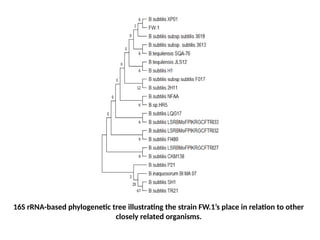
    16S rRNA-based phylogenetictree illustrating the strain FW.1’s place in relation to other closely related organisms.
  • 22.
    Analysis of saccharidesin coconut water C, F and food waste 1 by HPLC: Refractive index Retention time ( Min ) Fructose Glucose Sucrose Chromatographic separation of fructose, glucose and sucrose in comparison with CW.C, CW.F and FW.1, comprising 0.9 mg/mL Inulin as a standard.
  • 23.
    Fructose Sucrose Retention time (Min ) Refractive index CW.C Chromatogram showing the presence of Fructose and Sucrose in CW.C
  • 24.
    CW.F Sucrose Fructose Retention time (Min ) Refractive index Chromatogram showing the presence of Fructose and Sucrose in CW.F
  • 25.
    FW.1 Fructose Retention time (Min ) Refractive index Chromatogram showing the presence of Fructose in FW.1
  • 26.
    Conclusion • This workhas developed a step-by-step methodology for the screening and optimization of Bacillus and Klebsiella strains for the synthesis of inulinases, using food waste and coconut water samples. • Bacillus subtilis (FW.1) and Klebsiella pneumonia (CW.C and CW.F) were the two strains that were found to have good potential for producing inulinase from food waste and coconut water residues during screening experiments. • This study demonstrated the high efficiency of Bacillus subtilis (FW.1) in the production of inulinase using locally accessible food waste sources, and Klebsiella pneumonia (CW.C and CW.F) growing on coconut water which can be very helpful in the commercial production of inulinase from such locally available sources and can be economically profitable. • Enzyme activity for these samples were conducted using DNS and found out that FW.1 had the highest concentration of fructose compared to the rest. • And by TLC, Endoinulinase activity was observed in CW.C, CW.F and FW.1. TLC showed the hydrolysis of inulin to produce Fructose and Glucose
  • 27.
    Future perspective • Bioconversionof inulin to 2,3-butanediol using inulinase- producing Klebsiella pneumoniae
  • 28.
    References • Chi, Z.-M.,Zhang, T., Cao, T.-S., Liu, X.-Y., Cui, W., & Zhao, C.-H. (2011). Biotechnological potential of inulin for bioprocesses. Bioresource Technology, 102(6), 4295–4303. https://doi.org/10.1016/j.biortech.2010.12.086 • de Paula, F. C., Cazetta, M. L., Monti, R., & Contiero, J. (2008). Sucrose hydrolysis by gelatin-immobilized inulinase from Kluyveromyces marxianus var. Bulgaricus. Food Chemistry, 111(3), 691–695. https://doi.org/10.1016/j.foodchem.2008.04.039 • Gao, L., Chi, Z., Sheng, J., Ni, X., & Wang, L. (2007). Single-cell protein production from Jerusalem artichoke extract by a recently isolated marine yeast Cryptococcus aureus G7a and its nutritive analysis. Applied Microbiology and Biotechnology, 77(4), 825–832. https://doi.org/10.1007/s00253-007-1210-7 • Kuzuwa, S., Yokoi, K., Kondo, M., Kimoto, H., Yamakawa, A., Taketo, A., & Kodaira, K.-I. (2012). Properties of the inulinase gene levH1 of Lactobacillus casei IAM 1045; cloning, mutational and biochemical characterization. Gene, 495(2), 154–162. https://doi.org/10.1016/j.gene.2011.12.004 • Liebl, W., Brem, D., & Gotschlich, A. (1998). Analysis of the gene for β-fructosidase (invertase, inulinase) of the hyperthermophilic bacterium Thermotoga maritima, and characterisation of the enzyme expressed in Escherichia coli. Applied Microbiology and Biotechnology, 50(1), 55–64. https://doi.org/10.1007/s002530051256