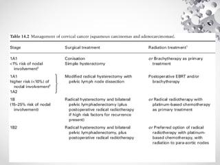
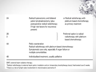
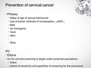
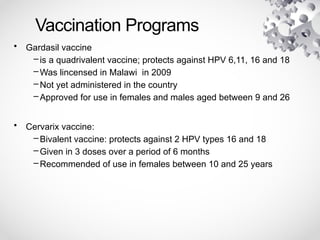
CERVICAL CANCER
CHN
Outline
• Epidemiology
• Pathophysiology
• Risk factors
• Presentation
• Investigations
• Staging
• Management
• Prevention
• Screening methods
Epidemiology
• About 2314 cervical cancer deaths occur
annually in Malawi (2012 estimations) (1)
• Cervical cancer ranks as first cause of female
cancer deaths(1)
• Cervical cancer is the first leading cause of
cancer deaths in Malawi aged between 15-44
years in Malawi (1)
Pathophysiology of cervical cancer
Pathophysiology continued…
• Ectocervix covered by squamous stratified
epithelium
• Canal of cervix lined by columnar epithelium;
one cell thick
• The two meet at Squamocolumnar Junction
(SCJ)
• Position of SCJ varies; lying in cervical os during
infancy but rolls onto ectocervix in puberty,
exposing it to delicate acid environment of
vagina ---> squamous cell metaplasia
Pathophysiology continued…
• Transformation zone
PATHOPHYSIOLOGY continued…
• Dysplasia
–Metaplasia can be interrupted by other factors
such as HPV infection coupled with other risk
factors  dysplastic epithelium
–Dysplastic epithelium has immature cells with
large nuclei, variable size and shape; and
actively dividing
• Dysplasias referred to as Cervical
Intraepithelial Neoplasia (CIN)- histological
diagnosis.
AETIOLOGY OF CERVICAL CANCER
• Human Papilloma Virus (HPV), types 16,18, 33,
35, 45, 52, 58 associated and proven to cause
cervical cancer
• HPV-DNA detected in 95% to 100% of adequate
specimens of cervical cancer, supporting the
claim that HPV is the necessary cause. (J Natl
Cancer Inst Monogr. 2003;(31):3-13)
TYPES OF CERVICAL CANCERS
• Two main types of cervical cancer:
–Squamous cell carcinoma: most common
–Adenocarcinoma
–Adenosquamous
–Small cell undifferentiated
Risk factors
• HPV infection; necessary but not sufficient
cause
• Other factors:
–Smoking
–Tobacco smoking,
–High parity
–Sex with multiple partners, early sexual debut,
promiscuous partner
–Long-term hormonal contraceptive use
–Co-infection with HIV
–Immunosuppresion
–Genetic
Presentation
• Abnormal vaginal bleeding (postcoital,
intermenstrual, postmenopausal or
menorrhagia)
• Vaginal discharge, sometimes
malodorous.
• Symptoms and signs of anaemia
• Dysuria
Physical Exam
• General exam
• Pelvic mass
• Bimanual palpation
• Speculum exam
• Rectal exam
Pretreatment and staging interventions
• Bimanual pelvic examination
• Speculum examination.
• Colposcopy, endocervical curettage, hysteroscopy.
• Conisation of the cervix is regarded as a clinical
examination.
• Cystoscopy or proctoscopy (confirmed by biopsy)
• Intravenous urography.
• X-ray examination of the lungs and skeleton.
• Other investigations;
• full blood count, urea and electrolytes and liver function tests
• computerised tomography scan
MAKING THE DIAGNOSIS
• A tissue biopsy is required to confirm
malignancy and to identify the histological type
of tumour.
• Endocervical curettage (ECC) should be
done as well, to detect any lesions that may be in
the endocervical canal.
FIGO CLASSIFICATION OF CERVICAL CANCER
• Stage 0: Pre- invasive carcinoma
• Stage I: Carcinoma confined to cervix
– Ia: microscopically identified, depth <5mm and <7mm wide
• Ia1: invasion < 3mm and < 7mm wide
• Ia2: Invasion >3cm < 7mm wide
– Ib: Confined to cervix, lesion greater than Ia
• Ib1:lesion < 4cm
• Ib2: lesion >4cm
• Stage II: Carcinoma beyond cervix
– IIa: involving vagina but not lower third,
• IIa1: 4cm or less
• IIa2: > 4cm
– IIb: infiltrating parametrium but not pelvic side wall
FIGO classification continued…
• Stage III: Involving lower third of vagina
and extending to pelvic wall side
–IIIa: Lower third involved
–IIIb: extending to pelvic wall,
hydronephrosis/nonfunctioning kidneys
• Stage IV: Involving other organs
–IVa: Mucosa of bladder or rectum; extension
beyond true pelvis
–IVb: Distant organs
Management of cervical dysplasia
• Ablative methods
–Cryotherapy,
–Electrocautery.
–Laser ablation
• Excional methods
–LEEP
–cone biopsy
–hysterectomy
–Radical hysterectomy plus BLND
Bishop A, et al. Program for Appropriate Technology in Health. 1995
Prevention of cervical cancer
• Primary
• Delay of age of sexual intercourse.
• Use of barrier methods of contraception _x0001_
• Bilat
• ral monogamy
• Vacci
• ation
•
• Seco
ary
• Educa
• ion for cervical screening to target under-screened populations.
• Impro
• ement of sensitivity and specificity of screening for the precursors
Vaccination Programs
• Gardasil vaccine
– is a quadrivalent vaccine; protects against HPV 6,11, 16 and 18
– Was lincensed in Malawi in 2009
– Not yet administered in the country
– Approved for use in females and males aged between 9 and 26
• Cervarix vaccine:
– Bivalent vaccine: protects against 2 HPV types 16 and 18
– Given in 3 doses over a period of 6 months
– Recommended of use in females between 10 and 25 years
CERVICAL
SCREENING
METHODS
What is ‘screening’?
• Definition:
–‘the systematic application of a test or enquiry
to identify individuals in a defined population
who are at sufficient risk of a specific disorder,
but have not sought medical attention on
account of symptoms, to enable them to
benefit from further investigation or direct
preventive action’
Criteria for a good screening Program
• Affordable
• Sensitive
• Specific
• Acceptable (socially and morally)
• Time and resource factors taken into account: Is
screening going to make any difference
Screening methods
• Visual inspection with acetic acid (VIA)
• Visual inspection with acetic acid magnification
• Visual inspection with Lugol’s iodine
• Pap smear
• Human Papilloma Virus DNA
• Speculoscopy
–Inspection with chemiluminiscent light & a low
power magnification following application of
acetic acid
• Cervicography
–Photographs taken by a 35mm specialised
1.Visual inspection with acetic acid
• Promising approach in resource limited
settings
• Application of 3-5% dilute acetic acid on
the cervix
• Detection of opaque acetowhite stain
constitutes a positive test
–Reversible coagulation of intracellular proteins
• Possible results;
–Positive
–
VIA Positive
2.Visual inspection with lugol’s iodine
(vili)
• Similar to Schiller iodine test of 1930s
• VILI findings depend on interaction between iodine &
glycogen
• Glycogen abundant in normal, mature squamous
epithelium
– Little or no glycogen in abnormal epithelium
• A dark mahogany brown cervix equates a negative test
• Colourless, pale or mustard yellow cervix equates a
positive test
Negative VILI
4. Cytology
• Convetional
–Pap smear
• Liquid based
• Cytology + hpv dna testing
–Uses hybrid capture 2 kit to detect high risk
HPV Serotypes in cervical sample
References
• Msyamboza et al. “Burden of cancer in Malawi;
common types, incidence and trends: National
population-based cancer registry”BMC Research
Notes 2012, 5:149
• Mishra G. “An overview of prevention and early
detection of cervical cancer”
• WHO cervical cancer prevention pilot project in 6
African countries
• Handbook of Gynaecology Management. Sylvia
K. Rosevear.
• Clinical gynaecology, TF Kruger, MH Botha

CERVICAL_CANCER.power point presentation.

  • 1.
  • 2.
    Outline • Epidemiology • Pathophysiology •Risk factors • Presentation • Investigations • Staging • Management • Prevention • Screening methods
  • 3.
    Epidemiology • About 2314cervical cancer deaths occur annually in Malawi (2012 estimations) (1) • Cervical cancer ranks as first cause of female cancer deaths(1) • Cervical cancer is the first leading cause of cancer deaths in Malawi aged between 15-44 years in Malawi (1)
  • 4.
  • 5.
    Pathophysiology continued… • Ectocervixcovered by squamous stratified epithelium • Canal of cervix lined by columnar epithelium; one cell thick • The two meet at Squamocolumnar Junction (SCJ) • Position of SCJ varies; lying in cervical os during infancy but rolls onto ectocervix in puberty, exposing it to delicate acid environment of vagina ---> squamous cell metaplasia
  • 6.
  • 7.
    PATHOPHYSIOLOGY continued… • Dysplasia –Metaplasiacan be interrupted by other factors such as HPV infection coupled with other risk factors  dysplastic epithelium –Dysplastic epithelium has immature cells with large nuclei, variable size and shape; and actively dividing • Dysplasias referred to as Cervical Intraepithelial Neoplasia (CIN)- histological diagnosis.
  • 8.
    AETIOLOGY OF CERVICALCANCER • Human Papilloma Virus (HPV), types 16,18, 33, 35, 45, 52, 58 associated and proven to cause cervical cancer • HPV-DNA detected in 95% to 100% of adequate specimens of cervical cancer, supporting the claim that HPV is the necessary cause. (J Natl Cancer Inst Monogr. 2003;(31):3-13)
  • 9.
    TYPES OF CERVICALCANCERS • Two main types of cervical cancer: –Squamous cell carcinoma: most common –Adenocarcinoma –Adenosquamous –Small cell undifferentiated
  • 10.
    Risk factors • HPVinfection; necessary but not sufficient cause • Other factors: –Smoking –Tobacco smoking, –High parity –Sex with multiple partners, early sexual debut, promiscuous partner –Long-term hormonal contraceptive use –Co-infection with HIV –Immunosuppresion –Genetic
  • 11.
    Presentation • Abnormal vaginalbleeding (postcoital, intermenstrual, postmenopausal or menorrhagia) • Vaginal discharge, sometimes malodorous. • Symptoms and signs of anaemia • Dysuria
  • 12.
    Physical Exam • Generalexam • Pelvic mass • Bimanual palpation • Speculum exam • Rectal exam
  • 13.
    Pretreatment and staginginterventions • Bimanual pelvic examination • Speculum examination. • Colposcopy, endocervical curettage, hysteroscopy. • Conisation of the cervix is regarded as a clinical examination. • Cystoscopy or proctoscopy (confirmed by biopsy) • Intravenous urography. • X-ray examination of the lungs and skeleton. • Other investigations; • full blood count, urea and electrolytes and liver function tests • computerised tomography scan
  • 14.
    MAKING THE DIAGNOSIS •A tissue biopsy is required to confirm malignancy and to identify the histological type of tumour. • Endocervical curettage (ECC) should be done as well, to detect any lesions that may be in the endocervical canal.
  • 15.
    FIGO CLASSIFICATION OFCERVICAL CANCER • Stage 0: Pre- invasive carcinoma • Stage I: Carcinoma confined to cervix – Ia: microscopically identified, depth <5mm and <7mm wide • Ia1: invasion < 3mm and < 7mm wide • Ia2: Invasion >3cm < 7mm wide – Ib: Confined to cervix, lesion greater than Ia • Ib1:lesion < 4cm • Ib2: lesion >4cm • Stage II: Carcinoma beyond cervix – IIa: involving vagina but not lower third, • IIa1: 4cm or less • IIa2: > 4cm – IIb: infiltrating parametrium but not pelvic side wall
  • 16.
    FIGO classification continued… •Stage III: Involving lower third of vagina and extending to pelvic wall side –IIIa: Lower third involved –IIIb: extending to pelvic wall, hydronephrosis/nonfunctioning kidneys • Stage IV: Involving other organs –IVa: Mucosa of bladder or rectum; extension beyond true pelvis –IVb: Distant organs
  • 17.
    Management of cervicaldysplasia • Ablative methods –Cryotherapy, –Electrocautery. –Laser ablation • Excional methods –LEEP –cone biopsy –hysterectomy –Radical hysterectomy plus BLND Bishop A, et al. Program for Appropriate Technology in Health. 1995
  • 20.
    Prevention of cervicalcancer • Primary • Delay of age of sexual intercourse. • Use of barrier methods of contraception _x0001_ • Bilat • ral monogamy • Vacci • ation • • Seco ary • Educa • ion for cervical screening to target under-screened populations. • Impro • ement of sensitivity and specificity of screening for the precursors
  • 21.
    Vaccination Programs • Gardasilvaccine – is a quadrivalent vaccine; protects against HPV 6,11, 16 and 18 – Was lincensed in Malawi in 2009 – Not yet administered in the country – Approved for use in females and males aged between 9 and 26 • Cervarix vaccine: – Bivalent vaccine: protects against 2 HPV types 16 and 18 – Given in 3 doses over a period of 6 months – Recommended of use in females between 10 and 25 years
  • 22.
  • 23.
    What is ‘screening’? •Definition: –‘the systematic application of a test or enquiry to identify individuals in a defined population who are at sufficient risk of a specific disorder, but have not sought medical attention on account of symptoms, to enable them to benefit from further investigation or direct preventive action’
  • 24.
    Criteria for agood screening Program • Affordable • Sensitive • Specific • Acceptable (socially and morally) • Time and resource factors taken into account: Is screening going to make any difference
  • 25.
    Screening methods • Visualinspection with acetic acid (VIA) • Visual inspection with acetic acid magnification • Visual inspection with Lugol’s iodine • Pap smear • Human Papilloma Virus DNA • Speculoscopy –Inspection with chemiluminiscent light & a low power magnification following application of acetic acid • Cervicography –Photographs taken by a 35mm specialised
  • 26.
    1.Visual inspection withacetic acid • Promising approach in resource limited settings • Application of 3-5% dilute acetic acid on the cervix • Detection of opaque acetowhite stain constitutes a positive test –Reversible coagulation of intracellular proteins • Possible results; –Positive –
  • 27.
  • 28.
    2.Visual inspection withlugol’s iodine (vili) • Similar to Schiller iodine test of 1930s • VILI findings depend on interaction between iodine & glycogen • Glycogen abundant in normal, mature squamous epithelium – Little or no glycogen in abnormal epithelium • A dark mahogany brown cervix equates a negative test • Colourless, pale or mustard yellow cervix equates a positive test
  • 29.
  • 31.
    4. Cytology • Convetional –Papsmear • Liquid based • Cytology + hpv dna testing –Uses hybrid capture 2 kit to detect high risk HPV Serotypes in cervical sample
  • 33.
    References • Msyamboza etal. “Burden of cancer in Malawi; common types, incidence and trends: National population-based cancer registry”BMC Research Notes 2012, 5:149 • Mishra G. “An overview of prevention and early detection of cervical cancer” • WHO cervical cancer prevention pilot project in 6 African countries • Handbook of Gynaecology Management. Sylvia K. Rosevear. • Clinical gynaecology, TF Kruger, MH Botha