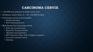
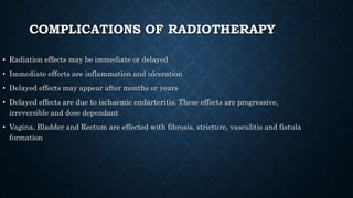
CLASSIFICATION AND DETECTION OF
CERVICAL CANCER BASED ON DEEP
LEARNING ALGORITHMS
Chandran Venkatesan
INTRODUCTION
• Cervical cancer begins on the surface of your cervix. It happens when the cells on your
cervix begin to change to precancerous cells.
• Not all precancerous cells will turn to cancer, but finding these problematic cells and
treating them before they can change is critical to preventing cervical cancer.
• Cervical cancer is the third most common cancer in women worldwide, affecting over
500,000 women and resulting in approximately 275,000 deaths every year.
• cervical cancer is potentially preventable and curable.
• Cervical cancer can be prevented through early administration of the HPV(Human
papillomavirus Vaccines) and regular pap smear screenings, which indicate the presence
of precancerous cells.
• Screening and early treatment can lead to potential cures in about 95% of women at risk
for cervical cancer.
• There are two main types of cancer of the cervix: squamous cell
carcinomas and adenocarcinomas. About 80% to 90% of cervical cancers are squamous
cell carcinomas, while 10% to 20% are adenocarcinomas.
CARCINOMA CERVIX
• 1,25,000 new patients in India every year
• Incidence varies from 15 – 48 / 100,000 women
• Carcinoma cervix is preventable
• Health education
• Screening programmes
• Risk factors for Carcinoma cervix
• Early age at intercourse
• Repeated / Frequent births
• Multiple sexual partners
• HPV infections (Type 16 & 18 highly oncogenic)
• Low socio-economic status
• Smoking
STAGES
Stage I: Cancer is found only in your cervix. It hasn't spread and is small.
Stage II: Cancer has spread beyond your cervix and uterus but hasn't yet spread to your pelvic
wall (the tissues that line the part of the body between your hips) or your vagina.
Stage III: Cancer has spread to the lower part of your vagina and may have spread to your
pelvic wall, ureters (tubes that carry urine) and nearby lymph nodes.
Stage IV: Cancer has spread to your bladder, rectum or other parts of the body like your bones
or lungs
INVESTIGATIONS
• For confirmation of diagnosis
• Biopsy
• From obvious growth or abnormal area
• Directed biopsy in very early lesions
• Cone biopsy
• For staging of disease
• Intravenous Urography
• Abdominal Ultrasonography
• Cystoscopy
• Proctosigmoidoscopy
• Examination under anaesthesia (EUA)
• CT / MRI
• Base line investigations of general condition
COMPLICATIONS OF RADIOTHERAPY
• Radiation effects may be immediate or delayed
• Immediate effects are inflammation and ulceration
• Delayed effects may appear after months or years
• Delayed effects are due to ischaemic endarteritis. These effects are progressive,
irreversible and dose dependant
• Vagina, Bladder and Rectum are effected with fibrosis, stricture, vasculitis and fistula
formation
TREATMENT
• Factors
• Stage of disease
• Age of patient
• General condition / Associated problems
• Tumor configuration
• Modalities
• Surgery
• Radiotherapy
• Combined
• Chemo-radiation

cervical cancer.pptx

  • 1.
    CLASSIFICATION AND DETECTIONOF CERVICAL CANCER BASED ON DEEP LEARNING ALGORITHMS Chandran Venkatesan
  • 2.
    INTRODUCTION • Cervical cancerbegins on the surface of your cervix. It happens when the cells on your cervix begin to change to precancerous cells. • Not all precancerous cells will turn to cancer, but finding these problematic cells and treating them before they can change is critical to preventing cervical cancer. • Cervical cancer is the third most common cancer in women worldwide, affecting over 500,000 women and resulting in approximately 275,000 deaths every year. • cervical cancer is potentially preventable and curable. • Cervical cancer can be prevented through early administration of the HPV(Human papillomavirus Vaccines) and regular pap smear screenings, which indicate the presence of precancerous cells. • Screening and early treatment can lead to potential cures in about 95% of women at risk for cervical cancer. • There are two main types of cancer of the cervix: squamous cell carcinomas and adenocarcinomas. About 80% to 90% of cervical cancers are squamous cell carcinomas, while 10% to 20% are adenocarcinomas.
  • 3.
    CARCINOMA CERVIX • 1,25,000new patients in India every year • Incidence varies from 15 – 48 / 100,000 women • Carcinoma cervix is preventable • Health education • Screening programmes • Risk factors for Carcinoma cervix • Early age at intercourse • Repeated / Frequent births • Multiple sexual partners • HPV infections (Type 16 & 18 highly oncogenic) • Low socio-economic status • Smoking
  • 4.
    STAGES Stage I: Canceris found only in your cervix. It hasn't spread and is small. Stage II: Cancer has spread beyond your cervix and uterus but hasn't yet spread to your pelvic wall (the tissues that line the part of the body between your hips) or your vagina. Stage III: Cancer has spread to the lower part of your vagina and may have spread to your pelvic wall, ureters (tubes that carry urine) and nearby lymph nodes. Stage IV: Cancer has spread to your bladder, rectum or other parts of the body like your bones or lungs
  • 5.
    INVESTIGATIONS • For confirmationof diagnosis • Biopsy • From obvious growth or abnormal area • Directed biopsy in very early lesions • Cone biopsy • For staging of disease • Intravenous Urography • Abdominal Ultrasonography • Cystoscopy • Proctosigmoidoscopy • Examination under anaesthesia (EUA) • CT / MRI • Base line investigations of general condition
  • 6.
    COMPLICATIONS OF RADIOTHERAPY •Radiation effects may be immediate or delayed • Immediate effects are inflammation and ulceration • Delayed effects may appear after months or years • Delayed effects are due to ischaemic endarteritis. These effects are progressive, irreversible and dose dependant • Vagina, Bladder and Rectum are effected with fibrosis, stricture, vasculitis and fistula formation
  • 7.
    TREATMENT • Factors • Stageof disease • Age of patient • General condition / Associated problems • Tumor configuration • Modalities • Surgery • Radiotherapy • Combined • Chemo-radiation