This document provides an overview of cells as the fundamental units of life, highlighting their types, growth, and organelle functions crucial for biotechnological applications. It emphasizes the distinction between prokaryotic and eukaryotic cells, the importance of the cell theory, and the development of micro-electromechanical systems (MEMS) for cell cultivation. Additionally, it explains the potential of these technological advancements in health diagnostics and biotechnology.


![The Cell Theory
In the 1830's, Matthias Schleiden
and Theodor Schwann proposed
a set of hypotheses that came to
be called the cell theory. The cell
theory states
(1) all organisms are composed
of one or more cells
(2) the cell is the structural unit of
life
(3) cells can arise only by division
from a preexisting cell Plant Cell
[Image courtesy of Mariana RuizVillarreal]](https://image.slidesharecdn.com/cellsbuildingblockspresentation-220816061348-af7c81dc/85/Cells_Building_Blocks_Presentation-pdf-9-320.jpg)
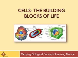
![Prokaryotic and Eukaryotic Cells
Cells vary in the types and complexity of structures found both
internally and externally. However, they can be categorized into two
broad types: prokaryotic and eukaryotic. The structurally simpler
prokaryotic cells include bacteria (Fig 1). The more complex
eukaryotic cells include protists, fungi, animals (Fig 2) and plants (Fig
3). [Bacteria and plant cell graphics courtesy of Mariana Ruiz Villarreal]
Bacteria Animal Cell Plant Cell](https://image.slidesharecdn.com/cellsbuildingblockspresentation-220816061348-af7c81dc/85/Cells_Building_Blocks_Presentation-pdf-10-320.jpg)


![Organelles
Two organelles,
mitochondria and
chloroplasts, function
specifically to supply
energy to cells.
Nearly all eukaryotic cells
contain mitochondria.
Chloroplasts are found only
in photosynthetic cells such
as plants and algae.
Plant Cell showing organelles, Cytoplasm,
Plasma Membrane and Cell Wall
[Image courtesy of Mariana Ruiz Villarreal]](https://image.slidesharecdn.com/cellsbuildingblockspresentation-220816061348-af7c81dc/85/Cells_Building_Blocks_Presentation-pdf-17-320.jpg)
![Organelles
Organelles are found
within the cytoplasm of
the cell.
For animal cells, the
boundary of the cell is
defined by the plasma
membrane.
For plant cells, the
boundary is defined by
the plasma membrane
plus a cell wall composed
of cellulose (see graphic).
Plant Cell showing organelles, Cytoplasm,
Plasma Membrane and Cell Wall
[Image courtesy of Mariana Ruiz Villarreal]](https://image.slidesharecdn.com/cellsbuildingblockspresentation-220816061348-af7c81dc/85/Cells_Building_Blocks_Presentation-pdf-18-320.jpg)

![Using MEMS for Cell Cultivation
MEMS are being develop and testing for use in cell cultivation.
BioPOETS at the University of California, Berkeley has
developed a MEMS cell culture array (left image). The array uses
microfluidics to create an optimal micro-environment for cell
cultivation (inset). The right images are cells that have been
cultivated in this array. [Images courtesy of BioPOETS Lab, UC-Berkeley]](https://image.slidesharecdn.com/cellsbuildingblockspresentation-220816061348-af7c81dc/85/Cells_Building_Blocks_Presentation-pdf-20-320.jpg)


